On electrophoresis or HPLC, beta thalassemia trait has a raised HbA2 (above 3.5%, typically 4–6%), while adult alpha thalassemia trait looks normal and needs DNA testing. Severe forms show their own patterns: HbH (β4) in HbH disease, Hb Bart's (γ4) in newborns and hydrops, and HbF up to 95% in beta thalassemia major.
The genetics explain the difference. Alpha thalassemia is usually caused by deletions of some of the four alpha-globin genes (HBA1 and HBA2, two on each chromosome 16); beta thalassemia by point mutations and other small variants in the two HBB genes, one on each chromosome 11. Both traits cause microcytosis with a normal or high red cell count and normal iron studies.
Symptoms follow the missing chain. Alpha chains are part of both fetal (α2γ2) and adult (α2β2) hemoglobin, so the most severe alpha form, Hb Bart's hydrops fetalis, begins before birth. Beta chains matter once fetal hemoglobin gives way to HbA, so beta thalassemia major appears at 6–24 months. Carriers of either have little or no anemia.
Alpha vs beta thalassemia electrophoresis: patterns by type and age
HPLC and capillary electrophoresis are now the usual methods; read HbA, HbA2 (α2δ2), HbF (α2γ2) and any extra band.
| Condition | HbA | HbA2 | HbF | Extra band |
|---|---|---|---|---|
| Normal adult | 96–98% | 2–3% | Under 1% | None |
| α silent carrier (–α/αα) | Normal | Normal | Normal | Hb Bart's 1–3%, at birth only |
| α trait (– –/αα or –α/–α) | 96–98% | Normal or low (1.5–3.0%) | Under 1% | Hb Bart's 3–8%, at birth only |
| HbH disease (– –/–α) | 60–90% | Low (under 2%) | Under 1% | HbH, usually 3–30%; Hb Bart's above 15% at birth |
| Hb Bart's hydrops (– –/– –) | None | None | None | Hb Bart's 80–90%, Hb Portland 10–15% |
| β trait (minor) | 92–95% | Above 3.5% (typically 4–6%) | 0.5–4% | None |
| β intermedia (NTDT) | 10–50% | Above 4% | 10–50% | None |
| β major (TDT) | None (β0/β0) to about 30% | Variable, often raised | Up to 95% | None |
- Beta minor vs major: in trait, HbA still dominates and the clue is HbA2. In major, HbA is absent or scant and HbF carries the load (92–95% in β0/β0).
- HbA2 can mislead: HbE, HbC and HbO-Arab run with HbA2 on alkaline electrophoresis, and an "HbA2" above 10% suggests a variant. B12 or folate deficiency (megaloblastic anemia) and hyperthyroidism raise HbA2; a coinherited δ-thalassemia can normalize a carrier's HbA2.
- Newborns: HbF dominates at birth and HbA takes over by about 6 months. On the newborn screen, Hb Bart's flags alpha thalassemia (more Bart's, more genes lost), and absent HbA is diagnostic of β0 thalassemia. Beta patterns apply after 12 months; before that, β+ disease overlaps the normal infant range, so DNA testing is used.
Alpha thalassemia trait vs beta thalassemia trait
| Feature | Alpha thalassemia trait | Beta thalassemia trait |
|---|---|---|
| Genotype | Two α genes lost: – –/αα (cis, α0) or –α/–α (trans) | One HBB variant (β0 or β+) |
| MCV and MCH | About 72 fL and 23 pg | Often 60–70 fL and 19–23 pg |
| RBC count | Normal or high | High for the degree of anemia |
| Smear | Microcytes, hypochromia, mild anisopoikilocytosis | Microcytes, hypochromia, target cells, basophilic stippling |
| HbA2 | Normal or low (1.5–3.0%) | Above 3.5% |
| HbF | Under 1% | 0.5–4%; mildly raised in about 30% |
| Confirmed by | DNA testing (gap-PCR, then MLPA or sequencing) | Hemoglobin quantitation; DNA not required |
Target cells are not specific: they also occur in HbC disease, liver disease and after splenectomy.
Why alpha trait electrophoresis is normal in adults
- Two working α genes still make enough α chains: HbA stays at 96–98%, and HbA2, which also needs α chains, is normal or slightly low.
- The excess-chain tetramers don't show. Hb Bart's (γ4) is a newborn-period finding that fades as γ chains give way to β chains; HbH (β4) is 0% in trait and needs three missing genes.
- Analyzers can't reliably detect deletional alpha carriers, and silent carriers often aren't even microcytic. So alpha trait is suspected from microcytosis with normal iron studies and HbA2, then confirmed by DNA.
- The Thalassaemia International Federation (TIF) advises DNA testing for α0 trait when both partners have an MCH under 25 pg after iron deficiency is excluded; beta trait in one partner can mask α0 trait.
Alpha vs beta thalassemia for USMLE: genes, symptoms and timing
| Feature | Alpha thalassemia | Beta thalassemia |
|---|---|---|
| Genes | Four: HBA1 and HBA2 on each chromosome 16 | Two: HBB on each chromosome 11 |
| Usual defect | Deletions; non-deletional variants (such as Hb Constant Spring) are rarer and more severe | Point mutations and small variants (almost 300 known); large deletions are rare |
| Unpaired chain | γ4 (Hb Bart's) in the fetus and newborn; β4 (HbH) after birth | Free α chains precipitate in red cell precursors: ineffective erythropoiesis and hemolysis |
| Severe form appears | Before birth (hydrops); HbH disease usually in the first years | 6–24 months, as HbF falls |
| Symptoms | Trait: none. HbH: hemolytic anemia, jaundice, splenomegaly, gallstones, crises with infection or oxidant drugs | Trait: none. Major: pallor, poor growth, jaundice, hepatosplenomegaly, bone changes. Intermedia: later, milder |
| Common in | Southeast Asia (cis deletions), Africa (trans), Mediterranean, Middle East, India | Mediterranean, Middle East, India, Southeast Asia, Africa |
| Trait electrophoresis | Normal | HbA2 above 3.5% |
Both are autosomal recessive: two β carriers have a 25% risk of an affected child in each pregnancy, while the alpha risk depends on cis vs trans (below). Memory hook: Bart's for babies, H for hemolysis.
Alpha thalassemia: count the missing genes
| α genes lost | Genotype | Name | Hemoglobin pattern | Clinical picture |
|---|---|---|---|---|
| 1 | –α/αα | Silent carrier | Normal | Healthy; indices normal or near normal |
| 2 | – –/αα (cis) or –α/–α (trans) | α-thalassemia trait (minor) | Normal adult pattern | Mild microcytosis, little or no anemia |
| 3 | – –/–α | HbH disease | HbH 3–30%, HbA2 under 2% | Hemolytic anemia (Hb around 9.5–11 g/dL), splenomegaly, gallstones, iron overload even without transfusion; crises with infection or oxidant drugs |
| 4 | – –/– – | Hb Bart's hydrops fetalis | Hb Bart's 80–90%, no HbA or HbF | Hydrops (edema, effusions, heart failure), hepatosplenomegaly; large hypochromic red cells with many nucleated red cells; maternal preeclampsia and preterm delivery |
- Why tetramers fail: Hb Bart's and HbH have very high oxygen affinity, so they deliver little oxygen (see the oxygen-hemoglobin curve). HbH is also unstable and precipitates as inclusions in 5–80% of red cells on brilliant cresyl blue stain.
- Non-deletional HbH (for example with Hb Constant Spring) is more severe: it is often diagnosed at 1.5–3.5 years, and about a third of patients become transfusion dependent.
- Hydrops can be treated: two or more intrauterine transfusions, started as early as possible (about 18 weeks at most centers), can resolve it; survivors stay transfusion dependent unless transplanted.
Cis vs trans: the counseling question
- Cis (– –/αα, α0): common in Southeast Asia (the – –SEA deletion), where Hb Bart's syndrome occurs in 0.5–5 per 1,000 births; – –MED is the usual α0 deletion around the Mediterranean and Middle East. Two cis carriers have a 25% risk of Hb Bart's hydrops in each pregnancy.
- Trans (–α/–α): the –α3.7 allele reaches frequencies of 0.30–0.40 in equatorial Africa, while cis deletions are reported only very rarely in North Africa and African Americans. Two trans carriers can pass on only –α chromosomes, so every child is –α/–α and none can have Hb Bart's hydrops.
Beta thalassemia: minor, intermedia, major, and NTDT vs TDT
| Type | Genetics | Hb and electrophoresis | Clinical picture |
|---|---|---|---|
| Minor (trait) | One HBB variant | Hb normal or mildly low; HbA2 above 3.5%; HbF under 5% | No symptoms; often mistaken for iron deficiency |
| Intermedia (NTDT) | Two variants, at least one mild (β+), or a severe genotype softened by modifiers | Hb 7–10 g/dL; HbF 10–50% | Presents later; not regularly transfused; gallstones, splenomegaly, leg ulcers, pulmonary hypertension, thrombosis, extramedullary masses, iron overload from absorption |
| Major (TDT, Cooley anemia) | Two severe variants (for example β0/β0) | Hb under 7 g/dL; HbF up to 95%; HbA very low or absent | Onset at 6–24 months; smear with target and teardrop cells, basophilic stippling, many nucleated red cells; lifelong transfusion |
- β0 vs β+: nonsense, frameshift and some splice variants abolish β-chain output (β0); promoter, intron, poly(A)-signal and untranslated-region variants reduce it (β+). How these break mRNA processing is in the RNA processing cheat sheet.
- Modifiers: coinherited alpha thalassemia or hereditary persistence of fetal hemoglobin makes beta thalassemia milder, because fewer α chains are left unpaired; extra α genes (triplication) make it worse.
NTDT vs TDT: the terms the guidelines now use
- TIF classifies by transfusion need. TDT: regular, lifelong transfusion is needed for survival (usually β-thalassemia major and severe HbE/β-thalassemia). NTDT: it is not, though transfusions may be needed for a time (β-thalassemia intermedia, mild to moderate HbE/β-thalassemia; GeneReviews adds HbH disease).
- TIF counts a patient as TDT after 6 or more red cell units in 6 months (gaps of 6 weeks or less) or frequent transfusions for over a year. The labels are fluid, trait is not NTDT, and "intermedia" fell out of favor because it can make disease sound milder than it is.
Thalassemia trait vs iron deficiency: the Mentzer index and its limits
| Clue | Thalassemia trait | Iron deficiency |
|---|---|---|
| RBC count | Normal or high | Reduced |
| Mentzer index (MCV ÷ RBC in millions/µL) | Below 13 | 13 or above |
| RDW | Normal or mildly high | High, with overlap |
| Ferritin | Normal or high | Low (the best initial test) |
| Serum iron, transferrin saturation | Normal | Low; zinc protoporphyrin high |
| Iron therapy | Doesn't fix the microcytosis | Corrects the anemia |
- A screen, not a diagnosis. None of the 40-plus such indices is fully sensitive and specific; a meta-analysis of 99 studies found they work better in adults than children, with RDW alone the weakest. In 434 Saudi children, the Mentzer index was 74% sensitive and 63% specific for beta trait. Both conditions can also coexist.
- The HbA2 pitfall: iron deficiency can lower HbA2 in a beta carrier, though it usually stays in the carrier range; in exceptional cases very severe deficiency pushes it into the normal range, and then TIF advises correcting the iron before repeating HbA2. One HPLC study found no such effect; sources disagree, but treat-then-retest is the safe answer.
- No empiric iron: give iron only for proven deficiency; GeneReviews lists inappropriate iron therapy among things to avoid in HbH disease.
- Other microcytic anemias: anemia of chronic disease, sideroblastic anemia (ring sideroblasts, no raised HbA2 or HbF) and lead poisoning.
Complications: marrow expansion, crew-cut skull, extramedullary hematopoiesis and iron overload
- Marrow expansion (untreated or undertransfused TDT, and NTDT): frontal bossing, malar prominence, a depressed nasal bridge and maxillary hypertrophy exposing the upper teeth (the "chipmunk" face), long-bone deformities, osteoporosis and fractures.
- Skull X-ray: a widened diploic space and vertical trabeculae give the "hair-on-end" or "crew-cut" look (Merck calls it "sun-ray"). One review puts it at about 8% of patients; it is rare before age 5 and prevented by adequate early transfusion. Sickle cell disease can cause it too.
- Extramedullary hematopoiesis: hepatosplenomegaly first, then paraspinal masses, mostly thoracic, which can compress the cord. It affects up to 15–20% of patients with NTDT but 1–5% with TDT; new masses are an indication to start regular transfusion.
- Iron overload: from transfusion in TDT; from absorption in NTDT and HbH disease, because ineffective erythropoiesis releases erythroferrone, which suppresses hepcidin (compare hemochromatosis). It damages the heart (arrhythmias, dilated cardiomyopathy; once blamed for 71% of deaths in β-thalassemia major), liver (fibrosis, cirrhosis, hepatocellular carcinoma) and endocrine glands (growth failure, hypogonadism, diabetes, hypothyroidism, hypoparathyroidism). TIF advises ferritin at least every 3 months and yearly MRI of liver iron and cardiac T2* from age 8–10.
- Others: pigment gallstones, leg ulcers, pulmonary hypertension and venous thrombosis (more common in NTDT and after splenectomy), and parvovirus B19 aplastic crises.
Treatment and FDA-approved therapies (October 2026)
- Trait: no treatment; offer genetic counseling, and give iron only for proven deficiency.
- TDT: red cell transfusions every 2–5 weeks, keeping the pretransfusion Hb at 9.5–10.5 g/dL (10–11 g/dL with heart disease or extramedullary masses) and the post-transfusion Hb below 13–15 g/dL. Chelation usually starts after the first 10–20 transfusions or as ferritin approaches 1,000 ng/mL.
- NTDT and HbH disease: folic acid, transfusion when needed (infection, pregnancy), chelation for absorptive overload, cholecystectomy for biliary colic, and in HbH disease no oxidant drugs (the G6PD deficiency list). Splenectomy is for selected patients only, because it raises the risk of thrombosis and infection.
| Chelator | Route | Label warnings to know |
|---|---|---|
| Deferoxamine | Slow SC infusion over 8–12 hours, IM, or slow IV | Auditory and ocular toxicity, growth suppression in children at high doses, Yersinia and mucormycosis |
| Deferasirox | Oral, once daily | Boxed: renal failure, hepatic failure, GI hemorrhage; marrow suppression; also approved for NTDT from age 10 |
| Deferiprone | Oral | Boxed: agranulocytosis and neutropenia (monitor the ANC); zinc deficiency |
| Therapy | What it is | FDA status (October 2026) |
|---|---|---|
| Allogeneic stem cell transplant | Donor marrow or cord blood, ideally from an HLA-identical sibling, after myeloablative conditioning | Curative; offered early, before iron damage |
| Betibeglogene autotemcel (Zynteglo) | The patient's own stem cells, given a working β-globin gene (βA-T87Q) by a lentiviral vector | Aug 2022: adults and children with β-thalassemia who need regular transfusions |
| Exagamglogene autotemcel (Casgevy) | The patient's own stem cells, CRISPR-edited at the BCL11A erythroid enhancer to raise HbF; myeloablative (for example busulfan) conditioning | Jan 2024: TDT from age 12; July 2026: extended to ages 2 to 11 |
| Luspatercept (Reblozyl) | Erythroid maturation agent that traps TGF-β superfamily ligands; SC every 3 weeks | Since 2019: anemia in adults with β-thalassemia who need regular transfusions; not for urgent correction; not approved for NTDT; warnings for thrombosis, hypertension, extramedullary masses |
| Mitapivat (Aqvesme) | Oral pyruvate kinase activator | Dec 2025: anemia in adults with α- or β-thalassemia, transfusion dependent or not; boxed warning for hepatocellular injury; REMS only |
USMLE traps: classic teaching vs current evidence
| Trap | What's actually true |
|---|---|
| Adult alpha trait shows HbH or Hb Bart's | Its electrophoresis is normal; Bart's is a newborn finding, and HbH needs three missing genes |
| A normal electrophoresis rules out thalassemia | Not for alpha trait, silent β variants, δ-thalassemia coinheritance or severe iron deficiency |
| Mentzer index below 13 diagnoses trait | It's a screen; confirm with iron studies, HbA2 and DNA |
| Any raised HbA2 means beta trait | Consider HbE, HbC or HbO-Arab overlap, B12 or folate deficiency, hyperthyroidism, antiretroviral therapy |
| Two parents with alpha trait risk hydrops | Only if both carry cis (– –/αα) deletions |
| Beta thalassemia major presents at birth | It presents at 6–24 months; Hb Bart's hydrops is the one that starts in utero |
| "Intermedia" and "major" guide treatment | Guidelines classify by transfusion need (NTDT vs TDT), and patients can move between them |
More heme topics are on the Hematology and Oncology hub.
Worked USMLE vignettes
Vignette 1: the incidental CBC. A healthy 24-year-old man of Greek descent has Hb 11.4 g/dL, MCV 64 fL, RBC count 6.1 million/µL, normal RDW and ferritin 85 ng/mL; the smear shows target cells. HbA2 is 5.4% and HbF 1.6%. Which best explains his findings?
A. Impaired δ-aminolevulinate synthase; B. A variant in one HBB allele; C. Warm autoantibody hemolysis; D. Folate deficiency; E. Deletion of two α-globin genes
Answer: B, β-thalassemia minor. Marked microcytosis with mild anemia, a high RBC count (Mentzer 64 ÷ 6.1 ≈ 10.5), normal ferritin and HbA2 above 3.5% fit beta trait. A: sideroblastic anemia has ring sideroblasts and high iron, without a raised HbA2. C: warm autoimmune hemolysis has spherocytes and a positive direct Coombs test. D: folate deficiency is macrocytic. E: alpha trait has a normal or low HbA2.
Vignette 2: the preconception visit. A 27-year-old Vietnamese woman planning pregnancy has Hb 12.0 g/dL, MCV 70 fL, MCH 22 pg, RBC count 5.6 million/µL and ferritin 60 ng/mL. HPLC shows HbA2 2.3%, HbF under 1% and no abnormal bands. Her Vietnamese husband has similar indices. What is the best next step?
A. Oral iron for 3 months; B. Repeat HPLC in 6 months; C. Reassurance; D. DNA testing of both partners for α-globin deletions; E. Bone marrow biopsy
Answer: D. Microcytosis with normal iron studies and HbA2 suggests alpha trait, which HPLC can't confirm. If both carry a cis deletion such as – –SEA, each pregnancy has a 25% risk of Hb Bart's hydrops. A: she isn't iron deficient. B: a repeat won't reveal a deletion. C: reassurance misses the hydrops risk. E: the marrow adds nothing.
Vignette 3: the febrile crisis. A 7-year-old boy of Thai descent develops jaundice and dark urine during a febrile illness. He has splenomegaly; Hb is 7.9 g/dL (baseline 9.8), MCV 58 fL, reticulocytes 7%. Brilliant cresyl blue shows many inclusions, and HPLC shows an early-eluting peak of 14% with HbA2 1.4%. Which genotype is most likely?
A. – –/–α; B. – –/αα; C. β0/β0; D. Hemizygous G6PD variant; E. ANK1 variant
Answer: A, HbH disease. Three missing α genes leave excess β chains that form unstable HbH (the early peak and the inclusions); infection triggers hemolytic crises. B: alpha trait has a normal pattern and no hemolysis. C: β-thalassemia major presents in infancy with HbF up to 95%. D: G6PD deficiency gives Heinz bodies and bite cells with a normal MCV. E: hereditary spherocytosis gives spherocytes, not marked microcytosis.
Vignette 4: the pale infant. A 9-month-old of Cypriot parents has pallor, poor feeding and hepatosplenomegaly. Hb is 5.8 g/dL, MCV 59 fL; the smear shows target cells and many nucleated red cells. HPLC shows HbF 93% and no HbA. What is the most appropriate long-term management?
A. Oral iron; B. Splenectomy now; C. Hydroxyurea alone; D. Luspatercept; E. Regular transfusions to a pretransfusion Hb of 9.5–10.5 g/dL, with chelation later
Answer: E. This is β0/β0 thalassemia major (TDT), surfacing as HbF falls. Regular transfusion prevents marrow expansion and growth failure; chelation follows after 10–20 transfusions, and an HLA-identical sibling would make transplant an option. A: iron worsens overload. B: splenectomy is deferred in young children because of sepsis risk. C: hydroxyurea has a limited role. D: luspatercept is approved only for adults.
Key takeaways
- Electrophoresis: HbA2 above 3.5% means beta trait; adult alpha trait looks normal and needs DNA testing. HbH (β4) = three α genes lost; Hb Bart's (γ4) = newborn or hydrops; HbF up to 95% = beta major.
- Genetics and timing: alpha = mostly deletions of 4 genes on chromosome 16, with the most severe form in utero; beta = mostly point mutations in 2 genes on chromosome 11, with beta major at 6–24 months.
- Cis deletions (Southeast Asia) carry the hydrops risk; trans deletions (Africa) don't.
- Mentzer index below 13 favors trait but is only a screen; very severe iron deficiency can mask a raised HbA2, so treat it and retest.
- Treatment follows transfusion need (TDT vs NTDT): transfusion, chelation, transplant, gene therapy (Zynteglo; Casgevy from age 2), luspatercept (adults with TDT), mitapivat (adults, alpha or beta).
Sources
- Alpha-Thalassemia — GeneReviews (2026)
- Beta-Thalassemia — GeneReviews (2026)
- Genetic Basis, Pathophysiology, and Diagnosis — TIF TDT Guidelines, 5th ed. (2025)
- Iron Overload and Chelation — TIF TDT Guidelines, 5th ed. (2025)
- Other Complications — TIF TDT Guidelines, 5th ed. (2025)
- Musallam KM, et al. TIF Guidelines for Transfusion-Dependent β-Thalassemia — HemaSphere (2025)
- Guidelines for Non-Transfusion-Dependent β-Thalassaemia, 3rd ed. — TIF (2023)
- Epidemiology, Pathophysiology and Diagnosis of α-Thalassaemia — TIF α-Thalassaemia Guidelines (2023)
- Screening and Diagnosis for Haemoglobin Disorders — TIF, Prevention of Thalassaemias, Vol. 1 (2013)
- Thalassemias — Merck Manual Professional Edition (2026)
- Thalassemia — StatPearls (2023)
- Beta Thalassemia — StatPearls (2023)
- Baird DC, et al. Alpha- and Beta-thalassemia: Rapid Evidence Review — Am Fam Physician (2022)
- Hemoglobinopathies: Current Practices for Screening, Confirmation and Follow-up — APHL (2025)
- Hoffmann JJ, et al. Discriminant Indices for Thalassemia and Iron Deficiency: A Meta-Analysis — Clin Chem Lab Med (2015)
- Althumairi A, et al. Mentzer Index in Saudi Children With Microcytosis — Front Med (2024)
- Amid A, et al. Mentzer Index and Iron Deficiency Effect on HbA2 — Hemoglobin (2015)
- Gökçe E, Beyhan M. Radiological Findings of Calvarial Lesions — World J Radiol (2025)
- Sunil A, et al. Beta-Thalassemia With a Review of Skeletal Manifestations — Cureus (2023)
- ZYNTEGLO (betibeglogene autotemcel) — US FDA (2022)
- CASGEVY (exagamglogene autotemcel) — US FDA (2026)
- REBLOZYL (luspatercept-aamt) label — DailyMed (2026)
- AQVESME (mitapivat) label — DailyMed (2026)
- FDA Approves First Oral Treatment for Anemia in Thalassemia — US FDA (2026)
- EXJADE (deferasirox) label — DailyMed (2026)
- FERRIPROX (deferiprone) label — DailyMed (2026)
- DESFERAL (deferoxamine) label — DailyMed (2025)
Visual explanations from the StepGenie QBank
Infographics StepGenie drew to explain questions in its own QBank, matched to this article's topic. Each caption is the takeaway of the question it explains.
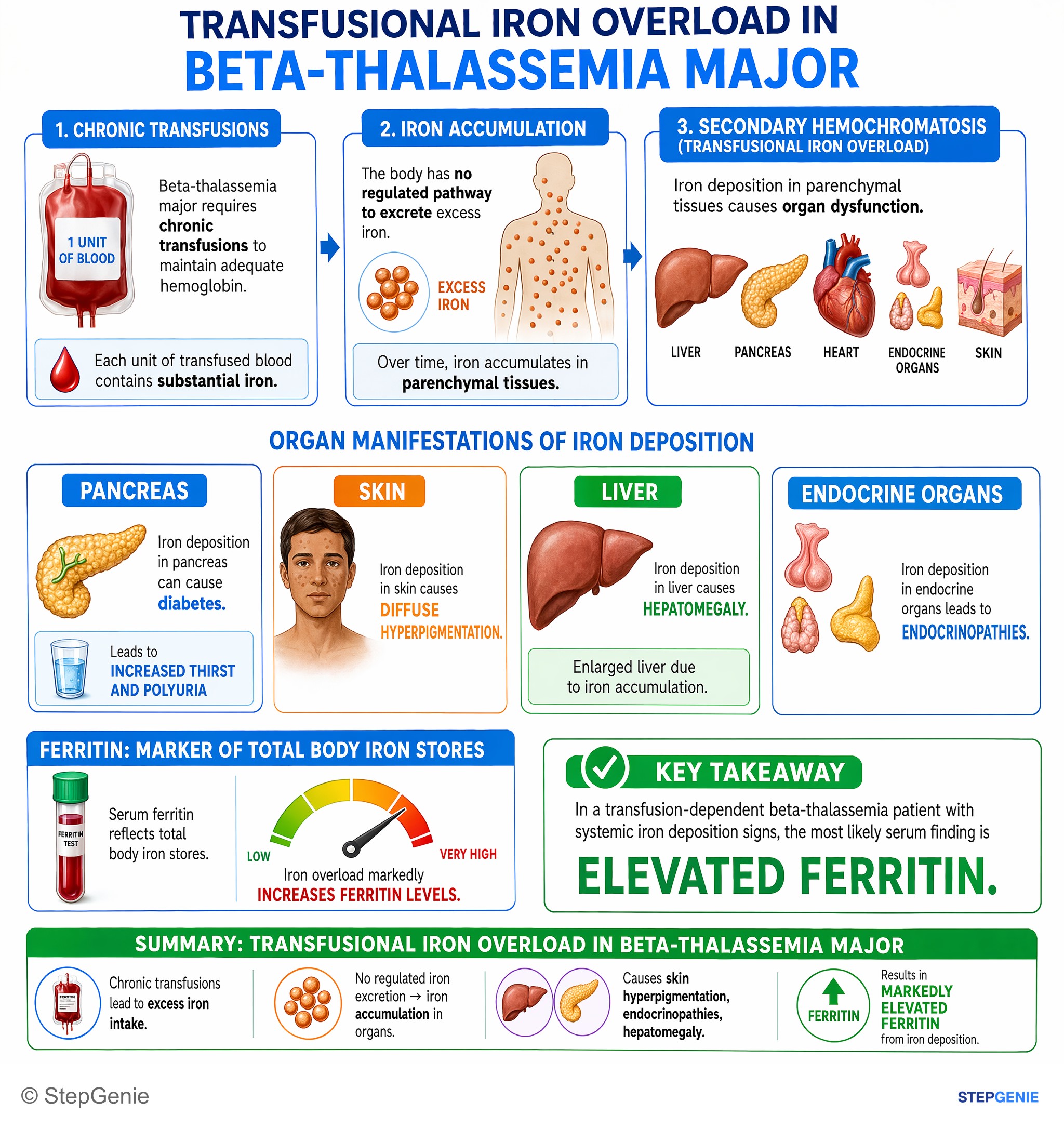
Transfusional iron overload in beta-thalassemia major causes skin hyperpigmentation, endocrinopathies, hepatomegaly and markedly elevated…
USMLE Step 1Frequently Asked Questions
How do alpha and beta thalassemia differ on hemoglobin electrophoresis?
Beta thalassemia trait shows a raised HbA2, above 3.5 percent and typically 4 to 6 percent, while HbA stays above about 90 percent. Adult alpha thalassemia trait usually shows a normal pattern with a normal or low HbA2, so it is confirmed by DNA testing. HbH disease shows a fast HbH band, Hb Bart's appears at birth or in hydrops fetalis, and beta thalassemia major shows HbF up to 95 percent.
How do you tell alpha thalassemia trait from beta thalassemia trait?
Both cause microcytosis with little or no anemia, a normal or high red cell count and normal iron studies. Beta thalassemia trait has an HbA2 above 3.5 percent, sometimes with a slightly raised HbF, and hemoglobin quantitation is enough to diagnose it. Alpha thalassemia trait has a normal or low HbA2 and no abnormal band in adults, so it needs DNA testing for alpha-globin deletions, such as gap-PCR.
What is the difference between alpha and beta thalassemia?
Alpha thalassemia is reduced alpha-globin production, usually from deletions of some of the four alpha-globin genes on chromosome 16. Beta thalassemia is reduced or absent beta-globin production, usually from point mutations in the two beta-globin genes on chromosome 11. Because alpha chains are part of both fetal and adult hemoglobin, the most severe alpha form starts before birth, while beta thalassemia major appears at 6 to 24 months as fetal hemoglobin falls.
What are the symptoms of alpha thalassemia vs beta thalassemia?
Carriers of either usually have no symptoms and at most a mild microcytic anemia. The most severe alpha form, Hb Bart's hydrops fetalis, starts before birth with fetal edema, effusions and heart failure. HbH disease usually appears in early childhood with hemolytic anemia, jaundice, splenomegaly and gallstones, plus crises during infections. Beta thalassemia major appears at 6 to 24 months with pallor, poor growth, jaundice and hepatosplenomegaly, plus bone changes from marrow expansion if transfusion is inadequate.
What does electrophoresis show in beta thalassemia minor vs major?
In beta thalassemia minor, HbA is still about 92 to 95 percent of the total, HbA2 is above 3.5 percent and HbF is under 5 percent. In beta thalassemia major, HbA is absent in beta-zero homozygotes or very low, HbF makes up most of the hemoglobin, up to about 95 percent, and HbA2 is variable. These patterns apply after 12 months of age; younger infants may need DNA testing.
How do you tell thalassemia trait from iron deficiency anemia?
Thalassemia trait typically shows marked microcytosis with a normal or high red cell count, a normal or mildly raised RDW and a normal ferritin, while iron deficiency shows a low ferritin and a reduced red cell count. A Mentzer index, the MCV divided by the red cell count in millions, below 13 favors trait, but it is only a screen. Very severe iron deficiency can push HbA2 into the normal range, so correct it and retest.