Sideroblastic anemia is an iron-utilization anemia: heme synthesis fails, so iron that cannot be inserted into protoporphyrin IX piles up in the mitochondria of marrow erythroblasts and forms ring sideroblasts on Prussian blue (Perls) iron stain. Iron studies are the mirror image of iron deficiency: high serum iron, transferrin saturation and ferritin with a normal TIBC. The most common inherited form is X-linked (ALAS2 mutations) and microcytic; acquired causes are myelodysplastic neoplasm with an SF3B1 mutation (usually normocytic or macrocytic), alcohol, isoniazid and other drugs, vitamin B6 or copper deficiency (for example from excess zinc), and lead.
The question
A 54-year-old man has 3 months of progressive fatigue and shortness of breath on exertion. He has alcohol use disorder and drinks 6-8 beers daily. Four months ago he started isoniazid for latent tuberculosis; pyridoxine was not prescribed. He is pale. Hemoglobin is 8.9 g/dL, MCV 72 fL and RDW increased. Serum iron and ferritin are increased, TIBC is normal and transferrin saturation is 78%. The blood smear shows a dimorphic population of hypochromic microcytes and normal-sized red cells, some containing small granules. Blood lead concentration is normal. A bone marrow aspirate stained with Prussian blue shows erythroid precursors with iron granules encircling the nucleus.
Which of the following best explains this patient's anemia?
A. Decreased synthesis of globin chains B. Depleted iron stores from chronic blood loss C. Hepcidin-mediated trapping of iron in macrophages D. Impaired δ-aminolevulinic acid synthesis due to pyridoxal phosphate deficiency E. Lead inhibition of δ-aminolevulinic acid dehydratase and ferrochelatase F. Impaired DNA synthesis from folate deficiency
Answer and every choice explained
Answer: D. This is acquired sideroblastic anemia. Pyridoxal phosphate (active vitamin B6) is the cofactor for ALA synthase, which joins glycine and succinyl-CoA to make δ-aminolevulinic acid (δ-ALA), the first and rate-limiting step of heme synthesis (ALAS2 is the red-cell form). Isoniazid inactivates pyridoxine, and alcohol use disorder both causes B6 deficiency and acts as a marrow toxin. Without heme, iron keeps entering erythroblast mitochondria with nowhere to go: ring sideroblasts in the marrow, iron overload on blood tests. Dimorphic red cells explain the high RDW.
Management: stop alcohol, give pyridoxine and stop or replace the offending drug; the anemia can recover once the cause is removed. Do not give iron. Most people taking isoniazid should receive pyridoxine anyway (Merck suggests 30-50 mg/day), to prevent neuropathy as well as anemia (see the tuberculosis breakdown).
- A. Globin chain synthesis (thalassemia): microcytic, often with a high red cell count and target cells; beta-thalassemia minor shows Hb A2 above 3.5%. Ring sideroblasts and iron overload are not features of thalassemia trait, and this anemia began in middle age with a clear trigger. Compare in the thalassemia table.
- B. Iron deficiency: ferritin below 30 ng/mL, transferrin saturation below 10%, high TIBC and absent marrow iron, the opposite of this patient. Microcytosis with a high ferritin is not iron deficiency (see the iron deficiency anemia breakdown).
- C. Hepcidin (anemia of chronic disease): inflammation raises hepcidin, so serum iron and transferrin saturation are low or normal while ferritin is above 100 ng/mL; the anemia is often normocytic. A high serum iron and a saturation of 78% rule it out (anemia of chronic disease).
- E. Lead: lead does block δ-ALA dehydratase and ferrochelatase, can produce basophilic stippling and ring sideroblasts, and causes a normocytic or microcytic anemia. But the blood lead level is normal, and the stem gives no exposure history, abdominal pain, constipation or neuropathy. Whole blood lead level is the diagnostic test.
- F. Folate deficiency: alcohol tempts you toward folate, but folate (or B12) deficiency causes a macrocytic, megaloblastic anemia and no ring sideroblasts.
Iron studies: the microcytic anemia grid
| Finding | Iron deficiency | Anemia of chronic disease | Thalassemia trait | Sideroblastic anemia |
|---|---|---|---|---|
| MCV | Low | Often normal, can be low | Low, with a high RBC count | Low in inherited forms; normal or high in MDS |
| RDW | High | Normal or slightly high | Often normal | High (dimorphic cells) |
| Serum iron | Low | Normal or low | Normal | High |
| TIBC | High | Normal or low | Normal | Normal |
| Transferrin saturation | Below 10% | Normal or low | Normal | Above 50% |
| Ferritin | Below 30 ng/mL | Above 100 ng/mL | Normal | Above 400 ng/mL |
| Marrow iron | Absent | Present | Present | Increased, ring sideroblasts |
| Smear clue | Hypochromic microcytes | Unremarkable | Target cells | Dimorphic cells, stippled cells, Pappenheimer bodies |
| Confirm with | Ferritin, then find the source | Clinical context | Hb electrophoresis (Hb A2 above 3.5% in beta trait); DNA testing for alpha trait | Marrow iron stain; SF3B1 testing in older adults |
Iron deficiency, anemia of chronic disease and sideroblastic values are from Merck's microcytic-anemia table; the thalassemia column is the standard exam pattern.
What a ring sideroblast is
- An erythroblast with 5 or more iron granules encircling at least one-third of the nucleus, seen on Prussian blue (Perls) stain of a marrow aspirate.
- The granules are iron-loaded mitochondria, which sit around the nucleus.
- Don't confuse it with Pappenheimer bodies (iron granules inside circulating red cells, called siderocytes) or basophilic stippling (fine blue dots on a routine smear; common but not specific in lead poisoning, and also seen in sideroblastic anemia and thalassemia).
Heme synthesis: where each cause hits
| Step | Enzyme | Disrupted by |
|---|---|---|
| Glycine + succinyl-CoA → δ-ALA (mitochondria, rate-limiting) | ALA synthase (ALAS2 in red cells), needs pyridoxal phosphate | ALAS2 mutation, B6 deficiency, isoniazid, alcohol |
| δ-ALA → porphobilinogen | δ-ALA dehydratase (porphobilinogen synthase) | Lead |
| Fe²⁺ + protoporphyrin IX → heme (mitochondria) | Ferrochelatase | Lead: zinc is inserted instead, raising zinc protoporphyrin |
Because lead blocks δ-ALA dehydratase, the enzyme that consumes δ-ALA (and less heme means less feedback restraint on ALA synthase), δ-ALA builds up and can be found in excess in the urine.
Causes of ring sideroblasts and the clue that gives each away
| Cause | Classic clue | Key point | Treatment |
|---|---|---|---|
| X-linked sideroblastic anemia (ALAS2) | Boy or young man with microcytic anemia and iron overload; carrier women can develop it later in life | Most common inherited form | Pyridoxine (often a partial response); treat iron overload |
| MDS with SF3B1 mutation | Older adult, normocytic or macrocytic anemia, usually lower-risk MDS; with thrombocytosis, think MDS/MPN with SF3B1 mutation and thrombocytosis | Somatic splicing-factor mutation | Transfusion, erythropoiesis-stimulating agents, luspatercept, iron chelation |
| Alcohol | Heavy drinking | Marrow toxin and cause of B6 deficiency | Abstinence |
| Drugs: isoniazid, linezolid, chloramphenicol, cycloserine, pyrazinamide | TB or long antibiotic course | Isoniazid and cycloserine inactivate pyridoxine | Stop or replace the drug; pyridoxine |
| Copper deficiency | Excess zinc, bariatric surgery or malabsorption; anemia with neutropenia and a B12-like myeloneuropathy | Zinc blocks copper absorption | Stop zinc, give oral copper (copper deficiency mnemonic) |
| Lead | Old paint, ceramic glaze, moonshine, retained bullets; abdominal pain, neuropathy, cognitive deficits | Blocks δ-ALA dehydratase and ferrochelatase | Remove the source; chelation for high levels |
Untreated iron overload damages the heart and liver, so patients who are iron-overloaded need chelation, or phlebotomy if the anemia allows.
MDS with SF3B1: the current names
- WHO 5th edition (2022) renamed myelodysplastic syndromes myelodysplastic neoplasms and defined MDS with low blasts and SF3B1 mutation (MDS-SF3B1): an SF3B1 mutation, fewer than 5% marrow and 2% blood blasts, and no 5q deletion, monosomy 7 or complex karyotype. It covers more than 90% of MDS with 5% or more ring sideroblasts.
- Ring sideroblasts of 15% or more can substitute for the mutation; "MDS with low blasts and ring sideroblasts" is the accepted term for SF3B1-wild-type cases. The old "MDS/MPN with ring sideroblasts and thrombocytosis" is now MDS/MPN with SF3B1 mutation and thrombocytosis.
- ICC (2022) uses "MDS with mutated SF3B1", defined by the mutation (variant allele fraction 10% or more, no RUNX1 co-mutation) rather than the ring sideroblast count.
- Luspatercept is an erythroid maturation agent that traps TGF-β superfamily ligands. Its FDA label covers anemia in very low- to intermediate-risk MDS in patients who have not had an erythropoiesis-stimulating agent and may need regular transfusions, and in MDS with ring sideroblasts (or MDS/MPN-RS-T) after such an agent fails, when 2 or more red cell units are needed over 8 weeks. It is also approved for transfusion-dependent beta-thalassemia.
More on MDS in the myelodysplastic syndromes breakdown.
How USMLE tests it
- "Microcytic anemia with high iron and high ferritin" is the classic exam signal for sideroblastic anemia. In practice, inherited forms are usually microcytic, while MDS-related forms are usually normocytic or macrocytic, often with a dimorphic smear.
- INH plus a new anemia or neuropathy: think pyridoxine.
- High ferritin alone is not enough: anemia of chronic disease also has high ferritin, but serum iron and saturation are low.
- Copper deficiency mimics B12 deficiency (anemia plus myeloneuropathy) and adds neutropenia; ask about zinc.
- Never treat ring sideroblasts with iron.
Key takeaways
- Sideroblastic anemia = defective heme synthesis with iron trapped in erythroblast mitochondria: ring sideroblasts on Prussian blue stain.
- Labs: high serum iron, transferrin saturation above 50%, high ferritin, normal TIBC, high RDW from dimorphic cells.
- Inherited: X-linked ALAS2 (microcytic, pyridoxine-responsive in part). Acquired: MDS with SF3B1 mutation (normocytic or macrocytic), alcohol, isoniazid and other drugs, B6 or copper deficiency (zinc excess), lead.
- ALA synthase needs pyridoxal phosphate; lead blocks δ-ALA dehydratase and ferrochelatase.
- Treat the cause: stop alcohol and drugs, give pyridoxine or copper, treat MDS (ESAs, luspatercept, transfusion); manage iron overload, and never give iron.
- WHO 2022: MDS with low blasts and SF3B1 mutation; ICC 2022: MDS with mutated SF3B1.
Sources
- Sideroblastic Anemias — Merck Manual Professional Edition (2025)
- Iron Deficiency Anemia — Merck Manual Professional Edition (2025)
- Thalassemias — Merck Manual Professional Edition (2026)
- Vitamin B6 Deficiency and Dependency — Merck Manual Professional Edition (2026)
- Copper Deficiency — Merck Manual Professional Edition (2025)
- Lead Poisoning — Merck Manual Professional Edition (2025)
- Toxicological Profile for Lead — Agency for Toxic Substances and Disease Registry (2020)
- X-linked sideroblastic anemia — MedlinePlus Genetics
- The 5th edition of the WHO Classification of Haematolymphoid Tumours: Myeloid and Histiocytic/Dendritic Neoplasms — Leukemia (2022)
- Comparison of the 2022 WHO classification and International Consensus Classification in myelodysplastic syndromes/neoplasms — Blood Cancer Journal (2024)
- Myelodysplastic Neoplasms (MDS) with Ring Sideroblasts or SF3B1 Mutations: The Improved Clinical Utility of World Health Organization and International Consensus Classification 2022 Definitions — Current Oncology (2024)
- Reblozyl (luspatercept) prescribing information — DailyMed (2026)
Visual explanations from the StepGenie QBank
Infographics StepGenie drew to explain questions in its own QBank, matched to this article's topic. Each caption is the takeaway of the question it explains.
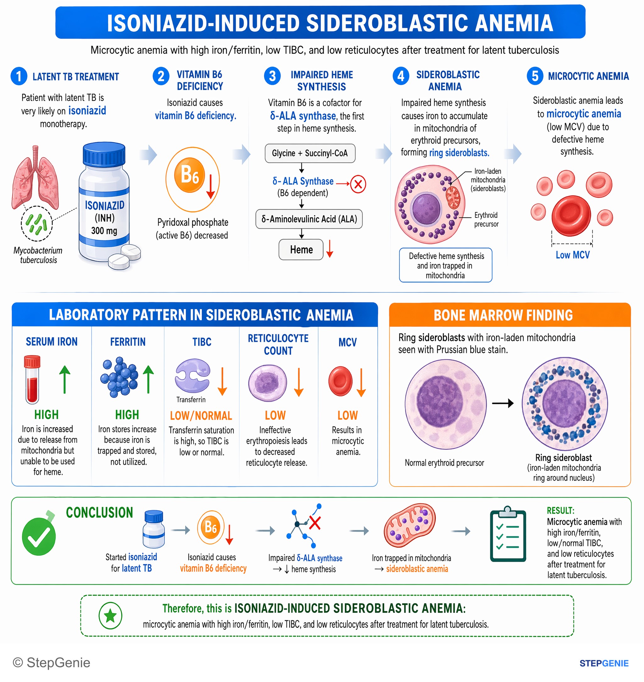
Isoniazid-induced sideroblastic anemia: microcytic anemia with high iron/ferritin, low TIBC, and low reticulocytes after treatment for…
USMLE Step 2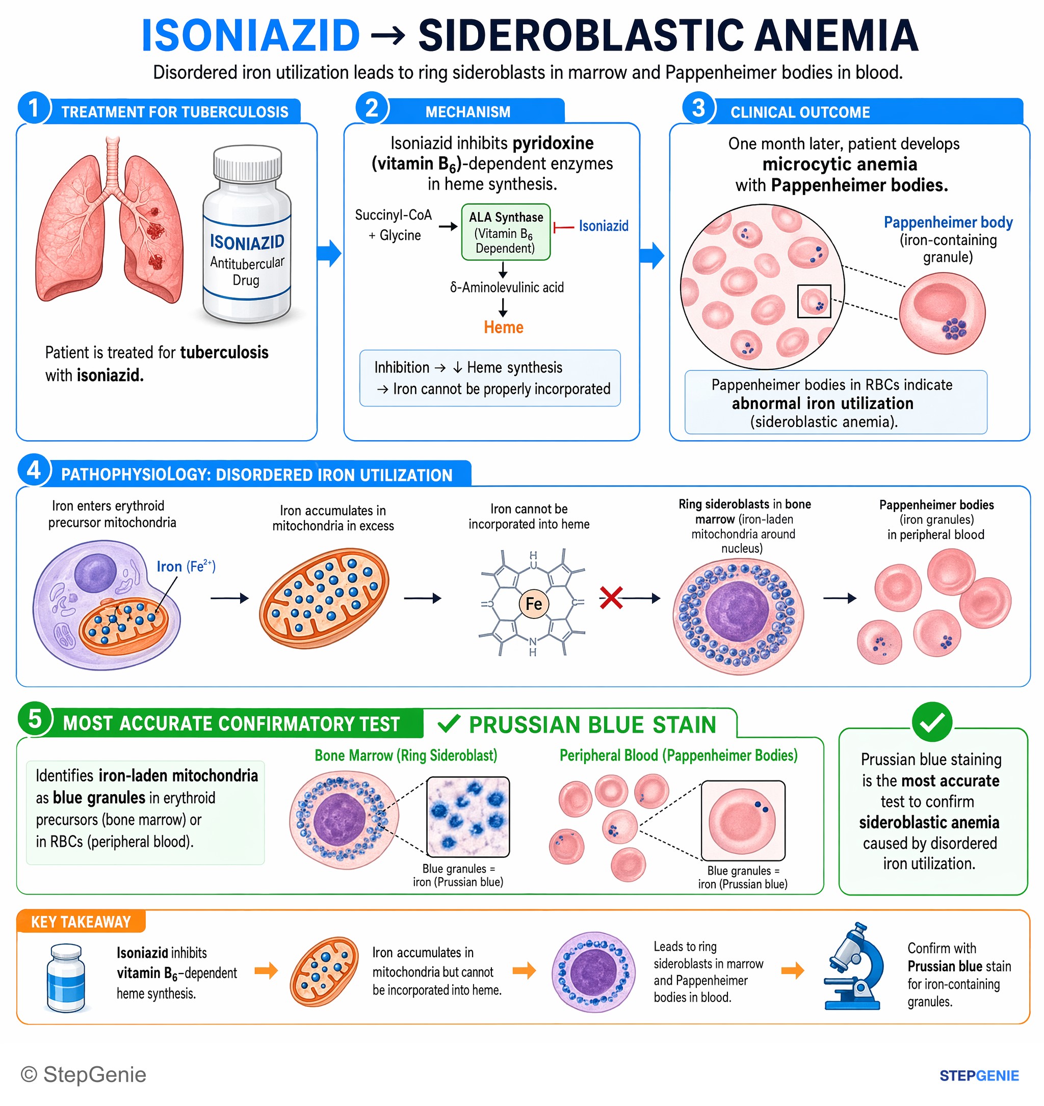
Sideroblastic anemia from disordered iron utilization shows Pappenheimer bodies; confirm with Prussian blue stain for iron-containing…
USMLE Step 1Frequently Asked Questions
What are ring sideroblasts?
Ring sideroblasts are red cell precursors (erythroblasts) in the bone marrow whose mitochondria are packed with iron that could not be built into heme. Because those mitochondria sit around the nucleus, a Prussian blue (Perls) iron stain shows a ring of iron granules; the formal definition is at least 5 granules encircling at least one-third of the nucleus. They define sideroblastic anemia and are absent in iron deficiency and anemia of chronic disease.
What do iron studies show in sideroblastic anemia?
Iron studies show iron overload rather than deficiency: a high serum iron, transferrin saturation above 50% and ferritin often above 400 ng/mL, with a normal TIBC. The RDW is high because small and normal-sized red cells circulate together. This separates it from iron deficiency (low ferritin, high TIBC, saturation below 10%) and from anemia of chronic disease, where serum iron and saturation are low or normal while ferritin is high.
What causes sideroblastic anemia?
Inherited forms are less common than acquired ones, and the most common inherited form is X-linked sideroblastic anemia from ALAS2 mutations. Acquired causes include myelodysplastic neoplasms with SF3B1 mutations in older adults, alcohol, drugs such as isoniazid, linezolid, chloramphenicol, cycloserine and pyrazinamide, vitamin B6 deficiency, copper deficiency (for example from excess zinc or bariatric surgery) and lead. Removing the toxin or drug, or replacing pyridoxine or copper, can reverse the acquired types.
Why does isoniazid cause sideroblastic anemia?
Isoniazid inactivates pyridoxine (vitamin B6). Its active form, pyridoxal phosphate, is the cofactor for ALA synthase, which makes delta-aminolevulinic acid from glycine and succinyl-CoA in the first, rate-limiting step of heme synthesis. With too little heme, iron accumulates in erythroblast mitochondria and forms ring sideroblasts. Pyridoxine treats it, and most people taking isoniazid are given pyridoxine routinely, which also prevents peripheral neuropathy.
Is sideroblastic anemia microcytic or macrocytic?
It depends on the cause. Inherited forms such as X-linked sideroblastic anemia are usually microcytic and hypochromic, which is why exam questions list sideroblastic anemia with the microcytic anemias. Acquired sideroblastic anemia from myelodysplastic neoplasms is usually normocytic or macrocytic. Many patients have two red cell populations, small hypochromic cells mixed with normal or large cells, so the RDW is high even when the MCV is normal.
What is MDS with SF3B1 mutation?
It is a myelodysplastic neoplasm, usually lower risk, defined by a mutation in the splicing-factor gene SF3B1 with fewer than 5% marrow blasts and no 5q deletion, monosomy 7 or complex karyotype. The 2022 WHO classification calls it MDS with low blasts and SF3B1 mutation and lets 15% or more ring sideroblasts substitute for the mutation; the ICC calls it MDS with mutated SF3B1. Anemia is treated with transfusion, erythropoiesis-stimulating agents or luspatercept.