Myelodysplastic syndromes (MDS) show up in Q-banks the same way they show up in clinic: subtle, confusing, and loaded with “almost right” answer choices. The trick isn’t just recognizing MDS—it’s knowing why each distractor is not MDS based on a few high-yield clues (age, cytopenias, smear, marrow cellularity, and key genetic associations).
Tag: Heme/Onc > WBC Disorders & Lymphomas
The Clinical Vignette (Q-bank style)
A 72-year-old man presents with progressive fatigue and occasional gum bleeding for 3 months. He has no recent infections. Past history includes treatment with chemotherapy and radiation for Hodgkin lymphoma 10 years ago. Exam shows conjunctival pallor and scattered petechiae.
Labs:
- Hemoglobin: 8.9 g/dL
- Platelets: 62,000/µL
- WBC: 2,400/µL (ANC low)
- MCV: 108 fL
- Reticulocyte count: low
Peripheral smear: macro-ovalocytes, hypogranular neutrophils, and neutrophils with bilobed nuclei.
Bone marrow biopsy: hypercellular marrow with dysplastic precursors.
Question: What is the most likely diagnosis?
Correct Answer: Myelodysplastic syndromes (MDS)
Why this is MDS
This vignette stacks classic MDS clues:
- Older adult (typically >60)
- Cytopenias in multiple cell lines (anemia + thrombocytopenia + leukopenia)
- Macrocytosis with low retic count (ineffective hematopoiesis)
- Dysplasia on smear
- Hypogranular neutrophils
- Pseudo–Pelger-Huët anomaly (bilobed neutrophils)
- Macro-ovalocytes
- Hypercellular marrow despite low peripheral counts → ineffective hematopoiesis
- History of chemo/radiation → therapy-related MDS (a high-yield association)
The core mechanism (Step-friendly)
MDS is a clonal stem cell disorder causing ineffective hematopoiesis → marrow makes cells, but they die off / fail maturation → cytopenias in the blood.
High-yield extras to know
- Major risk: progression to AML
- Common genetics: deletions like del(5q) (classically associated), del(7q), trisomy 8
- Therapy-related MDS: classically associated with prior exposure to alkylating agents and radiation, often years later
Why Every Other Answer Choice Is Wrong (and how they try to trick you)
Below are common distractors that Q-banks love because they share one feature with MDS (cytopenias, older age, abnormal smear, marrow changes)—but differ in a key discriminator.
Distractor 1: Aplastic anemia
Why it seems plausible
- Pancytopenia can look similar
- Low retic count also fits
Why it’s wrong here
The marrow in aplastic anemia is hypocellular and fatty, not hypercellular.
Key distinction:
- MDS: cytopenias + hypercellular marrow (ineffective hematopoiesis)
- Aplastic anemia: cytopenias + hypocellular marrow (hematopoietic failure)
Classic causes of aplastic anemia (high-yield):
- Autoimmune, drugs (e.g., chloramphenicol), viruses (parvovirus can cause pure red cell aplasia), benzene, radiation (can cause both, but marrow cellularity differentiates)
Distractor 2: Vitamin B12 deficiency (megaloblastic anemia)
Why it seems plausible
- Macrocytosis (MCV 108)
- Can cause pancytopenia in severe cases
- Hypersegmented neutrophils can be seen
Why it’s wrong here
The smear in MDS shows dysgranulopoiesis (hypogranular neutrophils) and Pseudo–Pelger-Huët (bilobed neutrophils), not classic B12 findings.
B12 deficiency smear clues:
- Hypersegmented neutrophils (≥5 lobes)
- Macro-ovalocytes (overlaps!)
- Often neurologic symptoms (posterior column + lateral corticospinal tract) depending on stem
Confirmatory testing (high-yield):
- B12 deficiency: ↑ methylmalonic acid, ↑ homocysteine
- Folate deficiency: normal methylmalonic acid, ↑ homocysteine
Exam trick:
If the stem emphasizes dysplasia + older age + prior chemo/radiation, lean MDS over nutritional deficiency.
Distractor 3: Acute myeloid leukemia (AML)
Why it seems plausible
- Can follow MDS (MDS → AML is a major concept)
- Cytopenias and marrow abnormalities can overlap
Why it’s wrong here
This stem points to dysplasia without mentioning blasts.
Diagnostic line in the sand:
- AML: myeloblasts in marrow (or blood) or defining genetic abnormalities
- MDS: dysplasia + cytopenias, typically <20% blasts
AML peripheral smear buzzwords (high-yield):
- Auer rods
- Myeloblasts
- DIC association with APL (t(15;17), PML-RARA)
Exam trick:
If you’re not given blast percentage, look for descriptions like “numerous blasts,” “Auer rods,” or severe acute presentation (infections/bleeding with blasts).
Distractor 4: Chronic lymphocytic leukemia (CLL)
Why it seems plausible
- Older patient
- Cytopenias can happen (marrow infiltration or autoimmune hemolysis)
Why it’s wrong here
CLL is a lymphoid malignancy and usually presents with lymphocytosis, lymphadenopathy, and smear findings like smudge cells—not dysplastic neutrophils and macrocytosis.
CLL high-yield:
- CD5+ CD23+ B cells
- Hypogammaglobulinemia → infections
- Autoimmune hemolytic anemia risk
- Smudge cells on smear
Distractor 5: Hairy cell leukemia
Why it seems plausible
- Can cause pancytopenia
- Older-ish demographic
Why it’s wrong here
Hairy cell leukemia is classically associated with:
- Massive splenomegaly
- “Dry tap” on marrow aspiration (fibrosis)
- Hairy cytoplasmic projections on smear
- TRAP positivity (older boards) and BRAF mutation associations (commonly taught)
This stem instead highlights dysplasia and therapy-related risk.
Quick Comparison Table: MDS vs Common Look-Alikes
| Condition | Typical Age | CBC Pattern | Marrow Cellularity | Smear Clues | Classic Associations |
|---|---|---|---|---|---|
| MDS | >60 | Cytopenias, often macrocytosis | Hypercellular (ineffective hematopoiesis) | Pseudo–Pelger-Huët, hypogranular neutrophils, dysplasia | Prior chemo/radiation; del(5q); AML risk |
| Aplastic anemia | Any | Pancytopenia | Hypocellular | No dysplasia (often bland smear) | Autoimmune, drugs, benzene, viral |
| B12 deficiency | Any (often older) | Macrocytic anemia ± pancytopenia | Hypercellular with megaloblasts | Hypersegmented neutrophils | Pernicious anemia, malabsorption |
| AML | Any (older common) | Cytopenias + blasts | Hypercellular | Blasts, Auer rods | blasts; APL → DIC |
| CLL | Older | Lymphocytosis | Infiltrative | Smudge cells | CD5+ CD23+; hypogammaglobulinemia |
USMLE High-Yield Takeaways (What to remember under time pressure)
-
Think MDS when you see:
- Older adult + cytopenias + macrocytosis + low retic
- Dysplastic neutrophils (Pseudo–Pelger-Huët, hypogranularity)
- Hypercellular marrow (ineffective hematopoiesis)
- Prior chemo/radiation history
-
The single best differentiator vs aplastic anemia: marrow cellularity
- MDS = hypercellular
- Aplastic = hypocellular
-
The single best differentiator vs B12 deficiency: type of neutrophil abnormality
- MDS = pseudo–Pelger-Huët, hypogranular
- B12 = hypersegmented
-
Always keep the progression in mind: MDS → AML (but AML requires blasts)
Visual explanations from the StepGenie QBank
Infographics StepGenie drew to explain questions in its own QBank, matched to this article's topic. Each caption is the takeaway of the question it explains.
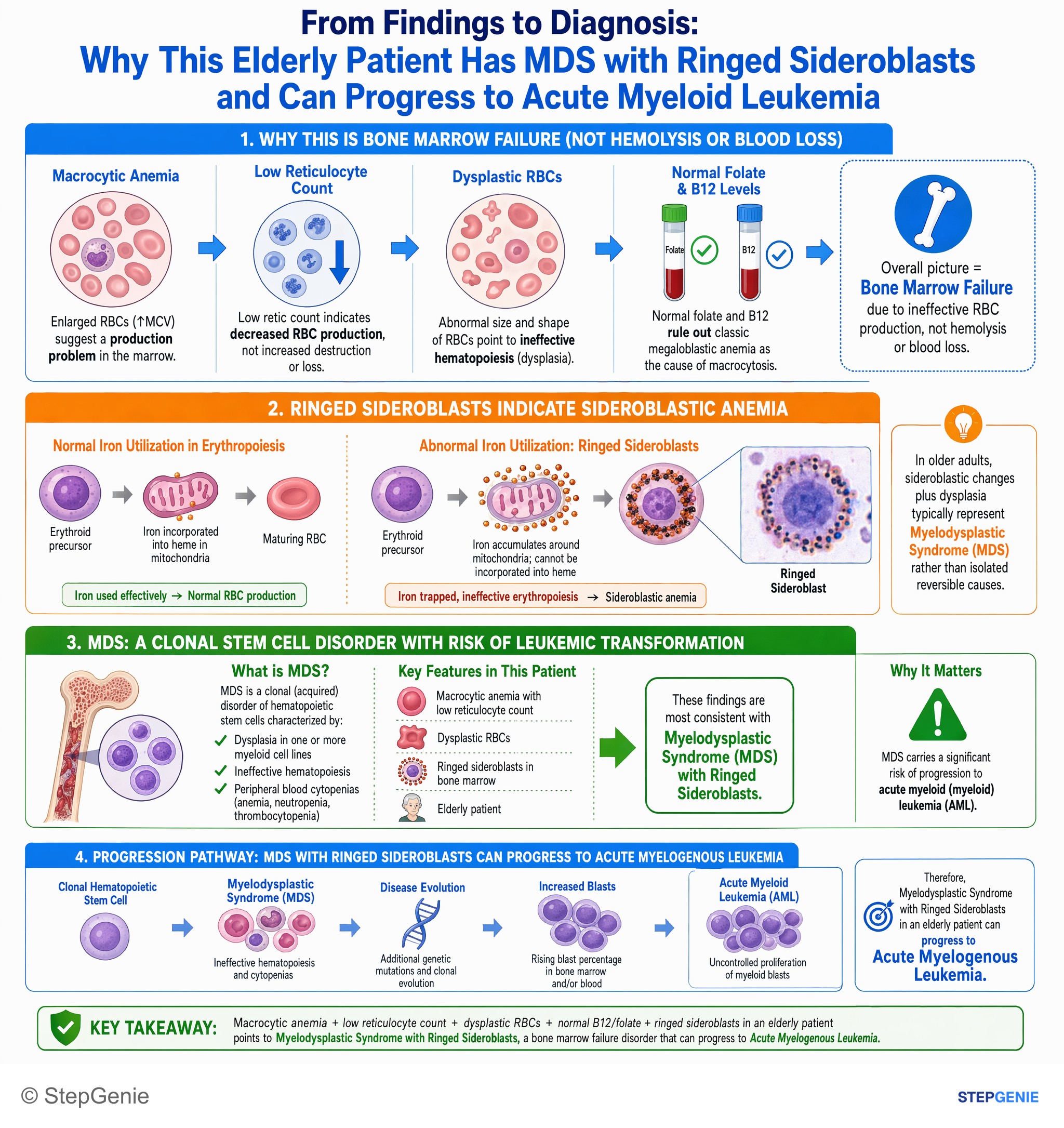
Myelodysplastic syndrome with ringed sideroblasts in an elderly patient can progress to acute myelogenous leukemia.
USMLE Step 1Frequently Asked Questions
How do you tell myelodysplastic syndromes from aplastic anemia on a question stem?
The key distinction is bone marrow cellularity. Myelodysplastic syndromes cause cytopenias with a hypercellular marrow because hematopoiesis is ineffective, so cells are produced but fail to mature properly or die off. Aplastic anemia can also cause pancytopenia and a low reticulocyte count, but the marrow is hypocellular and fatty, reflecting true hematopoietic failure rather than dysplastic production.
Why does myelodysplastic syndrome cause low blood counts even when the bone marrow is hypercellular?
Myelodysplastic syndrome causes cytopenias because it is a clonal stem cell disorder with ineffective hematopoiesis. The marrow is making cells, but the precursors are dysplastic and fail maturation or die off before reaching the peripheral blood. That is why patients can have anemia, thrombocytopenia, and leukopenia despite a hypercellular marrow on biopsy.
What smear findings point to myelodysplastic syndrome instead of vitamin B12 deficiency?
Hypogranular neutrophils and bilobed neutrophils consistent with pseudo-Pelger-Huët anomaly point toward myelodysplastic syndrome. Both conditions can show macro-ovalocytes and macrocytosis, which is why B12 deficiency is a common distractor. The stem favors myelodysplastic syndrome when it emphasizes dysgranulopoiesis, older age, and prior chemotherapy or radiation rather than classic hypersegmented neutrophils and neurologic findings.
How do you tell myelodysplastic syndrome from acute myeloid leukemia in a vignette?
Look for dysplasia without blasts to favor myelodysplastic syndrome, and look for blasts or Auer rods to favor acute myeloid leukemia. The diagnostic cutoff given is that AML has at least 20% myeloblasts in marrow or blood, or defining genetic abnormalities, whereas MDS usually presents with cytopenias and dysplasia with fewer than 20% blasts.
What history makes therapy-related myelodysplastic syndrome more likely?
A history of prior chemotherapy or radiation years earlier strongly supports therapy-related myelodysplastic syndrome. Prior exposure to alkylating agents and radiation is a classic association, often presenting long after treatment. In an older adult with macrocytosis, multilineage cytopenias, dysplastic smear findings, and hypercellular marrow, that treatment history is a major clue toward MDS.