The coagulation cascade turns an injury signal (tissue factor) into a burst of thrombin, which converts fibrinogen into a fibrin mesh that factor XIIIa then cross-links. For exams, split it into the extrinsic pathway (tissue factor + factor VII, measured by the PT/INR), the intrinsic pathway (factors XII, XI, IX and VIII, measured by the aPTT) and the common pathway (X, V, II, I). Inside the body, clotting actually follows the cell-based model: tissue factor-VIIa starts it and the IXa-VIIIa and Xa-Va complexes on activated platelets amplify it, which is why hemophilia bleeds and factor XII deficiency does not. Three quick tips turn this into points: learn the map, read PT vs aPTT as a locator, and pin each anticoagulant to its factor.
Tip 1: Learn the map, then the version that happens in a vessel
Most clotting factors circulate as inactive precursors that are switched on by cleavage, the same logic as the digestive enzymes in zymogen activation.
| Pathway | Factors | Test | Exam hook |
|---|---|---|---|
| Extrinsic | Tissue factor + VII | PT/INR | Factor VII has the shortest half-life, so the PT rises first with warfarin or vitamin K deficiency |
| Intrinsic (contact) | XII, XI, IX, VIII (plus prekallikrein and HMWK) | aPTT | Hemophilia A (VIII) and B (IX); heparin |
| Common | X, V, II (prothrombin), I (fibrinogen) | Both | A deficiency here prolongs both tests |
| Clot stabilization | XIII | Neither | Bleeding with a normal PT and aPTT |
Memory hooks: PT = "Play Tennis outside" (extrinsic) and PTT = "Play Table Tennis inside" (intrinsic). The intrinsic factors read like a price tag, "12 dollars, marked down to 11.98" (XII, XI, IX, VIII); extrinsic is lucky 7; the common pathway counts down like 10-, 5-, 2- and 1-dollar bills (X, V, II, I).
What actually happens in a vessel is the cell-based model (Hoffman and Monroe, 2001):
| Phase | Where | What happens |
|---|---|---|
| Initiation | Tissue factor-bearing cells at the injury | TF-VIIa activates X and IX; Xa with Va makes a small amount of thrombin |
| Amplification | Platelet surface | That thrombin activates platelets and factors V, VIII and XI |
| Propagation | Activated platelets | Tenase (IXa-VIIIa) and prothrombinase (Xa-Va) produce a thrombin burst → fibrin |
Why the model matters on exams:
- Hemophilia A and B bleed even though the extrinsic pathway works. TF-VIIa alone cannot make enough thrombin (Merck), so propagation through IXa-VIIIa is essential.
- Factor XII, prekallikrein and HMWK deficiencies prolong the aPTT but cause no bleeding. The contact factors matter in the test tube, not in the body.
- Factor XI deficiency causes milder bleeding, mainly after surgery or trauma.
Thrombin is the hub. It converts fibrinogen to fibrin, activates V, VIII and XI (positive feedback), activates XIII to cross-link fibrin, and activates platelets. Bound to thrombomodulin on endothelium, it switches sides and activates protein C.
The brakes:
- Antithrombin inhibits thrombin, Xa, XIa and IXa; heparin works by boosting it.
- Activated protein C with protein S cleaves Va and VIIIa.
- Tissue factor pathway inhibitor (TFPI) blocks Xa and the TF-VIIa complex.
- Fibrinolysis: tPA converts plasminogen to plasmin, which breaks fibrin into degradation products; a raised D-dimer reflects thrombin generation followed by fibrinolysis.
Tip 2: Read PT vs aPTT as a locator
Each test clots plasma through one arm of the map plus the common pathway (Merck):
- PT/INR: VII, X, V, II and fibrinogen.
- aPTT: XII, XI, IX, VIII (plus prekallikrein and HMWK), X, V, II and fibrinogen.
- INR standardizes the PT across laboratory reagents so warfarin can be monitored anywhere: , where ISI is the reagent's international sensitivity index.
| PT/INR | aPTT | Platelets | Think of |
|---|---|---|---|
| Long | Normal | Normal | Factor VII deficiency; early warfarin effect, early vitamin K deficiency or early liver disease |
| Normal | Long | Normal | Hemophilia A or B, factor XI deficiency, von Willebrand disease (low VIII), heparin; factor XII deficiency (no bleeding); lupus anticoagulant (clots rather than bleeds) |
| Long | Long | Normal | Deficiency of X, V, II or fibrinogen; liver disease; more marked warfarin effect or vitamin K deficiency |
| Long | Long | Low | DIC (sepsis, obstetric complications, cancer, shock); cirrhosis |
| Normal | Normal | Normal | Still bleeding? Von Willebrand disease, platelet function disorders, factor XIII deficiency, hereditary hemorrhagic telangiectasia |
Two more locator tricks:
- Mixing study: mix the patient's plasma 1:1 with normal plasma. If the long clotting time corrects, a factor is missing; if it does not correct, an inhibitor is present, such as a lupus anticoagulant or an acquired factor VIII antibody.
- Bleeding pattern: skin and mucosal bleeding points to platelets or vessels; deep bleeding (hemarthroses, muscle hematomas, retroperitoneal hemorrhage) points to a clotting factor. Von Willebrand disease bridges the two: mucosal bleeding, a normal PT and sometimes a slightly long aPTT from low factor VIII.
The two X-linked hemophilias are compared in the hemophilia A vs B mnemonic, and more lab patterns are drilled in PT/INR vs PTT.
Tip 3: Pin every anticoagulant to its factor
Vitamin K is needed for γ-carboxylation of factors II, VII, IX and X and proteins C and S (and Z); the carboxylated factors bind phospholipid surfaces through calcium bridges. Hook: "1972 plus C and S" (X, IX, VII, II).
| Drug | Hits | Test to follow | Reversal or key fact |
|---|---|---|---|
| Unfractionated heparin | Boosts antithrombin → inactivates IIa and Xa | aPTT (1.5-2 times normal) or anti-Xa | Protamine (1 mg neutralizes about 100 units) |
| LMWH (enoxaparin) | Antithrombin → mainly Xa | Standard tests unreliable; predictable dosing | Protamine, only partly |
| Fondaparinux | Antithrombin → Xa only | Fixed once-daily subcutaneous dose | Less likely than heparin to cause thrombocytopenia |
| Warfarin | Blocks VKORC1 → less γ-carboxylated II, VII, IX, X, C and S | PT/INR, target 2-3 for VTE and atrial fibrillation | Vitamin K; add 4-factor PCC for major bleeding |
| Dabigatran (oral); argatroban, bivalirudin (IV) | Thrombin (IIa) directly | No routine monitoring for dabigatran | Idarucizumab for dabigatran |
| Rivaroxaban, apixaban, edoxaban | Xa directly | No routine monitoring | 4-factor PCC may be considered; andexanet alfa is no longer sold in the US |
Why warfarin needs a heparin bridge. The warfarin label lists half-lives of 4-6 hours for factor VII, about 8 hours for protein C, 24 hours for IX, 30 hours for protein S, 48-72 hours for X and 60 hours for prothrombin. The INR therefore rises within about a day, while the full effect takes 72-96 hours. An idealized first-order sketch, assuming synthesis stopped completely, shows the gap: gives about 4% of factor VII left after 24 hours (using a 5-hour half-life) but about 76% of prothrombin. The early fall in protein C makes the first days relatively prothrombotic, the setup for warfarin skin necrosis in protein C or S deficiency. Hence the bridge: the warfarin label says to overlap full-dose heparin for 4-5 days and until the INR is therapeutic, and ACCP guidance summarized by AAFP asks for at least 5 days, with the INR in range for at least 24 hours.
Other high-yield anticoagulant facts:
- Warfarin is contraindicated in pregnancy (embryopathy with nasal hypoplasia and stippled epiphyses) except with mechanical heart valves; LMWH is used in pregnant patients.
- Mechanical heart valves: use warfarin, not a DOAC.
- Warfarin dose: CYP2C9 and VKORC1 variants explain the largest share of known variability.
- Heparin-induced thrombocytopenia: the heparin label says to monitor for it and to stop heparin if platelets fall below 100,000/mm³ or recurrent thrombosis develops.
- Vitamin K deficiency: newborns (hence the vitamin K shot at birth), broad-spectrum antibiotics and fat malabsorption. If IV vitamin K shortens the PT within 2-6 hours, liver disease is unlikely.
- Andexanet alfa (Andexxa) is no longer sold in the US. AstraZeneca ended US sales on December 22, 2025, after the FDA concluded that its risks, including more thrombosis in the ANNEXA-I trial, outweigh its benefits. Older resources that name it as the factor Xa inhibitor antidote are out of date for US practice.
A full drug-by-drug comparison is in the anticoagulants guide.
Practice question
A 30-year-old man has preoperative labs before an elective knee arthroscopy. The aPTT is 62 seconds (reference 25-35); the PT, INR and platelet count are normal. He had a tonsillectomy as a child and two tooth extractions last year without abnormal bleeding, and he has never had joint or muscle bleeds. A 1:1 mixing study with normal plasma corrects the aPTT. Which deficiency best explains these findings?
A. Factor VII B. Factor VIII C. Factor XII D. Factor XIII E. Von Willebrand factor
Answer: C. Factor XII. An isolated long aPTT that corrects on mixing means a missing intrinsic factor, and surviving a tonsillectomy and extractions without trouble means that factor is not needed for hemostasis in vivo. Factor XII, prekallikrein and HMWK deficiencies behave exactly this way.
- A. Factor VII: prolongs the PT, not the aPTT.
- B. Factor VIII: hemophilia A also gives an isolated long aPTT that corrects on mixing, but a man with an aPTT this long would be expected to bleed with surgery or extractions.
- D. Factor XIII: the PT and aPTT are both normal, because XIII acts after fibrin forms; patients bleed despite normal screening tests.
- E. Von Willebrand factor: mucosal bleeding would be expected, especially after tonsillectomy and dental work; the aPTT is normal or only slightly long.
Key takeaways
- Extrinsic = tissue factor + VII → PT/INR; intrinsic = XII, XI, IX, VIII → aPTT; common = X, V, II, I → both.
- In vivo, TF-VIIa initiates, thrombin amplifies on platelets, and IXa-VIIIa plus Xa-Va propagate. That is why hemophilia bleeds and factor XII deficiency does not.
- Thrombin makes fibrin, feeds back on V, VIII and XI, activates XIII and platelets, and activates protein C when bound to thrombomodulin.
- Normal PT and aPTT do not rule out a bleeding disorder: think von Willebrand disease, platelet function defects, factor XIII deficiency and HHT.
- Vitamin K-dependent: II, VII, IX, X, C and S. Warfarin is followed with the PT/INR; factor VII and protein C fall first, so bridge with heparin.
- Heparin (antithrombin → IIa and Xa) is followed with the aPTT and reversed with protamine; LMWH and fondaparinux mainly hit Xa; dabigatran hits thrombin (idarucizumab); the -xabans hit Xa.
Sources
- Overview of Hemostasis — Merck Manual Professional Edition (2025)
- Cell-Based Model of Coagulation — Society for the Advancement of Patient Blood Management (2026)
- Laboratory Tests of Hemostasis by Phase (table) — Merck Manual Professional Edition
- Screening Laboratory Test Results and Treatment of Inherited Blood Coagulation Deficiency (table) — Merck Manual Professional Edition
- Excessive Bleeding — Merck Manual Professional Edition (2025)
- Vitamin K Deficiency — Merck Manual Professional Edition (2026)
- Medications for Deep Venous Thrombosis — Merck Manual Professional Edition (2026)
- Anticoagulation: Updated Guidelines for Outpatient Management — American Family Physician (2019)
- The International Normalized Ratio: How Well Do We Understand This Measurement? — ADLM (2012)
- JANTOVEN (warfarin sodium) prescribing information — DailyMed
- Heparin Sodium Injection prescribing information — DailyMed (2025)
- Update on the Safety of Andexxa by AstraZeneca: FDA Safety Communication — FDA (2025)
Visual explanations from the StepGenie QBank
Infographics StepGenie drew to explain questions in its own QBank, matched to this article's topic. Each caption is the takeaway of the question it explains.
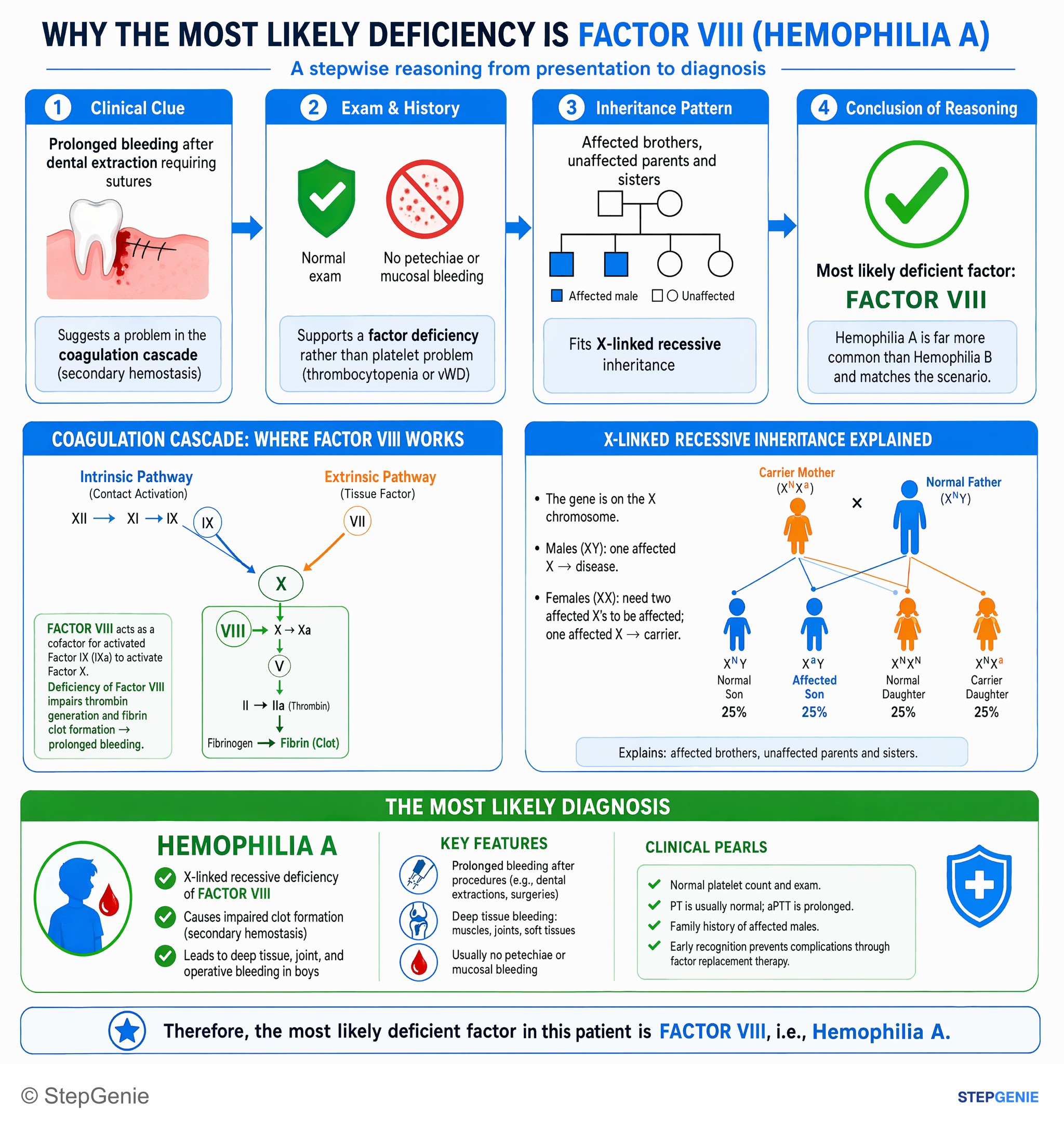
Hemophilia A is an X-linked recessive deficiency of factor VIII, causing deep tissue/operative bleeding in boys with affected male…
USMLE Step 1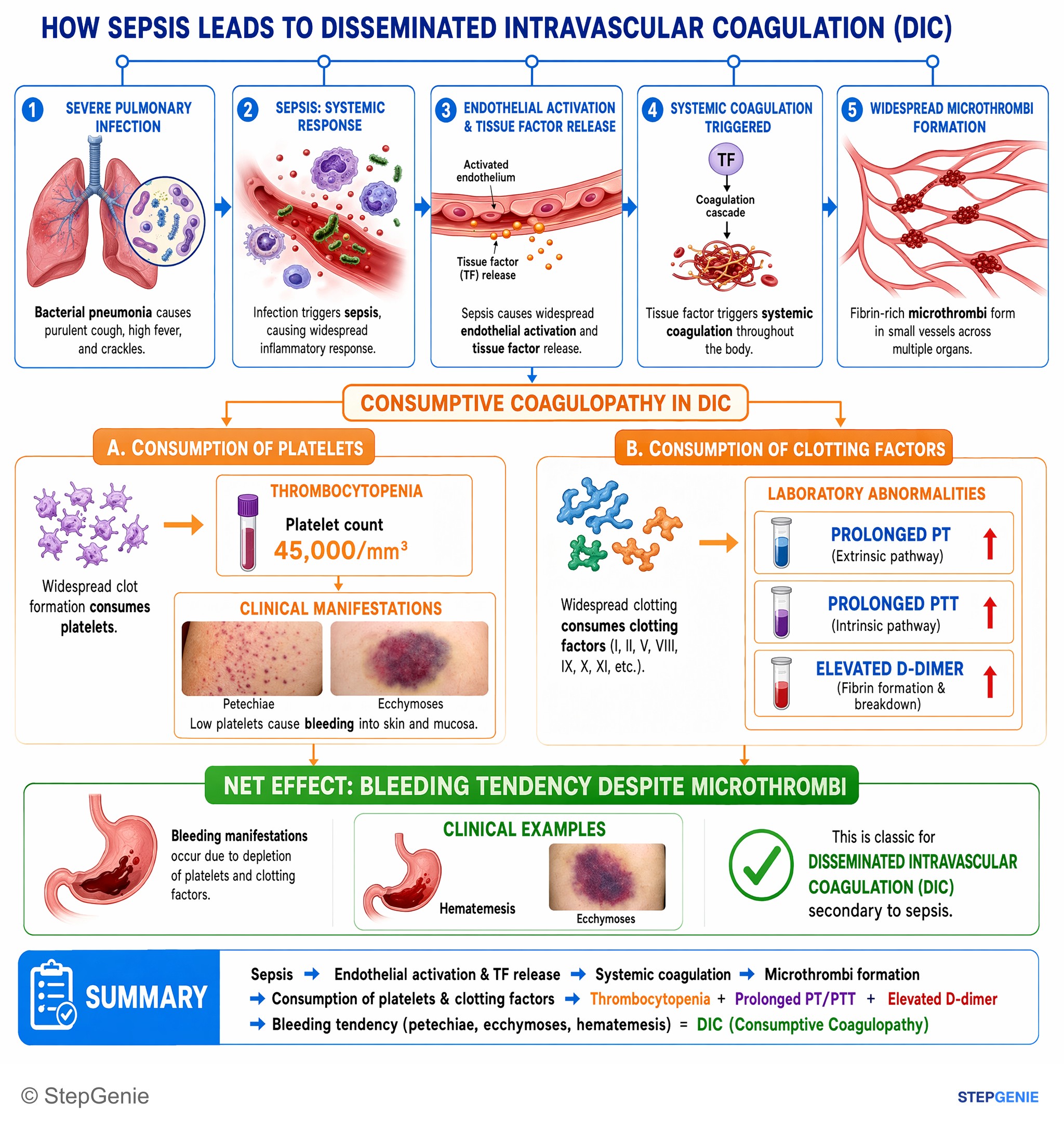
Disseminated intravascular coagulation (DIC) from sepsis causes consumptive coagulopathy with thrombocytopenia, prolonged PT/PTT, elevated…
USMLE Step 1Frequently Asked Questions
What is the difference between the intrinsic and extrinsic pathways?
The extrinsic pathway starts when tissue factor exposed by vessel injury binds factor VII, and it is measured by the PT/INR. The intrinsic pathway runs through factors XII, XI, IX and VIII and is measured by the aPTT. Both feed the common pathway (factors X, V, II and fibrinogen), where the Xa-Va complex makes thrombin that converts fibrinogen to fibrin.
What is the cell-based model of coagulation?
The cell-based model, proposed by Hoffman and Monroe in 2001, describes clotting as it happens on cell surfaces rather than as two separate test-tube pathways. Tissue factor-VIIa on cells at the injury makes a small amount of thrombin (initiation), that thrombin activates platelets and factors V, VIII and XI (amplification), and the IXa-VIIIa and Xa-Va complexes on activated platelets produce a burst of thrombin (propagation). It explains why hemophilia A and B bleed while factor XII deficiency does not.
Which clotting factors are vitamin K dependent?
Factors II, VII, IX and X and the anticoagulant proteins C and S (plus protein Z) need vitamin K for gamma-carboxylation, which lets them bind phospholipid surfaces through calcium. Warfarin blocks vitamin K epoxide reductase (VKORC1), so all of them fall. Factor VII and protein C have the shortest half-lives, so the INR rises within about a day and the first days of warfarin are relatively prothrombotic, which is why heparin is overlapped.
Does heparin prolong the PT or the aPTT?
Unfractionated heparin is monitored with the aPTT, or an anti-Xa level, because it boosts antithrombin to inactivate thrombin and factor Xa; the label notes it can also prolong the PT. Warfarin is followed with the PT/INR instead. Direct oral anticoagulants such as apixaban, rivaroxaban and dabigatran do not need routine monitoring.
Why does factor XII deficiency prolong the aPTT but not cause bleeding?
Factor XII, prekallikrein and high-molecular-weight kininogen drive contact activation in the aPTT test tube, so their absence lengthens the aPTT. In the body, clotting is started by tissue factor and factor VIIa, and thrombin activates factor XI directly, so the contact factors are not needed for hemostasis. An isolated long aPTT that corrects on mixing in a person who has never bled is the classic exam clue.
Why can a patient bleed with a normal PT and aPTT?
The PT and aPTT only measure fibrin formation by the clotting factors, so defects elsewhere are missed. Von Willebrand disease, platelet function disorders, hereditary hemorrhagic telangiectasia and factor XIII deficiency can all cause bleeding with normal results. Factor XIII cross-links fibrin after it forms, so its deficiency needs a clot stability test in urea.