PT (reported as the INR) screens the extrinsic and common pathways, factor VII plus factors X, V, II (prothrombin) and fibrinogen, and monitors warfarin. The aPTT (usually called the PTT) screens the intrinsic and common pathways, factors XII, XI, IX and VIII plus the same common factors, and monitors unfractionated heparin. An isolated long PT points to factor VII (warfarin, early vitamin K deficiency); an isolated long aPTT points to hemophilia A or B, von Willebrand disease, heparin or a lupus anticoagulant; both long means a common-pathway or multi-factor problem such as DIC, liver disease or severe vitamin K deficiency. A 1:1 mixing study then separates a factor deficiency (the clotting time corrects) from an inhibitor (it stays long).
PT/INR vs PTT at a glance
| PT / INR | aPTT (PTT) | |
|---|---|---|
| Pathway screened | Extrinsic + common | Intrinsic + common |
| Factors tested | VII, X, V, II, fibrinogen | Prekallikrein, HMWK, XII, XI, IX, VIII, X, V, II, fibrinogen |
| Blind spots | Intrinsic factors and factor XIII | Factor VII and factor XIII |
| Drug it monitors | Warfarin (INR) | Unfractionated heparin |
| Isolated prolongation | Warfarin, early vitamin K deficiency, inherited factor VII deficiency | Hemophilia A (VIII) or B (IX), factor XI or XII deficiency, von Willebrand disease (sometimes), heparin, lupus anticoagulant |
| Memory hook | WEPT: Warfarin, Extrinsic, PT | HIPTT: Heparin, Intrinsic, PTT |
- INR expresses the patient's PT as a ratio to the lab's control value, corrected for differences in reagents between labs. An INR above 1.5, or a PT at least 3 seconds longer than the control, is usually abnormal (Merck).
- aPTT reference ranges vary by lab; about 25-35 seconds is typical (StatPearls).
The cascade behind the two tests
- Extrinsic (PT): tissue factor binds factor VIIa, and the TF-VIIa complex activates factor X (and perhaps also IX).
- Intrinsic (aPTT): factor XII, prekallikrein, high-molecular-weight kininogen (HMWK) and XIa generate IXa; IXa plus its cofactor VIIIa activates factor X.
- Common (both tests): Xa plus Va (prothrombinase) turns prothrombin into thrombin; thrombin makes fibrin and activates factor XIII, which cross-links it. Most steps need calcium.
- The split is a test-tube construct. The body does not need factor XII, prekallikrein or HMWK to clot, so factor XII deficiency prolongs the aPTT without bleeding.
For the full cascade with drug targets, see coagulation cascade tips.
Reading the pattern: causes by lab result
| Platelets | PT/INR | aPTT | Think of | Clinchers |
|---|---|---|---|---|
| Normal | Long | Normal | Warfarin, early vitamin K deficiency, factor VII deficiency | Factor VII has the shortest half-life (4-6 hours) |
| Normal | Normal | Long, with bleeding | Hemophilia A or B, factor XI deficiency (Ashkenazi Jewish ancestry), von Willebrand disease, heparin, acquired factor VIII inhibitor | Deep bleeding = hemophilia; mucosal bleeding = vWD |
| Normal | Normal | Long, no bleeding | Factor XII (or prekallikrein, HMWK) deficiency; lupus anticoagulant | Lupus anticoagulant causes clots, not bleeding |
| Normal | Long | Long | Factor X, V or II deficiency, very low fibrinogen, severe vitamin K deficiency | Malabsorption, antibiotics, newborn who missed vitamin K |
| Low | Long | Long | DIC; advanced liver disease | DIC: falling fibrinogen, high D-dimer, schistocytes |
| Normal or low | Normal | Normal | Platelet disorders, mild vWD, factor XIII deficiency | XIII: umbilical stump bleeding, poor wound healing |
- The bleeding pattern still sorts the defect. Platelet and vessel problems cause petechiae, purpura, epistaxis, gum bleeding and menorrhagia; factor deficiencies cause deep bleeding (hemarthroses, muscle hematomas, retroperitoneal hemorrhage).
- Bleeding time survives in older questions, but Merck calls it too irreproducible for clinical decisions; platelet function is now studied with aggregometry.
The mixing study: deficiency or inhibitor?
When the PT or aPTT is long, the patient's plasma is mixed 1:1 with normal plasma, which supplies any missing factor.
- Corrects: a factor deficiency (hemophilia A or B, factor XI or XII deficiency, vitamin K deficiency).
- Does not correct, or corrects only partly: an inhibitor. Bleeding suggests a factor VIII antibody; clotting suggests a lupus anticoagulant. The mix is tested immediately and again after incubation.
- Factor VIII inhibitors: alloantibodies develop in about 30% of people with severe hemophilia A after factor replacement; autoantibodies (acquired hemophilia A) arise without hemophilia, for example in postpartum women, with autoimmune disease or in older adults with no other disorder.
- Lupus anticoagulant: antibodies against phospholipid-bound proteins (such as beta-2 glycoprotein I) prolong phospholipid-dependent tests. The aPTT does not correct with 1:1 or 4:1 mixing; the dilute Russell viper venom time (dRVVT) is more sensitive, and excess phospholipid corrects it. In the patient the antibodies are prothrombotic: venous or arterial thrombosis, recurrent pregnancy loss, thrombocytopenia. Positive tests are repeated after at least 12 weeks. See the antiphospholipid syndrome breakdown.
Warfarin vs heparin: what each does to the labs
| Warfarin | Unfractionated heparin | Enoxaparin (LMWH) | |
|---|---|---|---|
| Mechanism | Inhibits VKORC1, so less functional II, VII, IX, X and proteins C and S | Activates antithrombin, which inhibits mainly thrombin (IIa) and Xa | Antithrombin-mediated, far more anti-Xa than anti-IIa activity |
| Lab test | PT/INR: target 2-3 for VTE and AF; 2.5-3.5 for a mechanical mitral valve | aPTT: about 1.5-2 times normal | PT and aPTT not adequate; anti-Xa level if monitoring is needed |
| Reversal | Vitamin K; major bleeding: 4-factor PCC plus IV vitamin K (plasma if PCC is unavailable) | Protamine: 1 mg neutralizes about 100 units | Protamine only partly (at most about 60% of anti-Xa activity) |
| Classic trap | Skin necrosis, teratogen | Heparin-induced thrombocytopenia (HIT) | Anti-Xa monitoring in renal impairment |
- Why the PT rises first on warfarin: factor half-lives are VII 4-6 hours, IX 24 hours, X 48-72 hours and II about 60 hours. Factor VII falls first, so an effect appears within 24 hours, but the peak takes 72-96 hours.
- Why warfarin can clot early: protein C has a half-life of about 8 hours (protein S about 30 hours), so a natural anticoagulant drops early. Skin necrosis usually appears within a few days of starting and is linked to protein C or S deficiency; the label overlaps heparin with warfarin for 4-5 days and until the INR is therapeutic.
- DOACs break the old rules. Dabigatran tends to prolong the aPTT; the PT and aPTT are insensitive to apixaban. A normal PT and aPTT do not rule out a DOAC effect (for dabigatran, a normal thrombin time does); drug-calibrated anti-Xa assays measure the Xa inhibitors (ACC 2020).
Mechanisms, doses and antidotes for every agent are in the anticoagulants guide.
When both tests are long: vitamin K deficiency, liver disease, DIC
Vitamin K deficiency. Vitamin K is needed to make functional factors II, VII, IX and X and proteins C, S and Z. Causes: newborns (poor placental transfer, little vitamin K in breast milk, a sterile gut), fat malabsorption (biliary obstruction, cystic fibrosis, small-bowel resection) and broad-spectrum antibiotics (especially some cephalosporins); warfarin blocks vitamin K recycling, and hepatic insufficiency adds to the bleeding risk. Typically the PT/INR is long while the aPTT and platelets are normal (Merck); severe deficiency prolongs the aPTT too (StatPearls). Newborns get phytonadione 1 mg IM within 6 hours of birth (over 1500 g). The usual adult dose is 1-20 mg. Rarely, IV vitamin K causes anaphylaxis or anaphylactoid reactions, which can be fatal (a boxed warning on the injection label, which also covers IM use).
Liver disease. Hepatocytes make most clotting factors (factor VIII comes from liver sinusoidal and other endothelial cells), so severe cirrhosis, fulminant hepatitis or acute fatty liver of pregnancy prolongs the PT and aPTT. Splenic sequestration from portal hypertension lowers the platelet count, and less alpha-2-antiplasmin adds excess fibrinolysis.
DIC. Excess thrombin generation consumes platelets and factors while fibrinolysis runs. Rapidly evolving DIC gives low platelets, long PT and aPTT, rapidly falling fibrinogen, high D-dimer and schistocytes, and mainly bleeds; slowly evolving DIC causes mainly venous thrombosis. Triggers: obstetric complications (abruption, amniotic fluid embolism), gram-negative sepsis, adenocarcinomas, acute promyelocytic leukemia, shock, trauma and snake venom. Treat the cause; for bleeding, give platelets, cryoprecipitate if fibrinogen is below 100 mg/dL and fresh frozen plasma. More in the DIC guide.
How USMLE tests it
- Boy with hemarthroses and an isolated long aPTT: hemophilia A (factor VIII, about 80%) or B (factor IX, about 20%), X-linked, with normal PT and platelets; desmopressin can raise factor VIII in mild hemophilia A. Sort them with the hemophilia A vs B mnemonic.
- Menorrhagia, nosebleeds and bleeding after dental work with a normal platelet count and INR: von Willebrand disease, the most common inherited bleeding disorder. VWF anchors platelets (via glycoprotein Ib/IX) and stabilizes factor VIII, so the aPTT is normal or slightly long. See the von Willebrand disease guide.
- Long aPTT with DVT or pregnancy losses: lupus anticoagulant. Long aPTT on preoperative screening, never a bleed: factor XII deficiency; no treatment.
- Umbilical stump bleeding in a newborn, bleeding after surgery or minor trauma, poor wound healing, normal PT and aPTT: factor XIII deficiency (autosomal recessive); the clot, never cross-linked, dissolves in 5 M urea.
Practice question
A 31-year-old woman comes to the emergency department 10 weeks after an uncomplicated vaginal delivery because of large, painful bruises on her arms and thigh that appeared without trauma. She had no excessive bleeding at delivery or after previous dental extractions, and no family members have a bleeding disorder. She takes no medications. Hemoglobin is 9.8 g/dL, platelet count 265,000/mm³, PT 12 seconds (INR 1.0) and aPTT 68 seconds (reference 25-35). A 1:1 mix of her plasma with normal plasma stays prolonged both immediately and after incubation at 37°C. Which of the following is the most likely cause of her bleeding?
A. Antiphospholipid antibodies (lupus anticoagulant) B. Autoantibody against factor VIII C. Inherited factor IX deficiency D. Type 1 von Willebrand disease E. Vitamin K deficiency F. Disseminated intravascular coagulation
Answer: B. A normal PT and platelet count with a long aPTT localizes the problem to the intrinsic pathway, and failure to correct on mixing means an inhibitor, not a deficiency. New, spontaneous soft-tissue bleeding in a postpartum woman with no lifelong bleeding history is the classic setting for acquired hemophilia A (a factor VIII autoantibody). Acute bleeding is treated with recombinant factor VIIa or activated prothrombin complex concentrate, and immunosuppression (glucocorticoids, cyclophosphamide or rituximab) suppresses the antibody.
- A: a lupus anticoagulant also fails to correct, but it causes thrombosis and pregnancy loss, not spontaneous bleeding.
- C: hemophilia B is X-linked and lifelong, and a factor deficiency corrects on mixing.
- D: von Willebrand disease causes lifelong mucosal bleeding (her dental extractions and delivery were uneventful), and its mild aPTT prolongation corrects on mixing.
- E: vitamin K deficiency prolongs the PT first; her PT is normal.
- F: DIC lowers the platelet count, prolongs both PT and aPTT and needs a trigger; she has none of these.
Key takeaways
- PT/INR = extrinsic + common (VII, X, V, II, fibrinogen) and monitors warfarin; aPTT = intrinsic + common (XII, XI, IX, VIII, X, V, II, fibrinogen) and monitors unfractionated heparin. Neither detects factor XIII deficiency.
- Isolated long PT: warfarin, early vitamin K deficiency, factor VII deficiency. Isolated long aPTT: hemophilia A or B, factor XI or XII deficiency, vWD, heparin, lupus anticoagulant, factor VIII inhibitor.
- Both long: with low platelets, DIC or advanced liver disease; with normal platelets, a common-pathway deficiency or severe vitamin K deficiency.
- Mixing study: correction = deficiency; no correction = inhibitor (bleeding → factor VIII antibody; clotting → lupus anticoagulant).
- Factor VII (half-life 4-6 hours) makes the PT rise first on warfarin; protein C (about 8 hours) explains early hypercoagulability and skin necrosis.
- Reverse warfarin with vitamin K, adding 4-factor PCC for major bleeding; reverse heparin with protamine.
Sources
- Excessive Bleeding — Merck Manual Professional Edition (2025)
- Overview of Hemostasis — Merck Manual Professional Edition (2025)
- Coagulation Disorders Caused by Circulating Anticoagulants — Merck Manual Professional Edition (2025)
- Uncommon Hereditary Coagulation Disorders — Merck Manual Professional Edition (2025)
- Vitamin K Deficiency — Merck Manual Professional Edition (2026)
- Disseminated Intravascular Coagulation (DIC) — Merck Manual Professional Edition (2026)
- Von Willebrand Disease — Merck Manual Professional Edition (2026)
- Antiphospholipid Syndrome (APS) — Merck Manual Professional Edition (2025)
- Partial Thromboplastin Time — StatPearls, NCBI Bookshelf (2023)
- Warfarin Sodium Tablets: Prescribing Information — DailyMed (2025)
- Factor XIII Deficiency — MedlinePlus Genetics, National Library of Medicine
- 2020 ACC Expert Consensus Decision Pathway on Management of Bleeding in Patients on Oral Anticoagulants — Journal of the American College of Cardiology (2020)
Visual explanations from the StepGenie QBank
Infographics StepGenie drew to explain questions in its own QBank, matched to this article's topic. Each caption is the takeaway of the question it explains.
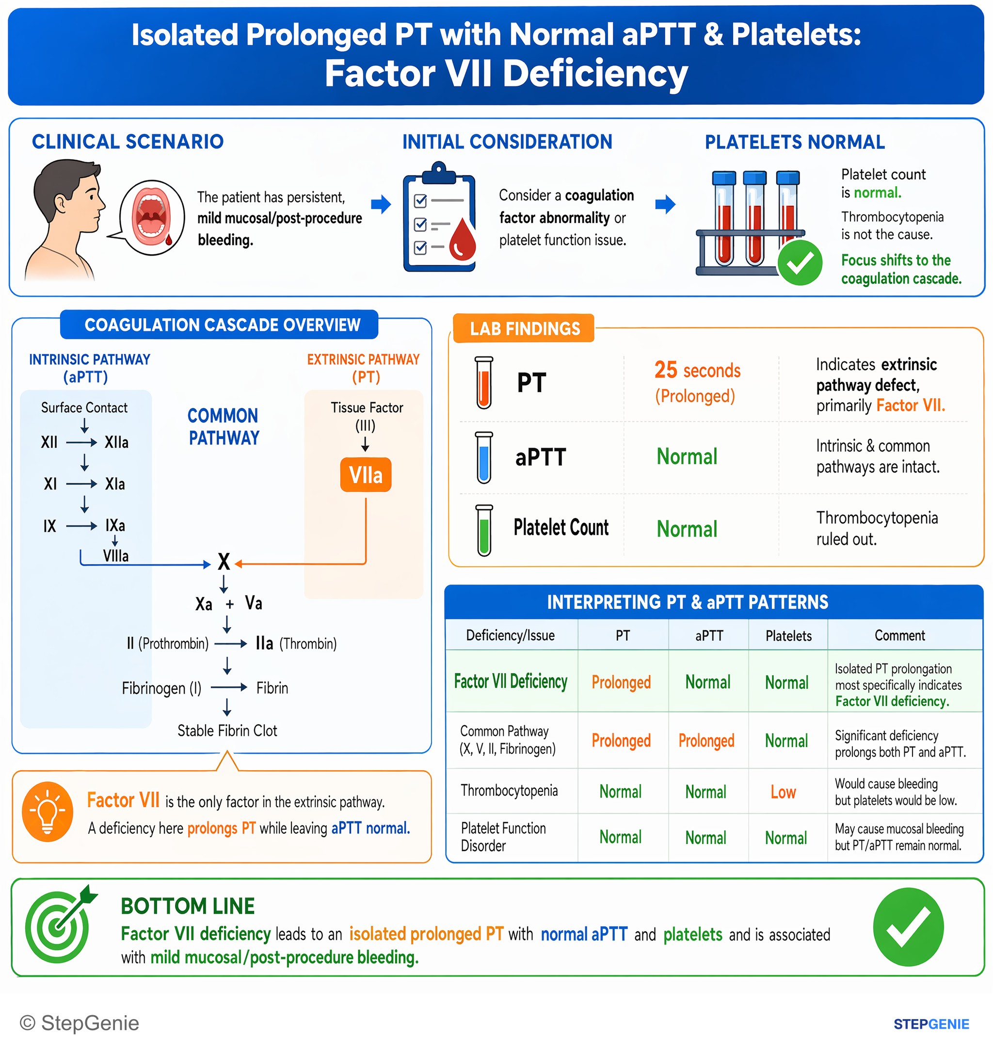
Factor VII deficiency → isolated prolonged PT with normal aPTT and platelets → mild mucosal/post-procedure bleeding.
USMLE Step 1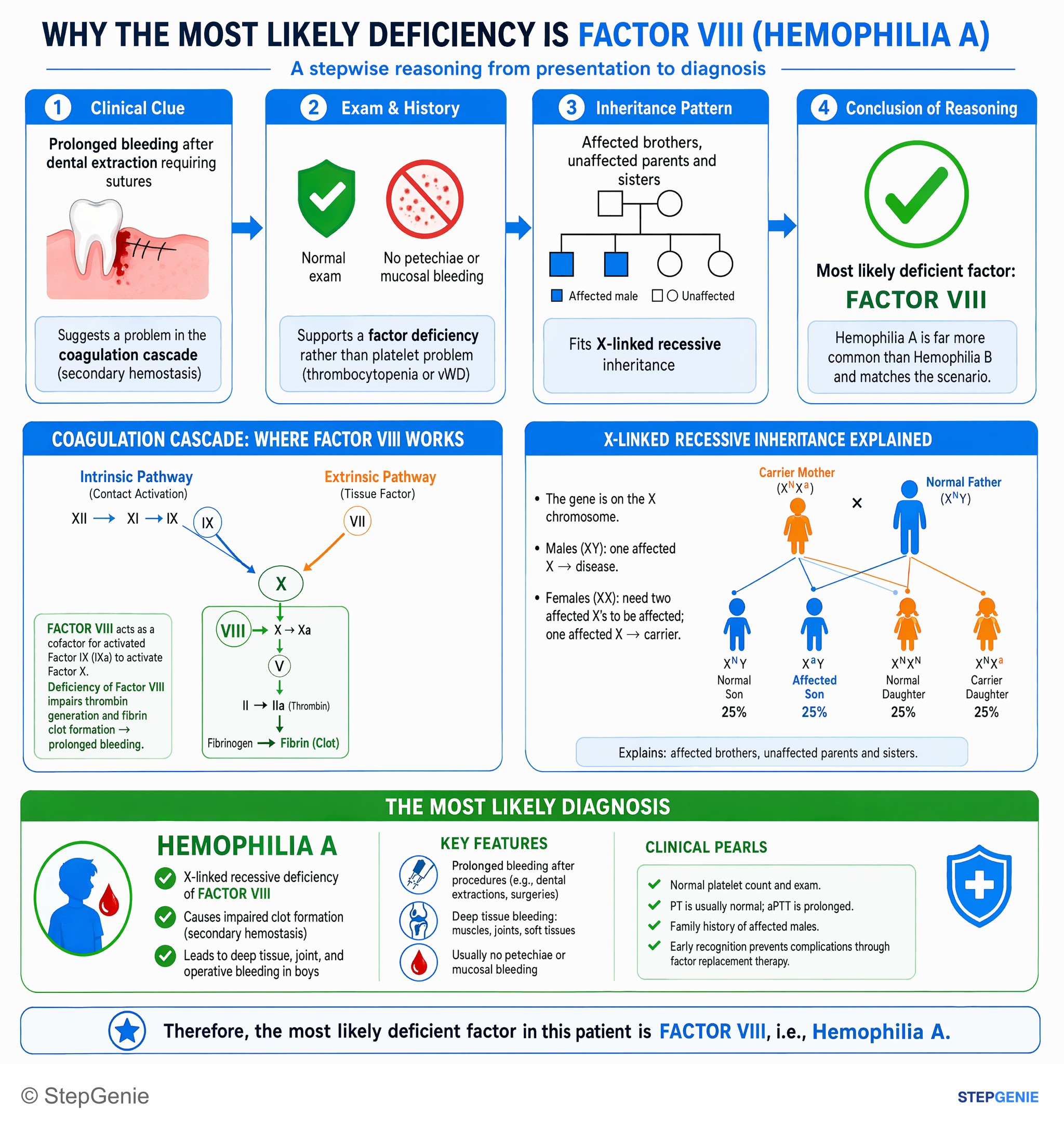
Hemophilia A is an X-linked recessive deficiency of factor VIII, causing deep tissue/operative bleeding in boys with affected male…
USMLE Step 1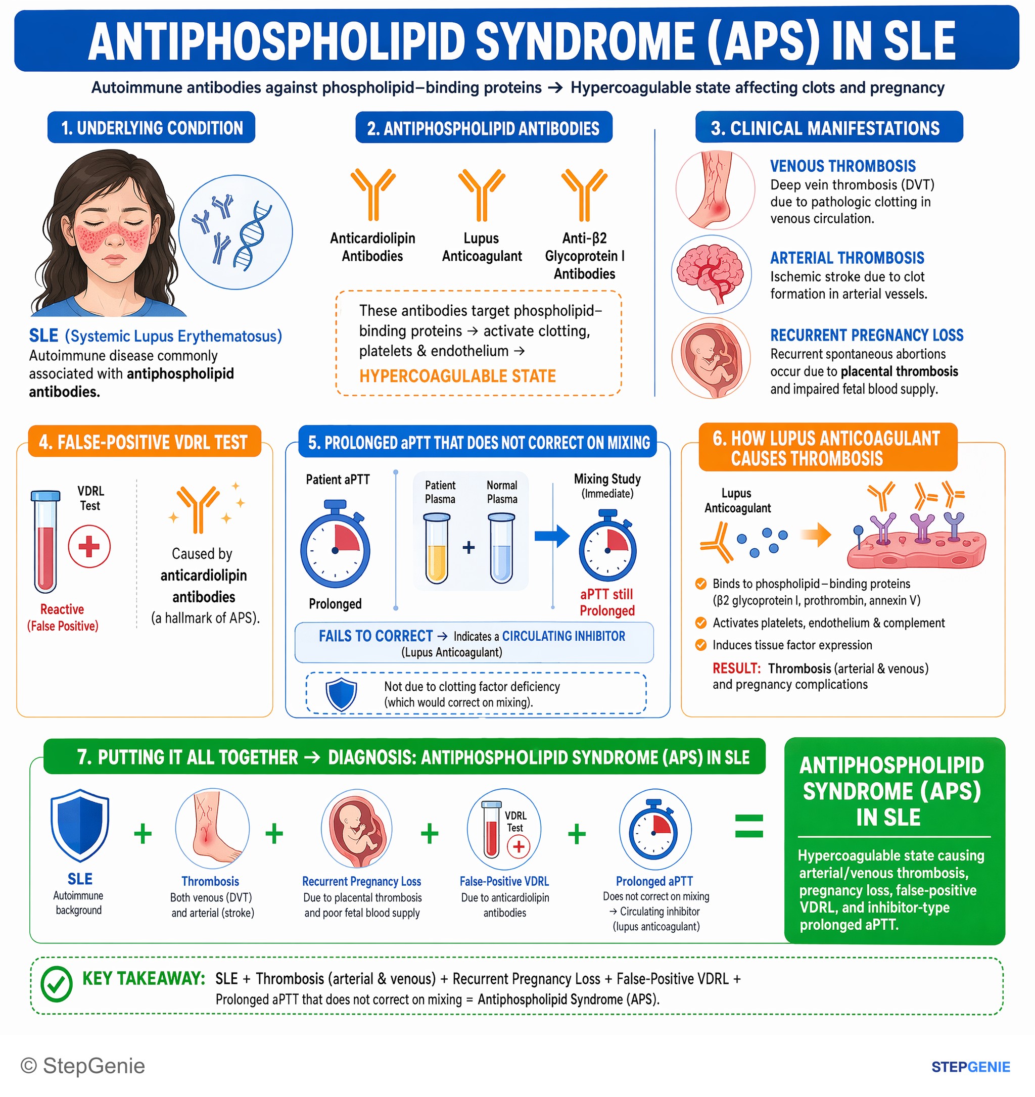
Antiphospholipid syndrome in SLE: arterial/venous thrombosis, recurrent pregnancy loss, false-positive VDRL, prolonged aPTT that does not…
USMLE Step 1Frequently Asked Questions
What is the difference between PT/INR and PTT?
The PT, reported as the INR, measures the extrinsic and common pathways: factor VII plus factors X, V, II and fibrinogen. The aPTT (usually called the PTT) measures the intrinsic and common pathways: factors XII, XI, IX and VIII plus the same common factors. The PT/INR is used to monitor warfarin, and the aPTT is used to monitor unfractionated heparin. Neither test detects factor XIII deficiency.
Why does warfarin raise the PT/INR first?
Warfarin inhibits vitamin K epoxide reductase (VKORC1), so the liver makes less functional factor II, VII, IX and X and proteins C and S. Factor VII has the shortest half-life, about 4 to 6 hours, so it falls first, and the PT/INR, which depends on factor VII, rises within a day. The full effect takes longer, peaking at 72 to 96 hours as the longer-lived factors fall.
What causes both PT and PTT to be prolonged?
Both tests lengthen when the common pathway or several factors are affected. With a low platelet count, think of disseminated intravascular coagulation (with falling fibrinogen and a high D-dimer) or advanced liver disease. With a normal platelet count, think of severe vitamin K deficiency or an inherited deficiency of factor X, V or II. Severe liver disease prolongs both because the liver makes most clotting factors.
What does a mixing study tell you?
A mixing study combines the patient's plasma 1:1 with normal plasma. If a prolonged PT or aPTT corrects, the patient is missing a clotting factor, as in hemophilia or vitamin K deficiency. If it does not correct, an inhibitor is likely. A bleeding patient most likely has a factor VIII antibody (acquired hemophilia A), whereas a patient with clots or pregnancy losses most likely has a lupus anticoagulant.
Why does lupus anticoagulant prolong the PTT but cause clots?
Lupus anticoagulant antibodies target phospholipid-bound proteins such as beta-2 glycoprotein I. In the test tube they interfere with the phospholipid the aPTT depends on, so the aPTT is prolonged and does not correct on mixing. In the body they promote clotting, causing venous or arterial thrombosis and recurrent pregnancy loss. The dilute Russell viper venom time is more sensitive, and positive tests are repeated after at least 12 weeks.
Can someone bleed with a normal PT and PTT?
Yes. The PT and aPTT measure clotting factors only, so they are normal in platelet disorders, often normal in mild von Willebrand disease, and always normal in factor XIII deficiency, which neither test detects. Factor XIII deficiency classically causes bleeding from the umbilical stump, bleeding after surgery or minor trauma and poor wound healing; the clot dissolves in 5 M urea because it was never cross-linked.