You’ve probably seen this annoying combo in Q-banks: a patient who keeps clotting… but their coag labs look like they “should” be bleeding. That cognitive dissonance is exactly what antiphospholipid syndrome (APS) is designed to test. The key to getting these questions right isn’t just knowing the diagnosis—it’s understanding why each answer choice is tempting and why it’s wrong.
The Vignette (Classic Q-Bank Style)
A 29-year-old woman presents to clinic after a second first-trimester miscarriage. She had a “blood clot in her leg” last year after a long flight. She has no personal history of heavy menstrual bleeding. She is not taking anticoagulants. Physical exam is unremarkable.
Labs:
- Platelets: 120,000/µL
- PT: normal
- aPTT: prolonged
- Mixing study: does not correct
- DRVVT: positive
Most likely underlying cause?
A. Factor VIII deficiency
B. Lupus anticoagulant (antiphospholipid antibodies)
C. Vitamin K deficiency
D. Von Willebrand disease
E. Heparin contamination
Correct Answer: B. Lupus anticoagulant (antiphospholipid antibodies)
Why it’s right (the “APS pattern”)
APS is an acquired autoimmune hypercoagulable state characterized by:
- Arterial or venous thrombosis
- Pregnancy morbidity (classically recurrent early miscarriages, fetal demise, preeclampsia/placental insufficiency)
- Often mild thrombocytopenia
Despite being prothrombotic in vivo, APS antibodies cause prolonged phospholipid-dependent clotting assays in vitro, especially aPTT.
The high-yield mechanistic core
- Antiphospholipid antibodies (including lupus anticoagulant) bind phospholipid-protein complexes (often β2-glycoprotein I).
- In the test tube: they interfere with phospholipid reagents → prolonged aPTT.
- In the body: they promote thrombosis via endothelial activation, platelet activation, complement, etc. → clots + pregnancy loss.
Why the mixing study matters
- Mixing study (patient plasma + normal plasma) helps differentiate:
- Factor deficiency → aPTT corrects
- Inhibitor (e.g., lupus anticoagulant) → aPTT does not correct
So: prolonged aPTT + no correction = inhibitor pattern → think APS/lupus anticoagulant.
Rapid APS Diagnostic Snapshot (USMLE-Friendly)
Clinical criteria (need ≥1)
- Vascular thrombosis (arterial/venous/small vessel)
- Pregnancy morbidity:
- ≥1 fetal death ≥10 weeks, or
- ≥1 premature birth <34 weeks due to preeclampsia/placental insufficiency, or
- ≥3 unexplained consecutive spontaneous abortions <10 weeks
Lab criteria (need ≥1, persistent)
Positive on two occasions ≥12 weeks apart:
- Lupus anticoagulant (functional assays like DRVVT)
- Anticardiolipin IgG/IgM (moderate-high titer)
- Anti–β2-glycoprotein I IgG/IgM
Why Each Distractor Is Wrong (and Why They Look Right)
A. Factor VIII deficiency (Hemophilia A)
Why it tempts you:
- Hemophilia A causes prolonged aPTT.
Why it’s wrong here:
- Hemophilia A causes bleeding, not thrombosis.
- Bleeding phenotype: hemarthroses, deep muscle bleeds, post-procedure bleeding.
- Mixing study should correct (factor deficiency).
USMLE tip:
- Hemophilia = corrects on mixing (unless an acquired inhibitor develops, which would present with severe bleeding).
C. Vitamin K deficiency
Why it tempts you:
- Coag disorder → abnormal labs.
Why it’s wrong here:
- Vitamin K deficiency affects II, VII, IX, X (and proteins C/S).
- Lab pattern: ↑ PT first (factor VII has the shortest half-life), and later ↑ aPTT.
- The vignette: PT is normal, aPTT prolonged, plus thrombosis + miscarriages strongly points away.
USMLE tip:
- PT is the canary for vitamin K deficiency and warfarin effect.
D. Von Willebrand disease
Why it tempts you:
- Can have prolonged aPTT (vWF stabilizes factor VIII).
Why it’s wrong here:
- vWD is a mucocutaneous bleeding disorder: epistaxis, gum bleeding, menorrhagia, easy bruising.
- Not a thrombotic pregnancy-loss picture.
- Mixing study in vWD is typically consistent with a deficiency state (should correct).
USMLE tip:
- vWD = platelet-type bleeding (mucosal) + possibly ↑ aPTT + abnormal platelet function tests (e.g., ↑ bleeding time / abnormal PFA-100).
E. Heparin contamination
Why it tempts you:
- Heparin prolongs aPTT and can cause “wonky” coag studies.
Why it’s wrong here:
- The patient is not on heparin; clinic outpatient setting; plus the clinical story (recurrent miscarriage + prior DVT) screams APS.
- Heparin effect can be suggested by prolonged thrombin time (TT) and can be confirmed with anti–factor Xa assay.
USMLE tip:
- If you’re worried about heparin contamination in a sample, check TT (often prolonged) and clinical context.
Quick Comparison Table: “Prolonged aPTT” Differential That USMLE Loves
| Condition | Bleeding vs Clotting | PT | aPTT | Mixing Study | Key Clue |
|---|---|---|---|---|---|
| APS (lupus anticoagulant) | Clotting | N | ↑ | No correction | Thrombosis + pregnancy loss, DRVVT+ |
| Hemophilia A (FVIII def) | Bleeding | N | ↑ | Corrects | Hemarthroses |
| vWD | Bleeding | N | N/↑ | Corrects | Mucosal bleeding, platelet dysfunction |
| Vitamin K deficiency | Bleeding | ↑ first | N/↑ | Corrects | Malnutrition, antibiotics, cholestasis |
| Heparin | Bleeding (med-related) | N/↑ | ↑ | Often no full correction | TT ↑, inpatient line draw |
High-Yield APS Pearls (Step 1 + Step 2)
1) “Lupus anticoagulant” is a misnomer
- It prolongs aPTT in vitro
- It causes thrombosis in vivo
2) APS can cause thrombocytopenia—but still clots
- Mild-moderate thrombocytopenia is common.
- Don’t let low platelets trick you into a bleeding diagnosis.
3) Don’t confuse APS with DIC
- DIC often has ↑ PT and ↑ aPTT, low fibrinogen, high D-dimer, schistocytes.
- APS usually has isolated aPTT prolongation and thrombosis history.
4) Pregnancy management is a Step 2 favorite
- Obstetric APS: commonly low-dose aspirin + heparin (LMWH) during pregnancy.
- Warfarin is teratogenic (especially early pregnancy).
5) Anticoagulation nuance (testable)
- APS patients with thrombosis often need long-term anticoagulation.
- Many exams still emphasize warfarin for long-term management after a thrombotic event (though real-world practice is nuanced; DOAC efficacy in high-risk APS is a known concern).
Take-Home Framework: How to Nail APS Questions Fast
- Clinical trigger: thrombosis + recurrent miscarriage (± livedo reticularis, thrombocytopenia)
- Lab trigger: isolated prolonged aPTT
- Confirmation logic: mixing study doesn’t correct → inhibitor
- Specific test: DRVVT positive or anticardiolipin/anti–β2GP1 antibodies
- Key paradox: prolonged aPTT but hypercoagulable
Visual explanations from the StepGenie QBank
Infographics StepGenie drew to explain questions in its own QBank, matched to this article's topic. Each caption is the takeaway of the question it explains.
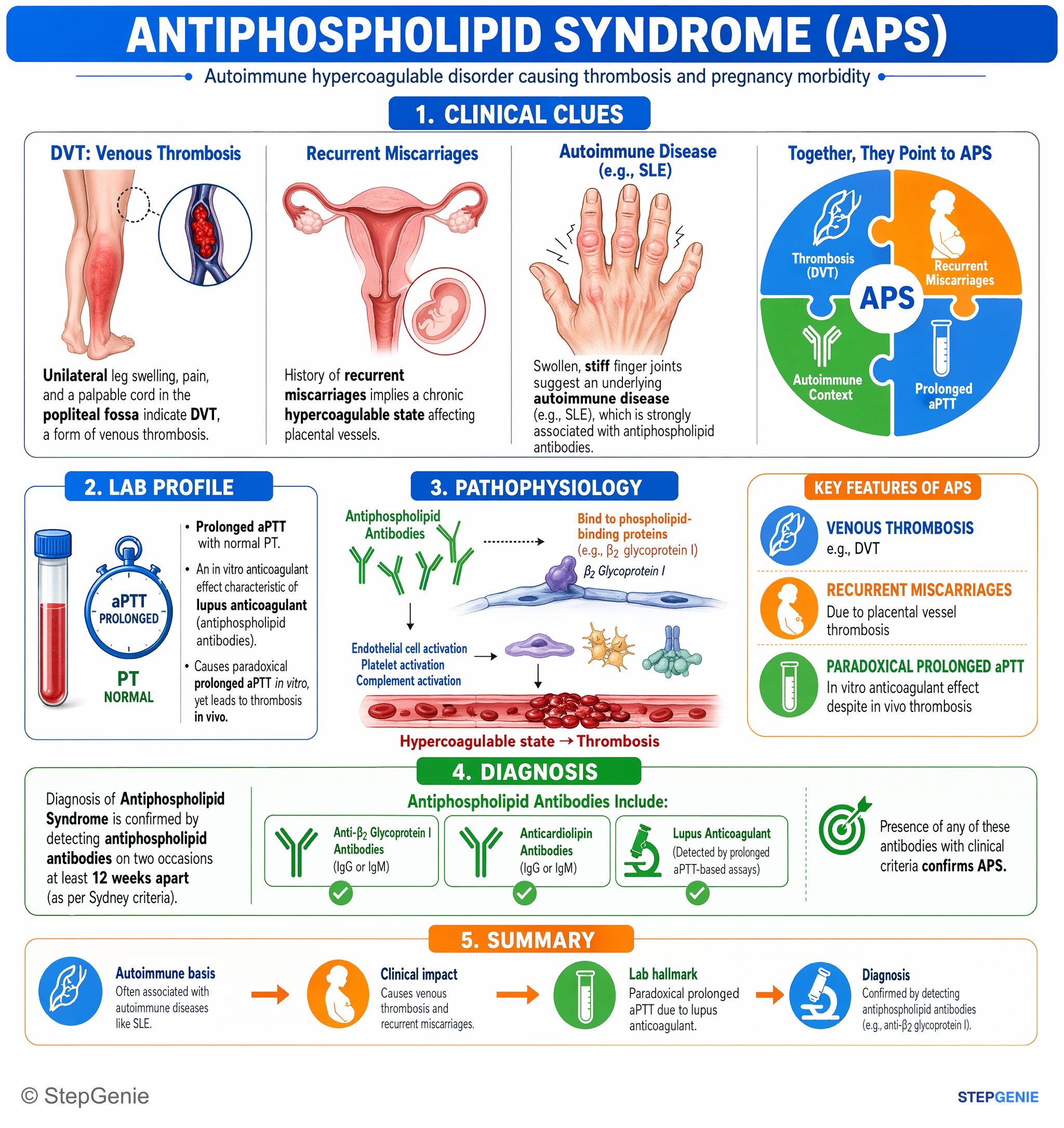
Antiphospholipid syndrome causes venous thrombosis, recurrent miscarriages, and paradoxical prolonged aPTT; diagnosis confirmed by…
USMLE Step 1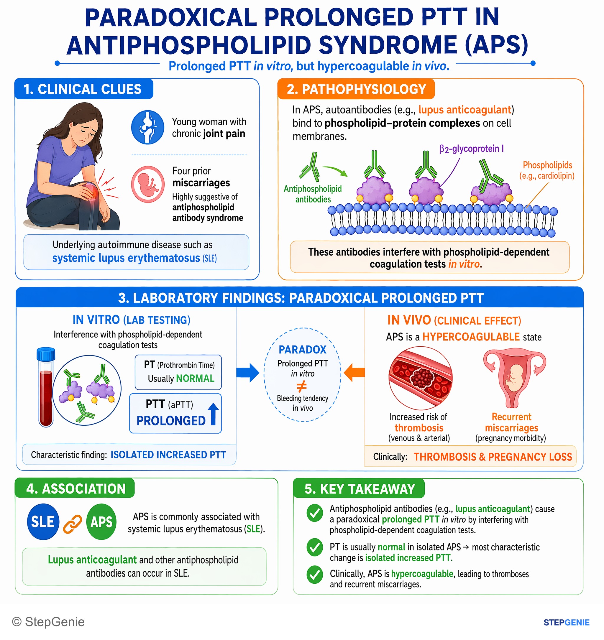
Antiphospholipid syndrome causes a paradoxical prolonged PTT in vitro but is clinically hypercoagulable with recurrent miscarriages and…
USMLE Step 1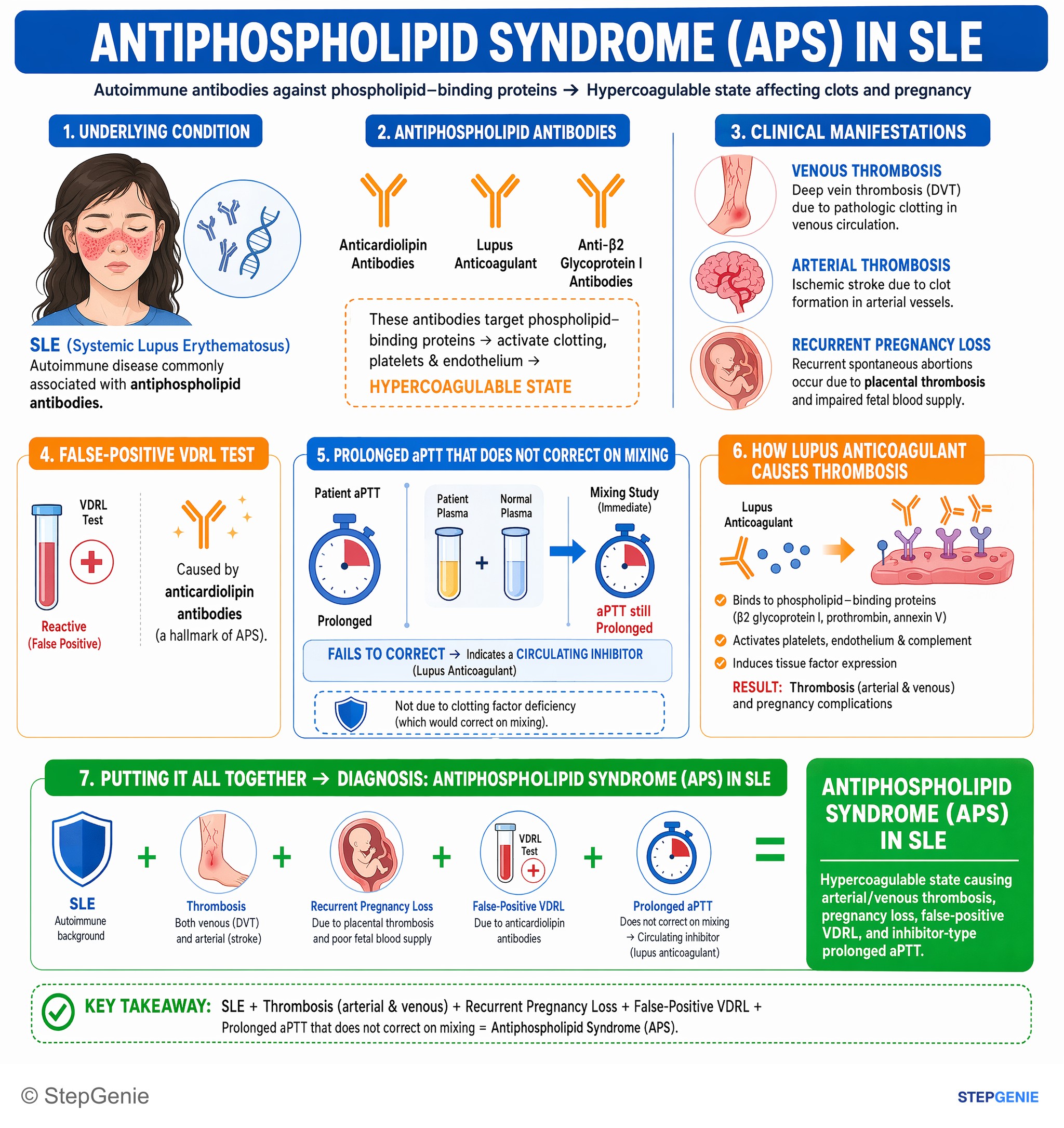
Antiphospholipid syndrome in SLE: arterial/venous thrombosis, recurrent pregnancy loss, false-positive VDRL, prolonged aPTT that does not…
USMLE Step 1Frequently Asked Questions
Why does antiphospholipid syndrome cause clotting even though the aPTT is prolonged?
Antiphospholipid syndrome is a hypercoagulable autoimmune disorder that causes thrombosis in vivo despite prolonging aPTT in vitro. The antibodies, including lupus anticoagulant, bind phospholipid-protein complexes such as β2-glycoprotein I. In the test tube they interfere with phospholipid-dependent clotting assays, which prolongs aPTT, but in the body they promote thrombosis through endothelial activation, platelet activation, and complement effects.
How do you tell lupus anticoagulant from a factor deficiency on a mixing study?
Lupus anticoagulant shows a prolonged aPTT that does not correct with a mixing study, whereas a factor deficiency corrects. Mixing patient plasma with normal plasma helps separate inhibitor patterns from deficiency patterns. If the abnormal aPTT corrects, think factor deficiency such as hemophilia A or von Willebrand-related factor VIII reduction. If it does not correct, think an inhibitor like lupus anticoagulant in antiphospholipid syndrome.
What clinical clues should make you think antiphospholipid syndrome instead of a bleeding disorder?
Recurrent pregnancy loss plus arterial or venous thrombosis should strongly suggest antiphospholipid syndrome rather than a bleeding disorder. A classic pattern is recurrent early miscarriages with a prior DVT, often with mild thrombocytopenia, normal PT, prolonged aPTT, and a positive DRVVT. Unlike hemophilia or von Willebrand disease, the patient does not have a mucosal or deep-tissue bleeding history.
How do you tell APS from vitamin K deficiency or vWD when aPTT is prolonged?
Antiphospholipid syndrome is favored when the patient has thrombosis and pregnancy morbidity with normal PT, prolonged aPTT, and no correction on mixing study. Vitamin K deficiency usually raises PT first, with aPTT becoming abnormal later, so a normal PT argues against it. Von Willebrand disease causes mucocutaneous bleeding such as epistaxis, gum bleeding, menorrhagia, and typically behaves like a deficiency state that corrects on mixing.
What lab tests support the diagnosis of antiphospholipid syndrome?
The diagnosis is supported by persistent antiphospholipid antibody testing along with compatible clinical features. Lab criteria include lupus anticoagulant detected by functional assays such as DRVVT, anticardiolipin IgG or IgM, or anti-β2-glycoprotein I IgG or IgM. These positive tests must be present on two occasions at least 12 weeks apart, alongside vascular thrombosis or defined pregnancy morbidity.