Disseminated intravascular coagulation (DIC) is one of those Step diagnoses that feels chaotic—because it is. The key is to remember that DIC is not a primary disease, but a final common pathway of systemic coagulation activation. Once you see it as “pathologic thrombin everywhere” → microthrombi + consumption of platelets/clotting factors, the labs, symptoms, and treatment all line up.
What DIC Is (Definition You Can Use on Test Day)
Disseminated intravascular coagulation (DIC) = widespread activation of coagulation leading to:
- Microvascular thrombi → ischemia/organ dysfunction
- Consumption of platelets + clotting factors (“consumption coagulopathy”) → bleeding
- Secondary fibrinolysis → elevated fibrin breakdown products (eg, D-dimer)
Classic Step phrasing: “Simultaneous thrombosis and bleeding.”
Pathophysiology: The Core Mechanism (Know This Cold)
At a high level, DIC is driven by excess thrombin generation.
Stepwise logic
- Trigger (eg, sepsis, obstetric catastrophe, malignancy, trauma) causes massive tissue factor exposure or endothelial injury.
- Tissue factor activates the extrinsic pathway → lots of factor VII activation → thrombin burst.
- Thrombin converts fibrinogen → fibrin, forming widespread microthrombi.
- Ongoing clot formation consumes:
- Platelets
- Fibrinogen
- Coagulation factors (especially V and VIII)
- The body tries to dissolve clots → plasmin activation → fibrinolysis → ↑ D-dimer and ↑ fibrin split products.
- Net result: both clotting (ischemia) and bleeding.
Microangiopathic hemolytic anemia (MAHA)
Microthrombi shear RBCs → schistocytes on smear (similar mechanism to TTP/HUS/HELLP).
First Aid cross-reference: DIC appears under Microangiopathic hemolytic anemia and coagulation disorders with schistocytes, ↑ PT/PTT, ↑ D-dimer, ↓ fibrinogen, ↓ platelets.
High-Yield Causes & Associations (Most Tested)
Think: “Things that dump tissue factor into blood” or “things that shred endothelium.”
The Step 1/2 greatest hits
- Sepsis (especially gram-negative, but any severe sepsis)
- Endotoxin/cytokines → tissue factor expression + endothelial activation
- Obstetric complications
- Placental abruption
- Amniotic fluid embolism
- Retained dead fetus
- Severe preeclampsia/HELLP (can overlap with MAHA picture)
- Malignancy
- Acute promyelocytic leukemia (APL; t(15;17)) is extremely high yield
- Also mucin-producing adenocarcinomas (can cause chronic DIC)
- Massive trauma / burns
- Severe pancreatitis
- Transfusion reactions (less common but testable)
Rapid associations table
| Trigger | Why it causes DIC | Step clue |
|---|---|---|
| Sepsis | Cytokine-driven tissue factor + endothelial activation | Sick ICU patient, oozing lines |
| APL (t(15;17)) | Procoagulant granules, hyperfibrinolysis | Leukemia + bleeding + DIC labs |
| Placental abruption / amniotic fluid embolism | Massive tissue factor release | Postpartum hemorrhage + abnormal coag |
| Trauma/burns | Endothelial damage, tissue factor exposure | Polytrauma with diffuse bleeding |
First Aid cross-reference: APL is paired with DIC; treatment with ATRA is emphasized (and can precipitate differentiation syndrome—separate entity).
Clinical Presentation: What You See at the Bedside
DIC is often a mix of bleeding, thrombosis, and organ dysfunction.
Bleeding findings (very common)
- Oozing from IV sites, venipuncture wounds, surgical incisions
- Petechiae/purpura, ecchymoses
- Mucosal bleeding (epistaxis, gingival bleeding)
- Hematuria, GI bleeding
- Postpartum hemorrhage (obstetric DIC)
Thrombotic/ischemic findings (don’t forget these)
- Acute kidney injury
- Altered mental status, stroke-like symptoms
- Respiratory failure (microthrombi in pulmonary circulation)
- Digital ischemia/skin necrosis (purpura fulminans in severe cases)
USMLE tip: If a vignette stresses bleeding + organ dysfunction in the setting of sepsis/obstetric emergency/cancer, your reflex should be DIC and then confirm with labs.
Diagnosis: Pattern Recognition with Coag Labs
The classic lab pattern
DIC consumes platelets and clotting factors and generates fibrin breakdown products:
- ↓ Platelets
- ↑ PT
- ↑ PTT
- ↓ Fibrinogen
- ↑ D-dimer (and fibrin split products)
- Schistocytes on peripheral smear (MAHA)
- Often ↓ factor V and VIII (consumption)
Quick “DIC vs TTP/HUS” differentiation (very high yield)
| Feature | DIC | TTP/HUS |
|---|---|---|
| Platelets | ↓ | ↓ |
| PT/PTT | ↑ | Normal |
| D-dimer | ↑ | Normal/slightly ↑ |
| Fibrinogen | ↓ | Normal |
| Smear | Schistocytes | Schistocytes |
| Core problem | Systemic coag activation | Platelet microthrombi (ADAMTS13 issue in TTP) |
Exam shortcut:
If PT/PTT are prolonged, think DIC (or severe liver disease/vit K deficiency)—but with high D-dimer + low fibrinogen + schistocytes, DIC jumps to the top.
Treatment: Treat the Cause + Support the Coagulation System
1) The most important step: treat the underlying trigger
- Sepsis: antibiotics + source control + supportive care
- Obstetric: deliver placenta/fetus, manage hemorrhage
- APL: treat leukemia (ATRA/arsenic regimens per oncology guidance)
- Trauma: damage control resuscitation
2) Supportive transfusion therapy (when bleeding or high-risk)
Use clinical status + labs; you’re not “normalizing numbers” in isolation.
- Platelets if:
- Active bleeding, or
- Platelets very low (commonly <50k with bleeding; thresholds vary by setting)
- Fresh frozen plasma (FFP) to replace clotting factors if bleeding with prolonged PT/PTT
- Cryoprecipitate to raise fibrinogen (key when fibrinogen is low and bleeding)
- Cryo contains fibrinogen, factor VIII, factor XIII, vWF
Step pearl: In DIC, fibrinogen is often low—and replacing it with cryo is a common test answer when there’s bleeding.
3) Anticoagulation (select cases)
This is nuanced and often Step-tested conceptually:
- Consider heparin in predominantly thrombotic DIC (eg, chronic DIC from malignancy, or severe clotting manifestations) if not actively bleeding.
- Do not give anticoagulation reflexively in a hemorrhaging patient.
4) What about vitamin K?
Not a primary DIC treatment. If the vignette suggests mixed etiologies (malnutrition/antibiotics/liver disease), vitamin K can be adjunctive, but DIC itself is consumption, not deficiency.
First Aid–Style High-Yield Facts (Rapid Review)
The “classic DIC labs” sentence
DIC = ↑ PT, ↑ PTT, ↓ platelets, ↓ fibrinogen, ↑ D-dimer, schistocytes.
High-yield triggers to memorize
- Sepsis
- Obstetric catastrophe (abruption, amniotic fluid embolism, retained dead fetus)
- Malignancy (especially APL t(15;17))
- Trauma/burns
Most common clinical clue
Diffuse bleeding/oozing + critically ill context (ICU, postpartum, leukemia, major trauma).
Common USMLE Pitfalls (How They Try to Trick You)
- Confusing DIC with TTP/HUS:
Both have thrombocytopenia + schistocytes, but DIC prolongs PT/PTT and lowers fibrinogen. - Assuming DIC is only bleeding:
Vignettes may focus on organ failure from microthrombi (kidney, lungs, CNS). - Missing APL:
If leukemia + bleeding + DIC labs → think APL (and remember t(15;17), Auer rods, ATRA). - Over-treating labs without context:
Management is guided by bleeding risk and cause, not just a slightly abnormal PT.
Mini Case Patterns (What You’ll See)
Pattern 1: Septic shock + oozing
ICU patient with hypotension, fever, oozing from IV sites → DIC (treat sepsis + blood products if bleeding).
Pattern 2: Postpartum hemorrhage after abruption
Severe vaginal bleeding + uterine tenderness + abnormal coag labs → obstetric DIC (stabilize + deliver/manage cause + cryo/FFP/platelets as needed).
Pattern 3: APL
Fatigue, bruising, pancytopenia + DIC labs → APL until proven otherwise.
Bottom Line (What to Write in the Margin)
If you remember nothing else:
- DIC = systemic coag activation → microthrombi + consumption bleeding
- Labs: ↑ PT/↑ PTT, ↓ platelets, ↓ fibrinogen, ↑ D-dimer, schistocytes
- Causes: sepsis, obstetric catastrophe, APL/malignancy, trauma
- Treat: underlying cause + supportive blood products; heparin only if clotting-predominant and not bleeding
Visual explanations from the StepGenie QBank
Infographics StepGenie drew to explain questions in its own QBank, matched to this article's topic. Each caption is the takeaway of the question it explains.
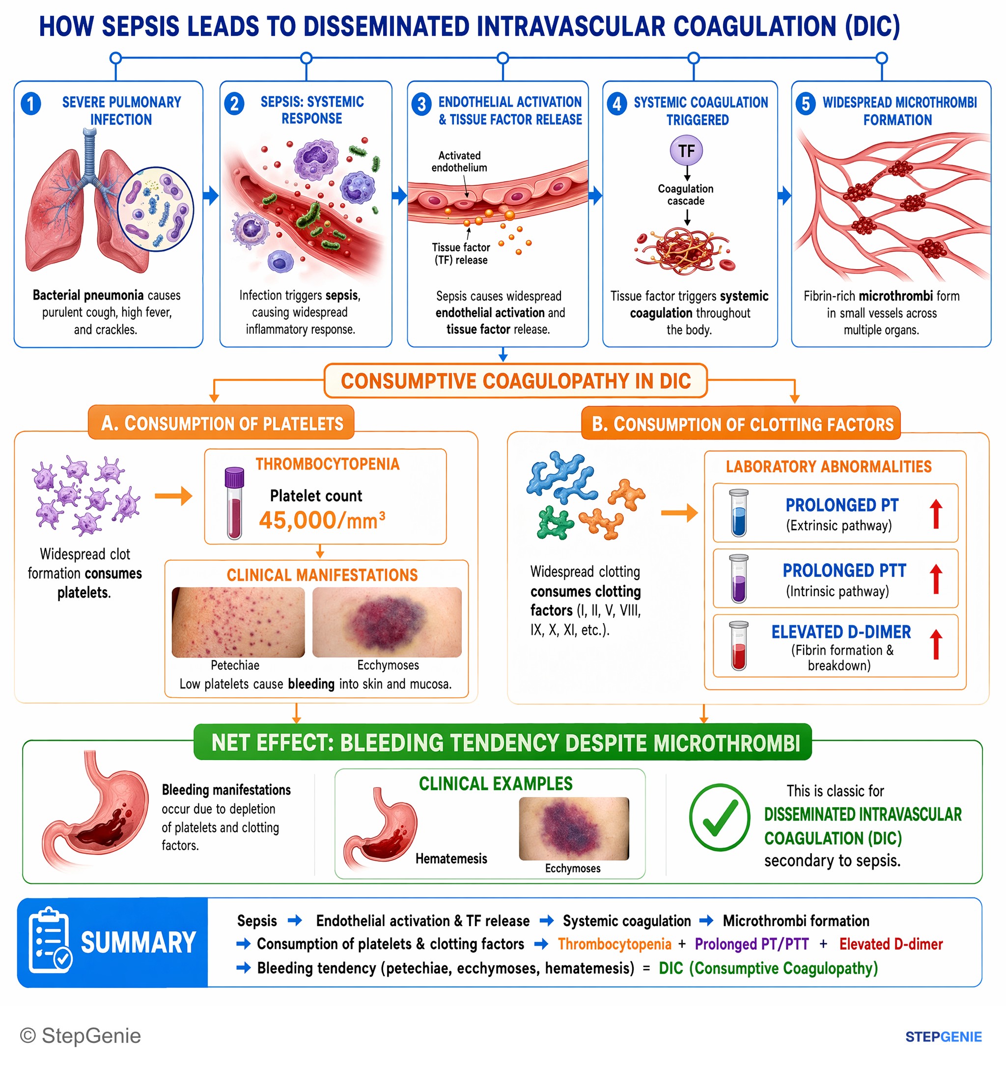
Disseminated intravascular coagulation (DIC) from sepsis causes consumptive coagulopathy with thrombocytopenia, prolonged PT/PTT, elevated…
USMLE Step 1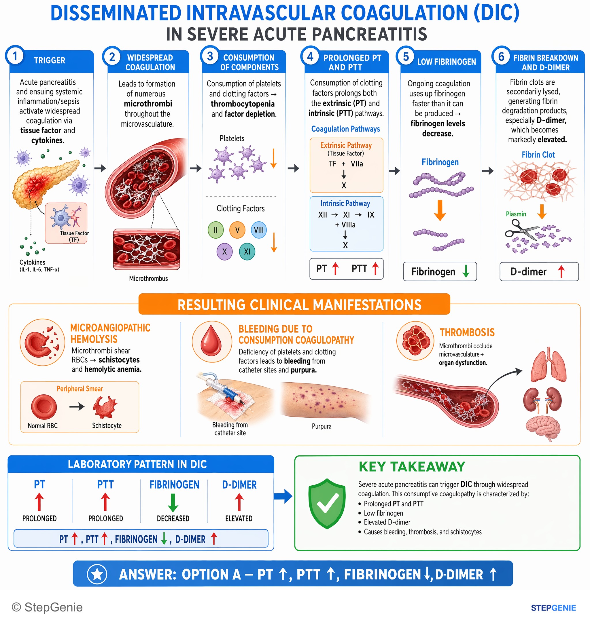
DIC from severe pancreatitis: consumptive coagulopathy with prolonged PT/PTT, low fibrinogen, and elevated D-dimer; causes bleeding…
USMLE Step 1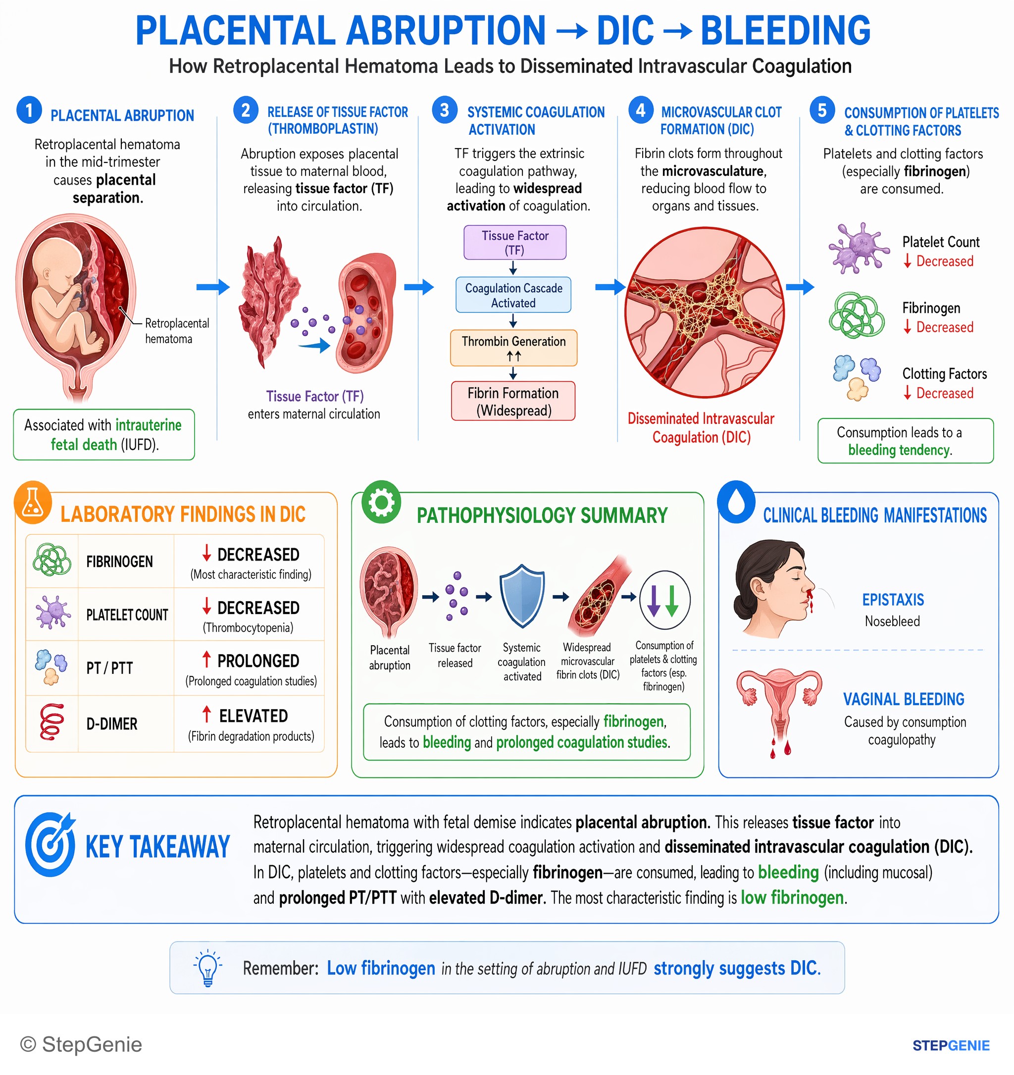
Placental abruption–induced DIC causes consumption of clotting factors, especially fibrinogen, leading to bleeding (including mucosal) and…
USMLE Step 2Frequently Asked Questions
Why does DIC cause both thrombosis and bleeding at the same time?
DIC causes both because it is widespread coagulation activation with excess thrombin generation. Thrombin forms diffuse microvascular fibrin thrombi that cause ischemia and organ dysfunction, while ongoing clot formation consumes platelets, fibrinogen, and other clotting factors. At the same time, plasmin-mediated fibrinolysis increases fibrin breakdown products such as D-dimer, so the net picture is simultaneous clotting and bleeding.
How do you tell DIC from TTP or HUS on labs?
The key distinction is that DIC prolongs PT and PTT, whereas TTP and HUS usually have normal PT and PTT. DIC also shows elevated D-dimer, low fibrinogen, thrombocytopenia, and schistocytes from microangiopathic hemolysis. TTP and HUS can also have low platelets and schistocytes, but the coagulation studies do not show the same consumptive pattern.
What lab pattern should make you think of DIC on a vignette?
Think of DIC when you see low platelets, increased PT, increased PTT, low fibrinogen, and increased D-dimer, often with schistocytes on peripheral smear. This pattern reflects consumption of platelets and clotting factors plus secondary fibrinolysis. If a patient also has bleeding and organ dysfunction in settings like sepsis, obstetric emergencies, malignancy, or trauma, DIC should move high on the differential.
What conditions commonly trigger DIC on Step 1?
Common high-yield triggers are sepsis, obstetric complications, malignancy, and major tissue injury. Specifically emphasized causes include sepsis, placental abruption, amniotic fluid embolism, retained dead fetus, severe preeclampsia or HELLP, acute promyelocytic leukemia with t(15;17), mucin-producing adenocarcinomas, massive trauma or burns, severe pancreatitis, and transfusion reactions. These conditions either expose tissue factor or cause endothelial injury that drives systemic coagulation activation.
What is the treatment approach for DIC?
The main treatment is to address the underlying cause and support coagulation when needed. That means treating sepsis with antibiotics and source control, managing obstetric causes, treating APL with leukemia-directed therapy such as ATRA-based regimens, or using damage control resuscitation in trauma. If there is active bleeding or high risk, supportive transfusion therapy may include platelets, fresh frozen plasma for prolonged PT and PTT, and cryoprecipitate to raise low fibrinogen.