In G6PD deficiency, Heinz bodies are clumps of oxidized, denatured hemoglobin that precipitate inside red cells once the cell runs short of NADPH and reduced glutathione; they show up only with a supravital stain such as crystal violet or new methylene blue, not on a routine Wright-Giemsa smear. Bite cells form when splenic macrophages pluck those Heinz bodies out, leaving a semicircular notch at the cell's edge, and bite cells and blister cells are what the routine smear shows.
Together they point to oxidative hemolysis: an X-linked enzyme defect, an oxidant trigger (infection most often, also fava beans or a drug such as primaquine, dapsone or rasburicase), a negative direct antiglobulin test (DAT) and an enzyme assay that can read falsely normal during the attack. Neither finding is unique to G6PD deficiency: unstable hemoglobins, dapsone (even when G6PD is normal) and thalassemias produce them too.
Heinz bodies, bite cells and blister cells at a glance
| Finding | What it is | How it forms | How you see it |
|---|---|---|---|
| Heinz body | A clump of denatured hemoglobin bound to the membrane | Oxidized hemoglobin denatures and precipitates | Supravital stain only (crystal violet, new methylene blue): round, dark blue-purple inclusions at the cell edge |
| Bite cell (degmacyte) | A red cell with one or more semicircular "bites," about 1 µm wide, missing from its edge | Splenic macrophages pit out the Heinz body with a piece of the cell periphery | Routine Wright-Giemsa smear |
| Blister cell | A red cell with a clear vacuole ("blister") at its edge | Heinz bodies attach to the membrane, which seals over a pocket; a ruptured blister can leave a bite | Routine Wright-Giemsa smear |
How Heinz bodies and bite cells form
G6PD catalyzes the rate-limiting first step of the pentose phosphate pathway, reducing NADP⁺ to NADPH. Red cells lack mitochondria and nuclei, so they have no other source of NADPH. NADPH drives glutathione reductase, which keeps glutathione in its reduced form (GSH), and glutathione peroxidase uses GSH to turn hydrogen peroxide into water.
Oxidant → NADPH falls → GSH runs out → sulfhydryl groups of hemoglobin are oxidized → denatured hemoglobin precipitates as Heinz bodies, and hemichromes damage the membrane → stiff cells are pitted or destroyed in the spleen, or lyse in the circulation.
Heinz bodies make red cells less deformable, so splenic macrophages remove ("pit") the inclusions, leaving bite cells. Blister cells form where Heinz bodies attach to the membrane. Without a spleen, pitting stops and Heinz bodies stay in circulating cells, which is why they appear after splenectomy.
Three points the exam uses:
- Hemolysis is intravascular and extravascular. Hemoglobinuria marks the intravascular part; hyperbilirubinemia and splenic removal mark the extravascular part.
- The oldest cells go first. Red cells lose G6PD as they age, so hemolysis starts with the oldest, most deficient cells and moves toward younger cells with more enzyme. In most episodes it destroys less than 25% of the red cell mass and stops on its own.
- The variant sets the severity. With primaquine, hemolysis was considerably more severe with G6PD Mediterranean (about 5% of normal activity) than with G6PD A− (about 13%).
The pathway is mapped in the pentose phosphate pathway visual hack, and the wider antioxidant relay in free radical injury.
Which stain shows Heinz bodies?
A supravital stain, such as crystal violet or new methylene blue. Heinz bodies are invisible on Wright and other Romanowsky stains, so the routine smear in G6PD deficiency shows bite and blister cells but no inclusions.
Absent Heinz bodies never rule out G6PD deficiency. The spleen clears them, a review in American Family Physician notes they are rarely seen in clinical practice, and newborns with G6PD-related jaundice often have none, because their jaundice reflects slow bilirubin clearance more than hemolysis.
| Inclusion | Made of | Seen on Wright stain? | Classic settings |
|---|---|---|---|
| Heinz bodies | Denatured hemoglobin | No; supravital stain only | G6PD deficiency, unstable hemoglobins, thalassemia, oxidant drugs, splenectomy |
| Howell-Jolly bodies | DNA remnant, usually one per cell | Yes | Splenectomy, thalassemia, megaloblastic anemia, myelodysplastic syndrome |
| Pappenheimer bodies | Iron granules | Yes; also Prussian blue | Sideroblastic anemia, splenectomy, thalassemia, sickle cell disease, hemochromatosis |
| Hemoglobin H inclusions | Precipitated HbH; many small dots ("golf ball" cells) | No; supravital stain (brilliant cresyl blue) | HbH disease, a form of alpha thalassemia |
Globin disorders are compared in the alpha vs beta thalassemia table.
What else causes Heinz bodies and bite cells?
| Cause | Clue |
|---|---|
| Unstable hemoglobin (e.g., Hb Köln) | Denatured variant hemoglobin forms Heinz bodies, with frequent bite and blister cells; G6PD activity is normal and HPLC shows an abnormal peak |
| Oxidant drugs, even with normal G6PD (dapsone) | Dapsone causes dose-related hemolysis with or without G6PD deficiency; a transplant series with normal G6PD found bite cells |
| Thalassemias | Excess globin chains precipitate, and HbH is itself unstable; microcytosis and target cells |
| Splenectomy | Heinz bodies persist; Howell-Jolly and Pappenheimer bodies alongside |
X-linked inheritance and common variants
The G6PD gene sits at Xq28, so a male with one deficient allele has only deficient red cells. Females are affected when homozygous or when heterozygous with skewed X inactivation (lyonization). Random X inactivation leaves a heterozygote with anywhere from nearly 0% to nearly 100% deficient cells, so her activity can be normal, deficient or in between, and genotype alone cannot predict it.
G6PD deficiency affects roughly 400 million (StatPearls) to 500 million (WHO) people, almost 5% of the world's population, and most never have symptoms. It is concentrated in the malaria belt: sub-Saharan Africa, the Mediterranean, the Middle East, and South and Southeast Asia. Prevalence reaches 30% or more in some groups and exceeds 10% in African American males. Pedigree logic is in the X-linked disorders flowchart.
| Variant | Common in | Residual activity | Old WHO class | 2022 WHO class | Notes |
|---|---|---|---|---|---|
| A− | People of African descent | About 13% in classic data; very variable (median 31.5%) in data WHO reviewed in 2022 | III | B | Less sensitive to fava beans, but not mild: in dapsone trials, 98% of deficient boys developed acute hemolysis and 11% of them needed a transfusion |
| Mediterranean | Mediterranean (most deficient Sardinians), Middle East, India | About 5% | II | B | More severe drug-induced hemolysis; more often severe neonatal jaundice |
Other variants, such as Canton, Kaiping, Mahidol and Viangchan, were also split between old classes II and III; every common variant is class B now.
WHO classes A, B, C and U vs the old classes I–V
In 2022, after a technical consultation, WHO replaced the 1985 Roman-numeral classes, mainly because activity in classes II and III overlapped across the arbitrary 10% cutoff and both classes carry the same kinds of risk: neonatal jaundice, favism and drug-induced hemolysis.
| 2022 class | Median activity (% of normal) | Clinical picture | Old class |
|---|---|---|---|
| A | <20% | Chronic nonspherocytic hemolytic anemia (CNSHA) | I (<10%, with CNSHA) |
| B | <45% | Acute hemolysis triggered by drugs, fava beans or infection; neonatal jaundice | II (<10%) and III (10–60%), merged |
| C | >60% (60–150% in the 2022 report) | No hemolysis | IV (60–150%) |
| U | Any | Uncertain clinical significance | New; V (more than twice normal) was dropped because it rested on a single case |
- The classes describe variants, measured at steady state in hemizygous males or homozygous females, not individual patients.
- Class A requires chronic hemolysis: a variant under 20% that causes only triggered hemolysis is class B.
Which will the exam use? Merck (2026) still lists classes I through V, and the 2023 CPIC guideline uses classes I to IV because alleles had not yet been mapped to the new classes. Know the Roman numerals (A− = III, Mediterranean = II) and recognize A, B, C and U as the current WHO system.
Triggers: infection, fava beans and oxidant drugs
Infection is the most common trigger of acute hemolysis; pneumonia, viral hepatitis, influenza A and Salmonella have all been implicated, possibly through oxidants released by phagocytes. Fever and diabetic ketoacidosis also trigger hemolysis.
Fava beans contain vicine and convicine, which are hydrolyzed to divicine and isouramil, reactive compounds that generate reactive oxygen species. Favism can be fatal, mostly in children, yet it strikes only some deficient people who eat the beans. Naphthalene (mothballs) is the classic household trigger.
Drugs. On a label, "contraindicated" means do not use; "caution" means use with monitoring. The 2023 CPIC guideline sorts drugs into high risk (avoid), medium risk (use with caution and close monitoring for anemia) and low-to-no risk (no reason to avoid at standard doses).
| Drug (use) | FDA label in G6PD deficiency | CPIC risk |
|---|---|---|
| Rasburicase (tumor lysis) | Contraindicated; boxed warning; screen patients of African or Mediterranean ancestry. Breaking down uric acid releases hydrogen peroxide | High |
| Pegloticase (refractory gout) | Contraindicated; boxed warning for hemolysis and methemoglobinemia | High |
| Methylene blue (methemoglobinemia) | Contraindicated; deficient cells may not reduce it to its active form, so it may also fail | High |
| Tafenoquine (P. vivax radical cure, prophylaxis) | Contraindicated with deficiency or unknown status; test everyone | High; safety established at ≥70% activity |
| Primaquine (P. vivax radical cure) | Contraindicated in severe deficiency; test first; weigh risks if mild to moderate | High at the daily 14-day dose; medium at weekly dosing for 8 weeks |
| Dapsone (leprosy, dermatitis herpetiformis) | Give with caution; dose-related hemolysis even without deficiency | High |
| Nitrofurantoin (cystitis) | Warning: "primaquine-sensitivity type" hemolysis; stop if it occurs | Medium |
| Trimethoprim-sulfamethoxazole | Precaution: hemolysis may occur, often dose-related | Low-to-no (sulfamethoxazole) |
CPIC also rates aspirin up to 1 g/day, chloroquine, ciprofloxacin, phenazopyridine and vitamin C as low-to-no risk at standard doses, although older lists still name several of them. Uricase drugs come back in the gout vs pseudogout guide.
Diagnosis: why the G6PD test can be falsely normal
Drug-induced hemolysis typically starts 24 to 72 hours after exposure; with rasburicase, severe hemolysis appeared within 2 to 4 days. Expect pallor, jaundice, dark urine, back or abdominal pain and sometimes splenomegaly; profound hemolysis can cause acute kidney injury. Tafenoquine's half-life of about 17 days can delay or prolong hemolysis, and a dose cannot be withdrawn once taken.
- Confirm hemolysis without antibodies: reticulocytosis, high LDH and indirect bilirubin, low haptoglobin, negative DAT.
- Smear: bite and blister cells; request a supravital stain for Heinz bodies.
- Measure activity. The fluorescent spot test is the most widely used screen, but it flags only samples at about 20% of normal or less, so it misses many heterozygous females. Quantitative spectrophotometry is the gold standard.
- Time it right. Recent hemolysis (reticulocytes and young cells carry more G6PD) and recent transfusion (donor cells are likely normal) can push a deficient result into the normal range. Repeat a quantitative assay after recovery: sources range from several weeks to several months, and one hematology review suggests about 2 months.
Test before high-risk drugs too: CDC says to start primaquine or tafenoquine only once a quantitative result is available.
Neonatal hyperbilirubinemia
G6PD deficiency raises the risk of neonatal hyperbilirubinemia about 1.6 to 2 times in pooled data, and about 20% of infants with kernicterus in a US registry analysis had it. The jaundice often comes with little overt hemolysis: reduced bilirubin conjugation contributes, and a coinherited UGT1A1 variant adds risk in deficient boys. Heterozygous girls can be affected and are easily missed.
The AAP recommends measuring G6PD activity in any infant with jaundice of unknown cause whose bilirubin rises despite intensive phototherapy, rises suddenly or rebounds after an initial decline, or who needs escalation of care. Treatment is phototherapy, with exchange transfusion in severe cases. G6PD deficiency is not on the US Recommended Uniform Screening Panel.
Malaria: protection and the primaquine problem
Exam answer: G6PD deficiency is common where P. falciparum is endemic because it gives some protection against malaria. Current evidence is more nuanced: StatPearls describes partial, variant- and sex-dependent protection against uncomplicated malaria, with protection against severe malaria uncertain or absent in pooled analyses. A 2017 meta-analysis found no significant overall association with uncomplicated falciparum malaria (OR 0.77), lower odds only in Africa (OR 0.59) and in heterozygotes (OR 0.70), and no association with severe malaria (OR 0.82, not significant), whereas the 2022 WHO report cites evidence that heterozygous females are protected from severe infection.
The twist: primaquine and tafenoquine, the only drugs that clear dormant P. vivax hypnozoites, are prime triggers. CDC: tafenoquine only with activity of at least 70% and never under age 16 or in pregnancy or breastfeeding; with intermediate activity, primaquine 45 mg weekly for 8 weeks with close monitoring; with deficiency, weekly chloroquine for a year instead. Primaquine is contraindicated in pregnancy because the fetus may be deficient. See the Plasmodium species guide.
Management
- Remove the trigger: stop the offending drug; avoid fava beans and naphthalene.
- Supportive care, including hydration; most episodes are self-limited.
- Transfuse only for severe anemia. One expert scheme transfuses below 7 g/dL, or below 9 g/dL with ongoing hemoglobinuria; between 7 and 9 g/dL without hemoglobinuria, hemolysis is probably subsiding, and close observation for at least 48 hours may suffice.
- Methemoglobinemia can accompany hemolysis, notably with rasburicase or pegloticase. Methylene blue is contraindicated on its FDA label and rated high risk by CPIC; exchange transfusion and hyperbaric oxygen are options, and high-dose ascorbic acid is generally ineffective. StatPearls' methemoglobinemia chapter argues it is not contraindicated because many deficient patients respond; on the exam, avoid it.
- Prevention: a list of high-risk drugs and foods, and testing before rasburicase, pegloticase, primaquine or tafenoquine.
G6PD deficiency vs other hemolytic anemias
| G6PD deficiency | Hereditary spherocytosis | Warm autoimmune hemolytic anemia | |
|---|---|---|---|
| Cause | X-linked enzyme deficiency | Usually autosomal dominant defect in spectrin, ankyrin, band 3 or protein 4.2 | IgG autoantibodies; some drug-induced (e.g., methyldopa) |
| Pattern | Episodic, after an oxidant trigger | Splenomegaly nearly always; gallstones common | Destruction mainly in the spleen |
| Smear | Bite and blister cells; Heinz bodies on supravital stain | Spherocytes; high MCHC | Microspherocytes |
| DAT | Negative | Negative | Positive (IgG, sometimes C3) |
| Confirmation | Quantitative G6PD assay after recovery | Osmotic fragility, EMA binding | DAT |
| Treatment | Avoid triggers; transfuse if severe | Splenectomy, after vaccination, when needed | Glucocorticoids first line |
Spherocytes are the dividing line; review them in the hereditary spherocytosis overview and the autoimmune hemolytic anemia guide. More red cell disorders are on the Hematology & Oncology hub.
USMLE traps
| Trap | What's actually true |
|---|---|
| No Heinz bodies on the smear rules it out | They need a supravital stain and are often cleared by the spleen; bite and blister cells are the routine-smear clue |
| A normal G6PD level during the attack excludes it | Recent hemolysis or transfusion raises measured activity; repeat after recovery |
| Only males are affected | Heterozygous females can hemolyze through skewed lyonization, and the spot test can miss them |
| Bite cells prove G6PD deficiency | Unstable hemoglobins, dapsone and thalassemias produce them too |
| Methylene blue for the methemoglobinemia | Contraindicated in G6PD deficiency: it can worsen hemolysis and may not work |
| Sulfa drugs are high-risk triggers | Classic lists include them and the TMP-SMX label carries a precaution, but CPIC rates sulfamethoxazole low-to-no risk; still accept a sulfa trigger in a vignette |
| A− is the mild variant | Severe hemolysis can occur with any class B variant |
Worked vignettes
Vignette 1: fava beans. A 7-year-old boy from a Sardinian family becomes pale and tired, with dark urine and scleral icterus, after a family meal that included fresh fava beans. Hemoglobin is 7.4 g/dL; reticulocytes, LDH and indirect bilirubin are high, haptoglobin is low and the DAT is negative. The Wright-Giemsa smear shows red cells with semicircular notches and a few cells with clear peripheral vacuoles, but no inclusions. A crystal violet preparation shows round, dark inclusions along the membrane. What are the inclusions, and how did the notched cells form?
Answer: Heinz bodies, denatured hemoglobin that only supravital stains reveal. Splenic macrophages pitted them out, leaving bite cells; the vacuolated cells are blister cells.
Reasoning: Divicine and isouramil from the beans generate oxidants that G6PD-deficient cells cannot neutralize, because they cannot regenerate reduced glutathione. Most deficient Sardinians carry G6PD Mediterranean (old class II, now class B). A negative DAT argues against immune hemolysis, and HbH inclusions would cover each cell with many small dots. Under one expert scheme, hemoglobin below 9 g/dL with ongoing hemoglobinuria calls for transfusion; confirm with a quantitative assay after recovery.
Vignette 2: rasburicase. A 63-year-old man of West African ancestry with newly diagnosed bulky lymphoma receives rasburicase before chemotherapy without a G6PD test. Two days later he has dark urine, dyspnea and cyanosis. Hemoglobin has fallen from 12.8 to 7.9 g/dL, haptoglobin is undetectable, the DAT is negative and methemoglobin is elevated. A G6PD assay sent today is normal. Which drug should be avoided for his methemoglobinemia, and what does the normal result mean?
Answer: Avoid methylene blue, which is contraindicated in G6PD deficiency and may not work. The normal level is probably false; repeat a quantitative assay after recovery.
Reasoning: Breaking down uric acid with rasburicase releases hydrogen peroxide, which overwhelms G6PD-deficient cells; the label calls for screening patients of African or Mediterranean ancestry first. Stop rasburicase permanently, give supportive care and transfuse as needed; exchange transfusion and hyperbaric oxygen are options for severe methemoglobinemia. Today's assay is unreliable because the oldest, most deficient cells are gone and reticulocytes carry more enzyme.
Key takeaways
- Heinz bodies: denatured hemoglobin, seen only with a supravital stain (crystal violet, new methylene blue).
- Bite cells: splenic macrophages pit out Heinz bodies; bite and blister cells are the routine-smear clue.
- Mechanism: low G6PD → low NADPH → low reduced glutathione → oxidized hemoglobin; oldest cells die first.
- X-linked; heterozygous females can be affected through lyonization.
- Triggers: infection (most common), fava beans, oxidant drugs. FDA labels contraindicate rasburicase, pegloticase, methylene blue, tafenoquine (deficient or unknown status) and primaquine (severe deficiency); dapsone and nitrofurantoin carry label cautions, but CPIC rates dapsone high risk and nitrofurantoin medium.
- 2022 WHO classes A, B, C and U replace I–V; A− and Mediterranean are both class B.
- The assay can be falsely normal after hemolysis or transfusion; retest after recovery.
Sources
- Glucose-6-Phosphate Dehydrogenase Deficiency — StatPearls, NCBI Bookshelf (2025)
- Glucose-6-Phosphate Dehydrogenase (G6PD) Deficiency — Merck Manual Professional Edition (2026)
- Gammal RS, et al. Expanded CPIC Guideline for Medication Use in the Context of G6PD Genotype — Clinical Pharmacology & Therapeutics (2023)
- Meeting Report of the Technical Consultation to Review the Classification of G6PD — World Health Organization (2022)
- Luzzatto L, et al. New WHO Classification of Genetic Variants Causing G6PD Deficiency — Bulletin of the World Health Organization (2024)
- Glucose-6-phosphate dehydrogenase deficiency — MedlinePlus Genetics (2023)
- Luzzatto L, Seneca E. G6PD Deficiency: A Classic Example of Pharmacogenetics — British Journal of Haematology (2014)
- Bussing K, et al. Educational Case: G6PD Deficiency Anemia — Academic Pathology (2026)
- Shean RC, et al. Unstable Hemoglobin, a Rare but Significant Cause of Hemolytic Anemia — International Journal of Laboratory Hematology (2026)
- Red Blood Cells: Abnormal RBC Inclusions — Villatoro & To, A Laboratory Guide to Clinical Hematology, LibreTexts (2026)
- Bite (Keratocyte) and Blister (Helmet) Cells — Villatoro & To, A Laboratory Guide to Clinical Hematology, LibreTexts (2023)
- Reticulocytes and Heinz Bodies 1 (new methylene blue) — University of Alberta Education and Research Archive (2018)
- Dewi M, et al. Quantifying Phenylhydrazine-Induced Heinz Body Formation — Biology Methods and Protocols (2026)
- Lee SM, Geetha D. Dapsone Induced Hemolysis in a Patient with Normal G6PD Level — SpringerPlus (2015)
- Lee HY, et al. G6PD Deficiency and Neonatal Hyperbilirubinemia — Frontiers in Pediatrics (2022)
- Espinosa K, Brown SR. Hyperbilirubinemia in Newborns: Updated Guidelines From the AAP — American Family Physician (2023)
- Mbanefo EC, et al. Association of G6PD Deficiency and Malaria: A Systematic Review and Meta-Analysis — Scientific Reports (2017)
- Frank JE. Diagnosis and Management of G6PD Deficiency — American Family Physician (2005)
- Malaria Diagnosis and Treatment Quick Reference — CDC (2024)
- Elitek (rasburicase) label — DailyMed (2026)
- Krystexxa (pegloticase) label — DailyMed (2026)
- ProvayBlue (methylene blue) label — DailyMed (2025)
- Primaquine phosphate label — DailyMed (2026)
- Krintafel (tafenoquine) label — DailyMed (2023)
- Arakoda (tafenoquine) label — DailyMed (2025)
- Dapsone tablets label — DailyMed (2026)
- Macrobid (nitrofurantoin) label — DailyMed (2025)
- Bactrim (sulfamethoxazole and trimethoprim) label — DailyMed (2024)
- Methemoglobinemia — StatPearls, NCBI Bookshelf (2025)
- Hereditary Spherocytosis and Hereditary Elliptocytosis — Merck Manual Professional Edition (2026)
- Autoimmune Hemolytic Anemia — Merck Manual Professional Edition (2026)
- Alpha thalassemia — MedlinePlus Genetics (2022)
Visual explanations from the StepGenie QBank
Infographics StepGenie drew to explain questions in its own QBank, matched to this article's topic. Each caption is the takeaway of the question it explains.
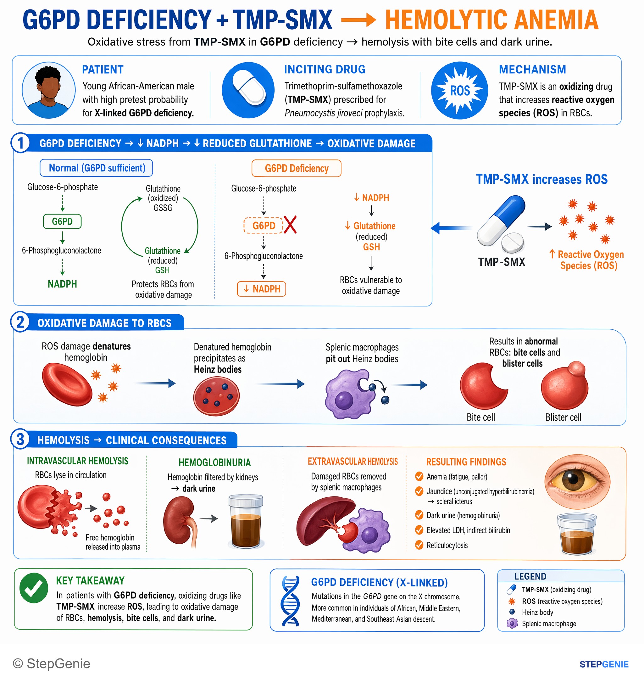
G6PD deficiency → oxidative stress from TMP-SMX causes hemolytic anemia with bite cells and dark urine in susceptible patients.
USMLE Step 1Frequently Asked Questions
What are Heinz bodies and bite cells in G6PD deficiency?
Heinz bodies are clumps of oxidized, denatured hemoglobin that precipitate inside red cells when G6PD deficiency leaves too little NADPH to keep glutathione reduced. They attach to the membrane and make the cell rigid. Bite cells form when splenic macrophages remove the Heinz bodies, leaving a semicircular notch at the cell edge. Blister cells, with a clear vacuole at the edge, often appear alongside them on the routine smear.
What stain is used to see Heinz bodies?
Heinz bodies are seen only with a supravital stain, such as crystal violet or new methylene blue, which shows them as round, dark inclusions at the edge of the red cell. They are invisible on a routine Wright or Wright-Giemsa smear, so the routine smear in G6PD deficiency shows bite and blister cells instead. Because the spleen clears Heinz bodies, they are often absent even with the right stain.
What else causes Heinz bodies and bite cells besides G6PD deficiency?
Unstable hemoglobin variants such as Hb Köln denature and form Heinz bodies, with bite and blister cells on the smear. Oxidant drugs, especially dapsone, cause dose-related hemolysis even in people with normal G6PD activity. Thalassemias can produce them because excess globin chains precipitate, and hemoglobin H is itself unstable. After splenectomy, Heinz bodies persist because no spleen remains to remove them from red cells.
Which drugs are contraindicated in G6PD deficiency?
FDA labels contraindicate rasburicase, pegloticase and methylene blue in G6PD deficiency, and tafenoquine in deficiency or unknown G6PD status; primaquine is contraindicated in severe deficiency and requires testing first. Dapsone, nitrofurantoin and trimethoprim-sulfamethoxazole carry cautions rather than contraindications. The 2023 CPIC guideline rates dapsone high risk (avoid), nitrofurantoin medium risk (use with caution) and sulfamethoxazole low-to-no risk at standard doses, although exam questions still treat sulfonamides as classic triggers.
Why can a G6PD test be normal during a hemolytic episode?
Hemolysis destroys the oldest red cells first, and they have the least G6PD activity. The marrow then releases reticulocytes and young cells that carry more enzyme, so a test during or just after an episode can read normal in a deficient patient. A recent transfusion has the same effect because donor cells are likely G6PD normal. Repeat a quantitative assay after recovery, for example about 2 months later.
Can females have G6PD deficiency?
Yes. The gene is X-linked, so males are much more likely to have significant hemolysis, but homozygous females are affected, and heterozygous females can be affected through skewed X inactivation (lyonization). A heterozygote's share of deficient red cells ranges from nearly 0% to nearly 100%, so her activity can be normal or deficient. The fluorescent spot test can miss heterozygotes, so a quantitative assay is preferred.