Gout and pseudogout both cause a sudden hot, swollen joint, and the crystals settle it: gout has needle-shaped, strongly negatively birefringent monosodium urate (MSU) crystals, while pseudogout (acute calcium pyrophosphate [CPP] crystal arthritis) has rhomboid or rod-shaped crystals that are weakly positively birefringent or not birefringent at all. Gout classically hits the first MTP joint (podagra) of a middle-aged man or postmenopausal woman; pseudogout favors the knee and wrist of an older adult, often with chondrocalcinosis and sometimes hyperparathyroidism, hemochromatosis or hypomagnesemia. Flares of both respond to colchicine, NSAIDs or glucocorticoids, but only gout has a crystal-clearing therapy: urate-lowering therapy to a serum urate below 6 mg/dL (ACR 2020).
The visual hack: Needles go Negative, Rhomboids are Positive
Picture the two crystals under the polarizing microscope:
- Gout = "Needles go Negative." Thin needles of urate, strongly negatively birefringent, floating free or swallowed by neutrophils.
- Pseudogout = "Rhomboids are Positive." Stubby rhomboids or rods of calcium pyrophosphate, weakly positively birefringent. CPP crystals can also show no birefringence at all, so a faint or non-birefringent crystal can still be CPP.
Then add the color rule, judged against the axis of the red compensator:
| Crystal | Lying parallel to the compensator axis | Lying perpendicular |
|---|---|---|
| Urate (gout) | Yellow | Blue |
| CPP (pseudogout) | Blue | Yellow |
Memory hook: YUP = Yellow when Urate is Parallel. CPP is the reverse.
Gout vs pseudogout at a glance
| Feature | Gout | Pseudogout (acute CPP crystal arthritis) |
|---|---|---|
| Crystal | Monosodium urate | Calcium pyrophosphate |
| Shape and birefringence | Needles, strongly negative | Rhomboids or rods, weakly positive or none |
| Typical patient | Man in middle age; woman after menopause (estrogen is partly protective) | Older adult: CPP deposition rises with age |
| Classic joint | First MTP; also instep, ankle, knee, wrist, elbow | Knee, wrist and other large joints; upper neck in crowned dens syndrome |
| Blood clue | Hyperuricemia, but urate is normal in at least 30% during a flare | Check calcium, PTH, magnesium, alkaline phosphatase and iron studies |
| X-ray | Punched-out erosions with overhanging edges ("rat bite"); joint space kept until late | Chondrocalcinosis: linear or punctate calcification of cartilage |
| Chronic form | Tophi, tophaceous arthritis, uric acid or calcium oxalate stones | RA-like chronic inflammatory arthritis, or OA with CPPD |
| Long-term fix | Urate-lowering therapy to a target below 6 mg/dL | No therapy removes CPP crystals |
Gout: too much urate
- Mechanism. Urate saturates body fluids above 6.8 mg/dL, and MSU crystals precipitate in and around joints. About 90% of filtered urate is reabsorbed, and renal underexcretion is by far the most common cause; pure overproduction is much less common.
- Overproduction: tumor lysis syndrome and cytotoxic therapy, leukemia, lymphoma, hemolytic anemia, psoriasis, and enzyme defects such as HGPRT deficiency (Lesch-Nyhan syndrome) and PRPP synthetase overactivity.
- Things that raise urate: thiazide and loop diuretics (see the thiazide mnemonic), low-dose aspirin, cyclosporine, tacrolimus, pyrazinamide and ethambutol; alcohol, especially beer (nonalcoholic beer too); purine-rich foods such as red meat, shellfish and organ meats; and high-fructose corn syrup.
- Flare triggers: trauma, surgery, infection, alcohol or purine binges, and any sudden change in serum urate (usually a fall), so flares cluster in the first months of allopurinol or febuxostat.
- Presentation: abrupt, often nocturnal, excruciating pain with redness, swelling and exquisite tenderness. Fever, chills and tachycardia sometimes occur. Early flares involve one joint and settle in 7-10 days; later flares can be polyarticular and last up to 3 weeks untreated.
- Chronic disease: tophi (chalky urate nodules on fingers, olecranon, Achilles tendon or ears), deforming tophaceous arthritis, and uric acid or calcium oxalate stones (see kidney stone types).
- Diagnosis: crystals in synovial fluid or a tophus aspirate. Ultrasound may show a double-contour sign (urate coating the cartilage), and dual-energy CT can map urate deposits when aspiration isn't possible.
Pseudogout: one face of CPPD disease
"Pseudogout" is the old name for acute CPP crystal arthritis. The 2023 ACR/EULAR criteria list it alongside chronic CPP crystal inflammatory arthritis, osteoarthritis with CPPD and crowned dens syndrome; asymptomatic chondrocalcinosis is also common.
- Associations: older age, hyperparathyroidism, hemochromatosis, hypomagnesemia (including Gitelman syndrome), hypophosphatasia, prior joint trauma or surgery, gout, and familial forms (usually autosomal dominant, involving the ANK pyrophosphate transporter).
- Acute flares look like gout in the knee or wrist but vary more in intensity and tend to last longer.
- Chronic patterns mimic RA (low-grade polyarthritis) or OA; OA-type changes in the wrist (radiocarpal) or the 2nd and 3rd MCPs, joints primary OA usually spares, point to CPPD. Hemochromatosis arthropathy also favors the 2nd and 3rd MCPs, so that pattern should prompt iron studies (see this hemochromatosis case).
- Crowned dens syndrome: CPP deposits around the odontoid cause acute or subacute, severe upper-neck pain, limited rotation, raised inflammatory markers and often fever; CT shows calcification of the transverse ligament behind the dens.
- Imaging: chondrocalcinosis in fibrocartilage and hyaline cartilage (knee menisci, wrist, symphysis pubis) supports the diagnosis but does not exclude gout or infection.
- Look for a cause: serum calcium, PTH, magnesium, alkaline phosphatase (low in hypophosphatasia) and iron studies, especially in younger patients with frequent flares. See the primary hyperparathyroidism review.
2023 ACR/EULAR CPPD classification criteria (built for research, but testable): CPP crystals in the fluid of a symptomatic joint or in joint tissue, or crowned dens syndrome, is sufficient on its own. Otherwise a weighted score above 56 points classifies CPPD; age over 60 at onset, typical flares, knee (+9) or wrist (+8) involvement, an associated metabolic disease and imaging evidence add points, while first MTP involvement subtracts 6. The big toe still says gout.
Don't miss septic arthritis
A hot, swollen joint needs arthrocentesis, and crystals do not exclude infection: the two can coexist. Gram stain and culture settle it; when you can't tell, treat for infection while cultures are pending.
| Synovial fluid | WBC per mcL | Clues |
|---|---|---|
| Noninflammatory (OA) | 2,000 or fewer | Clear and viscous |
| Gout flare | Usually 2,000-100,000, over 80% neutrophils | Crystals on polarized microscopy |
| Bacterial (septic) | Usually over 50,000, sometimes over 100,000 | Lower counts don't exclude it, especially early or after antibiotics |
Staphylococcus aureus is the most common cause in adults; consider Neisseria gonorrhoeae in sexually active patients. Send synovial fluid and blood cultures before antibiotics. More in septic arthritis.
Treatment: same flare drugs, different long game
Flares (gout and pseudogout)
- First line (ACR 2020, strong): colchicine, an NSAID or a glucocorticoid (oral, intra-articular or intramuscular).
- Low-dose colchicine is preferred over high-dose (similar efficacy, fewer adverse effects): 1.2 mg at the first sign, then 0.6 mg an hour later (label maximum 1.8 mg in that hour); it works best when started within 12-24 hours.
- NSAIDs at full anti-inflammatory doses (indomethacin, naproxen); peptic ulcer disease and CKD often rule them out.
- Glucocorticoids: an intra-articular injection for one large joint once infection is excluded, or oral prednisone about 0.5 mg/kg with a taper.
- IL-1 blockade if those fail or can't be used: canakinumab is FDA-approved for gout flares when NSAIDs and colchicine are contraindicated, not tolerated or ineffective and repeated steroid courses aren't appropriate; anakinra is off-label. Ice helps as an add-on.
- Pseudogout: drain the effusion and inject a glucocorticoid, or use NSAIDs, colchicine, oral glucocorticoids or anakinra; colchicine 0.6 mg once or twice daily may prevent recurrences. No drug dissolves CPP crystals.
Long-term gout care (ACR 2020)
- Start urate-lowering therapy (ULT) for at least one tophus, radiographic damage from gout, or 2 or more flares a year (strong); consider it after a first flare with CKD stage 3 or worse, urate above 9 mg/dL or urolithiasis. Asymptomatic hyperuricemia is not treated.
- Allopurinol is first line for everyone, even CKD stage 3 or worse: start at 100 mg/day or less (lower in CKD) and titrate to a serum urate below 6 mg/dL.
- Starting ULT during a flare is acceptable (ACR conditional, with anti-inflammatory cover), although older exam lore says wait; don't stop ULT that is already running.
- Prophylaxis with colchicine, an NSAID or low-dose prednisone for 3-6 months after starting ULT.
- HLA-B*58:01 testing before allopurinol in patients of Southeast Asian descent (Han Chinese, Korean, Thai) and African American patients (risk of severe skin reactions).
- Lifestyle (conditional): limit alcohol, purines and high-fructose corn syrup; lose weight if overweight; skip vitamin C. Swap hydrochlorothiazide for another antihypertensive when feasible (losartan preferred), but keep indicated low-dose aspirin.
| Drug | Mechanism | Key toxicity or exam trap |
|---|---|---|
| Colchicine | Blocks tubulin polymerization, so neutrophils can't activate, degranulate or migrate | Diarrhea, myelosuppression, myotoxicity; fatal toxicity with strong CYP3A4 or P-gp inhibitors such as clarithromycin |
| Allopurinol | Xanthine oxidase inhibitor (active metabolite oxypurinol) | Stop at the first rash (SJS/TEN, DRESS; HLA-B*58:01); cut azathioprine or mercaptopurine to about one-third to one-quarter of the usual dose |
| Febuxostat | Xanthine oxidase inhibitor | Boxed warning: more CV deaths than allopurinol in gout patients with CV disease; indicated only when allopurinol fails, isn't tolerated or isn't advisable; contraindicated with azathioprine or mercaptopurine |
| Probenecid | Blocks tubular reabsorption of urate (uricosuric) | Avoid with uric acid stones; loses effect as GFR falls; raises penicillin levels |
| Pegloticase | IV pegylated uricase that turns urate into allantoin | Boxed warning: anaphylaxis, infusion reactions, hemolysis and methemoglobinemia in G6PD deficiency; screen G6PD; give with weekly methotrexate; stop if urate climbs above 6 mg/dL |
How USMLE tests it
- A normal serum urate during a flare does not exclude gout. Measure it on more than one occasion, and aspirate the joint.
- A flare in the first months of allopurinol is the falling urate at work: keep the allopurinol, treat the flare.
- New gout after starting a thiazide or loop diuretic: blame the diuretic.
- Rhomboid crystals plus chondrocalcinosis plus diabetes, bronze skin or high calcium points to CPPD with hemochromatosis or hyperparathyroidism.
- Interaction pairs: allopurinol with azathioprine (myelosuppression), colchicine with clarithromycin (colchicine toxicity), probenecid with penicillin (higher penicillin levels).
Practice question
A 72-year-old man has 1 day of severe right knee pain and swelling and cannot bear weight. He has hereditary hemochromatosis and hypertension treated with hydrochlorothiazide. Temperature is 38.1°C (100.6°F). The knee is warm and tender with a large effusion. Radiographs show linear calcification of the menisci. Arthrocentesis yields cloudy fluid with 35,000 WBC/mcL (90% neutrophils), a negative Gram stain and rhomboid crystals that are weakly positively birefringent, appearing blue when parallel to the compensator axis. Culture is pending. Which is the most likely diagnosis?
A. Acute gout flare B. Acute CPP crystal arthritis (pseudogout) C. Septic arthritis D. Rheumatoid arthritis E. Osteoarthritis flare
Answer: B. Rhomboid, weakly positively birefringent crystals that are blue when parallel are CPP crystals; add chondrocalcinosis, age over 60, a knee and hemochromatosis (a listed CPPD-associated disease) and the picture is complete.
- A: the thiazide is a gout risk factor and is there to tempt you, but urate crystals are needles, strongly negatively birefringent and yellow when parallel.
- C: fever and a neutrophil-rich effusion fit infection, and crystals never exclude it, so follow the culture. But the Gram stain is negative, the count sits in the inflammatory range (septic fluid is usually above 50,000) and diagnostic crystals are present.
- D: RA is usually an insidious, symmetric polyarthritis of the wrists and MCPs with morning stiffness lasting over an hour, not an abrupt single hot knee with crystals.
- E: OA causes chronic mechanical pain with noninflammatory fluid (2,000 WBC/mcL or fewer), not 35,000 cells and fever.
Key takeaways
- Needles go Negative (gout); Rhomboids are Positive (pseudogout). Urate is yellow when parallel (YUP); CPP is blue when parallel.
- Gout: first MTP, underexcretion of urate, punched-out erosions with overhanging edges, tophi; urate can be normal in a flare.
- Pseudogout: knee and wrist of an older adult, chondrocalcinosis; screen for hyperparathyroidism, hemochromatosis, hypomagnesemia and hypophosphatasia.
- Crystals don't exclude septic arthritis: Gram stain and culture decide.
- Flares: low-dose colchicine, NSAIDs or glucocorticoids; canakinumab when those can't be used.
- Gout long term: allopurinol first line (even in CKD) to urate below 6 mg/dL with 3-6 months of prophylaxis; febuxostat and pegloticase carry boxed warnings. CPPD has no crystal-dissolving drug.
Sources
- Gout — Merck Manual Professional Edition (2025)
- Calcium Pyrophosphate Arthritis — Merck Manual Professional Edition (2025)
- 2020 American College of Rheumatology Guideline for the Management of Gout — American College of Rheumatology (2020)
- The 2023 ACR/EULAR Classification Criteria for Calcium Pyrophosphate Deposition Disease — American College of Rheumatology (2023)
- Gout — StatPearls, NCBI Bookshelf (2026)
- Acute Infectious Arthritis — Merck Manual Professional Edition (2024)
- Osteoarthritis (OA) — Merck Manual Professional Edition (2026)
- ULORIC (febuxostat) tablets: prescribing information — DailyMed, NLM
- KRYSTEXXA (pegloticase) injection: prescribing information — DailyMed, NLM (2026)
- COLCRYS (colchicine) tablets: prescribing information — DailyMed, NLM
- ILARIS (canakinumab) injection: prescribing information — DailyMed, NLM (2024)
- Allopurinol tablets: prescribing information — DailyMed, NLM (2024)
Visual explanations from the StepGenie QBank
Infographics StepGenie drew to explain questions in its own QBank, matched to this article's topic. Each caption is the takeaway of the question it explains.
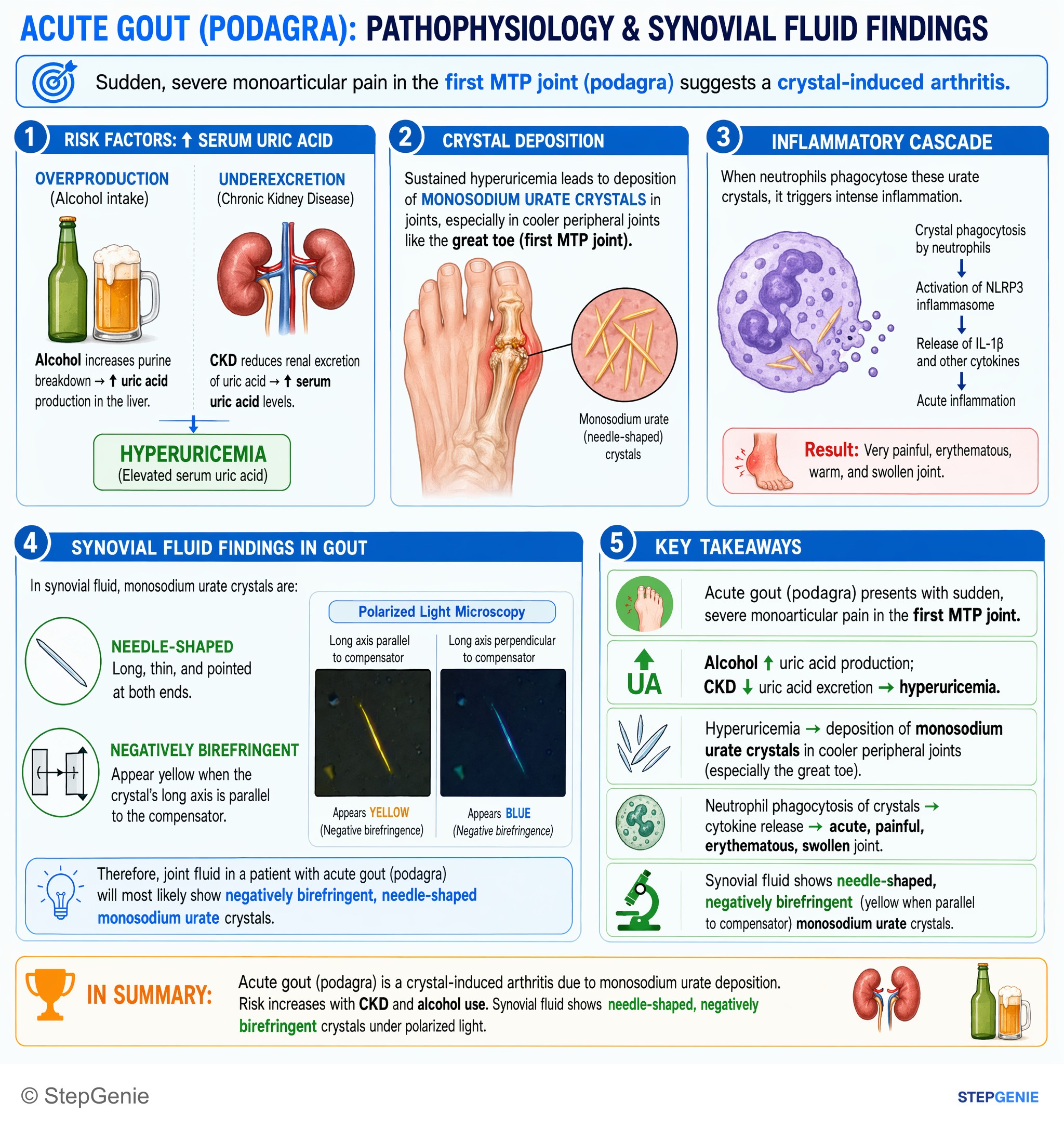
Acute gout (podagra) from monosodium urate deposition: needle-shaped, negatively birefringent crystals in joint fluid; risk ↑ with CKD and…
USMLE Step 1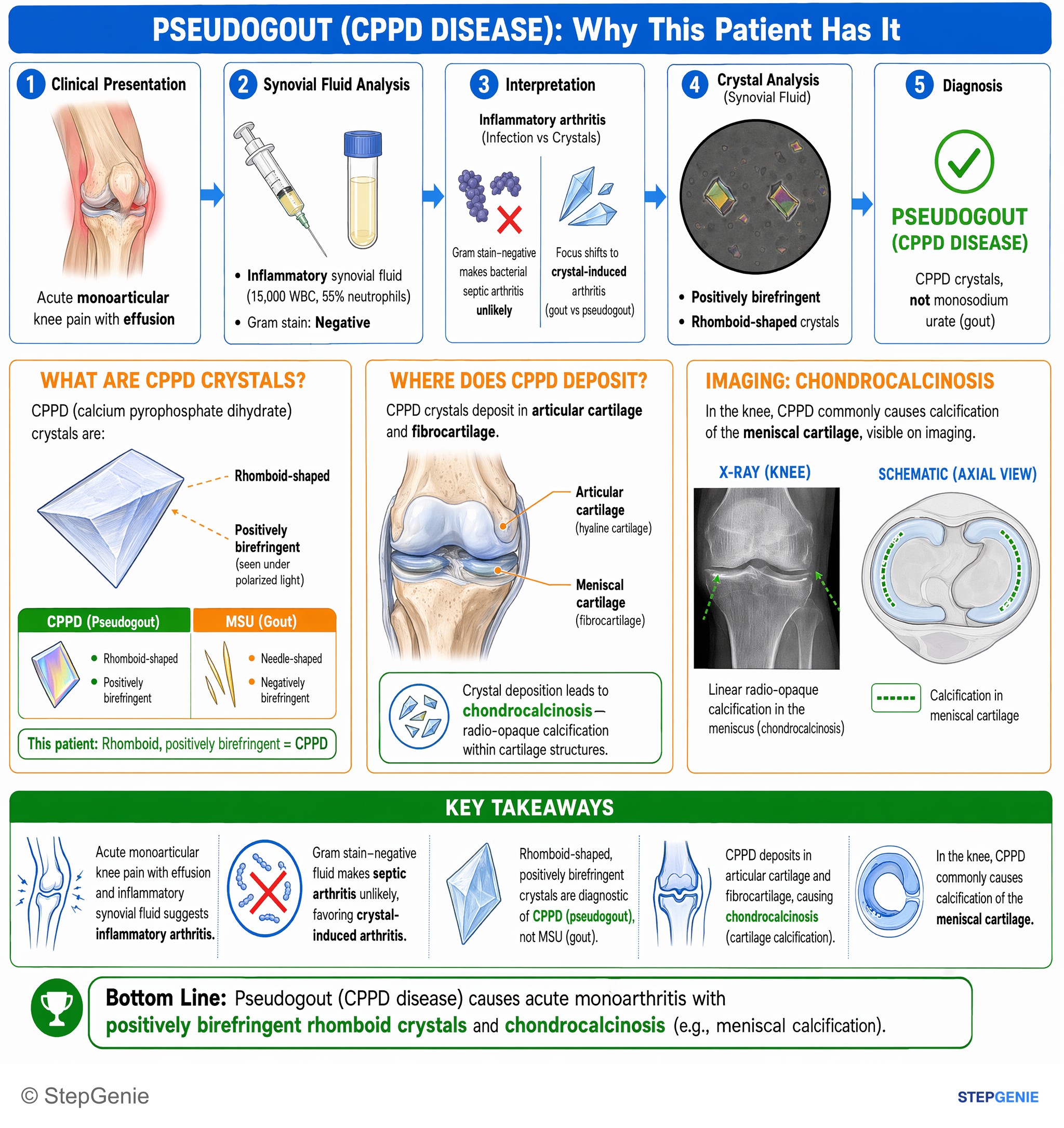
Pseudogout (CPPD disease) causes acute monoarthritis with positively birefringent rhomboid crystals and chondrocalcinosis (cartilage…
USMLE Step 1Frequently Asked Questions
How do you tell gout from pseudogout under polarized light?
Use shape, sign and color. Gout crystals are monosodium urate needles that are strongly negatively birefringent: yellow when lying parallel to the compensator axis and blue when perpendicular. Pseudogout crystals are calcium pyrophosphate rhomboids or rods that are weakly positively birefringent, or not birefringent at all: blue when parallel and yellow when perpendicular. A quick memory aid is Needles go Negative, Rhomboids are Positive, plus YUP: yellow when urate is parallel.
Which joints do gout and pseudogout usually affect?
Gout classically starts in the first metatarsophalangeal joint of the big toe (podagra) and also involves the instep, ankle, knee, wrist and elbow; early flares usually affect a single joint. Pseudogout, or acute calcium pyrophosphate crystal arthritis, usually affects the knee, wrist and other large joints of older adults. Deposits around the dens can cause crowned dens syndrome, with severe upper-neck pain and often fever. In the 2023 ACR/EULAR CPPD criteria, first MTP involvement subtracts points.
What is chondrocalcinosis and which conditions are linked to CPPD?
Chondrocalcinosis is calcification of cartilage on imaging, typically linear or punctate deposits in the knee menisci, wrist or symphysis pubis. It reflects calcium pyrophosphate deposition, which becomes more common with age. Linked conditions include hyperparathyroidism, hereditary hemochromatosis, hypomagnesemia (including Gitelman syndrome), hypophosphatasia, prior joint trauma or surgery, and familial forms. Checking calcium, PTH, magnesium, alkaline phosphatase and iron studies is reasonable, especially in younger patients with frequent flares.
Can uric acid be normal during a gout flare?
Yes. At least 30% of patients have a normal serum urate during an acute gout flare, partly because the inflammatory cytokine IL-6 increases urate excretion. A normal level therefore does not rule out gout. The diagnosis rests on finding needle-shaped, negatively birefringent urate crystals in synovial fluid or a tophus aspirate, and serum urate is best measured on more than one occasion before it is used to make decisions.
Can allopurinol be started during a gout flare?
Yes. The 2020 ACR guideline conditionally recommends starting urate-lowering therapy during a flare, when it is indicated, rather than waiting, as long as anti-inflammatory treatment covers the flare. Older teaching said to wait, so read exam questions carefully. Patients already taking allopurinol should keep taking it during the flare. When therapy starts, give colchicine, an NSAID or low-dose prednisone as prophylaxis for 3 to 6 months and titrate to a serum urate below 6 mg/dL.
Do crystals in joint fluid rule out septic arthritis?
No. Crystal arthritis and septic arthritis can coexist, and synovial fluid findings overlap, so crystals never exclude infection. Septic fluid usually has more than 50,000 white cells per microliter, sometimes more than 100,000, but lower counts occur early or after antibiotics. Gram stain and culture decide. If infection cannot be excluded, treat for it while cultures are pending. Staphylococcus aureus is the most common cause in adults; consider gonococcal arthritis in sexually active patients.