Dermatomyositis (DM) and polymyositis (PM) both cause symmetric proximal muscle weakness over weeks to months, usually with a raised CK, but DM adds a rash (heliotrope eyelids, Gottron papules) and is a complement-mediated microangiopathy with perifascicular atrophy and the strongest cancer link, while PM is a CD8+ T-cell attack on muscle fibers that express MHC class I, with no rash. Inclusion body myositis (IBM) is the look-alike in people over 50: slow weakness of the finger flexors and quadriceps that can be asymmetric, rimmed vacuoles on biopsy and little or no response to immunosuppression. The antibody sharpens the picture: anti-Jo-1 points to antisynthetase syndrome with interstitial lung disease (ILD), anti-MDA5 warns of rapidly progressive ILD, and anti-TIF1-γ or anti-NXP2 means a search for cancer.
Dermatomyositis vs polymyositis vs inclusion body myositis
| Feature | Dermatomyositis | Polymyositis | Inclusion body myositis |
|---|---|---|---|
| Who | Women more than men; adults 40–60 and children 5–15 | Adults; now an uncommon diagnosis that needs a biopsy to exclude mimics | Usually over 50; men more than women |
| Tempo | Weeks to months (can be acute in children) | Weeks to months | Months to years |
| Weakness | Symmetric proximal: shoulders, hips, neck flexors | Symmetric proximal | Proximal and distal, can be asymmetric: finger flexors, quadriceps; falls |
| Skin | Heliotrope rash, Gottron papules, shawl and V signs | None | None |
| CK | Normal to high | High | Normal or mildly raised |
| Antibodies | Anti-Mi-2, anti-MDA5, anti-TIF1-γ, anti-NXP2, anti-SAE | Anti-Jo-1 and other antisynthetase antibodies (antisynthetase syndrome can look like PM or DM) | Anti-cN1A (low specificity) |
| Biopsy | Perifascicular atrophy; perimysial and perivascular B cells and CD4+ T cells | Endomysial CD8+ T cells invading non-necrotic fibers | CD8+ invasion plus rimmed vacuoles and protein aggregates |
| Mechanism | Humoral, complement-mediated microangiopathy | Cell-mediated cytotoxicity | Inflammatory and degenerative |
| Cancer risk (IMACS) | Highest | Intermediate | Not increased |
| Response to treatment | Usually good | Usually good | Poor; no proven drug therapy |
Skin signs of dermatomyositis
| Sign | What it looks like |
|---|---|
| Heliotrope rash | Purplish discoloration of the upper eyelids, often with periorbital edema; relatively specific for DM |
| Gottron papules | Scaly, red to violaceous papules over the dorsal MCP and PIP joints; relatively specific |
| Gottron sign | Flat (non-palpable) erythematous to violaceous macules over the knuckles, elbows, knees or medial malleoli |
| Shawl and V signs | Photodistributed erythema over the upper back and shoulders (shawl) or anterior neck and chest (V) |
| Holster sign | Rash over the lateral thighs |
| Nailfolds | Periungual erythema and ragged, overgrown cuticles |
| Mechanic's hands | Hyperkeratotic, fissured skin on the radial sides of the fingers; a clue to antisynthetase syndrome |
| Calcinosis | Hard subcutaneous calcium nodules, most common in juvenile DM |
Skin disease can come before the weakness, and some patients never develop clinical muscle disease (clinically amyopathic DM), a pattern tied to anti-MDA5.
Myositis antibodies and what they predict
| Antibody | Clinical picture | Cancer risk (IMACS 2023) |
|---|---|---|
| Anti-Jo-1 (histidyl-tRNA synthetase; the most common antisynthetase antibody) | Antisynthetase syndrome: myositis, ILD, mechanic's hands, nonerosive arthritis, Raynaud phenomenon, fever | Low |
| Anti-Mi-2 | Classic DM with prominent skin disease; responds well to treatment | Intermediate (no significant link in the meta-analysis) |
| Anti-MDA5 | Amyopathic or mild DM, palmar papules, skin ulcers, rapidly progressive ILD | Intermediate |
| Anti-TIF1-γ | DM with striking skin disease and mild weakness | High; cancer risk about 4.7 times that of anti-TIF1-γ-negative myositis |
| Anti-NXP2 | DM with calcinosis, edema, dysphagia and myalgia | High |
| Anti-SRP | Necrotizing myopathy: severe weakness, dysphagia, very high CK, hard to treat | Low |
| Anti-HMGCR | Statin-associated necrotizing myopathy that can persist after the statin is stopped | Intermediate |
ANA is positive in up to 80% of patients with DM or PM, but a negative ANA does not exclude myositis.
Biopsy and mechanism: CD4 vs CD8
- Dermatomyositis: immune complexes and the membrane attack complex (C5b-9) are deposited on endomysial capillaries, capillaries drop out, and the fibers at the edge of each fascicle suffer most, producing the hallmark perifascicular atrophy. Inflammation is perimysial and perivascular, with B cells and CD4+ T cells rather than the endomysial CD8+ cells of PM; endothelial tubuloreticular inclusions appear on electron microscopy. The same vasculopathy explains the rash and the gut vasculitis of juvenile DM.
- Polymyositis: muscle fibers upregulate MHC class I, and CD8+ cytotoxic T cells surround and invade intact, non-necrotic fibers and kill them (cell-mediated cytotoxicity). The MHC class I and CD8 pairing is drawn out in MHC I vs MHC II.
- Inclusion body myositis: the same CD8+ invasion plus degeneration: rimmed (autophagic) vacuoles, congophilic β-amyloid and other protein aggregates, and cytochrome oxidase–negative fibers.
- Immune-mediated necrotizing myopathy: scattered necrotic, regenerating fibers with macrophages and little lymphocytic inflammation.
- When to biopsy: not usually needed when the DM rash is characteristic; recommended before treating suspected PM to exclude metabolic myopathies, necrotizing myopathy and other mimics. Choose a weak muscle or one that is edematous on MRI.
Workup
- Muscle enzymes: CK is the most specific; aldolase, AST, ALT and LDH also rise, so "transaminitis" in a weak patient may be muscle, not liver. CK can be normal in DM and in wasted muscle.
- Antibodies: a myositis-specific and myositis-associated antibody panel in every adult with new myositis, both for phenotype and for cancer risk.
- EMG and MRI: EMG separates myopathy from nerve disease and MRI shows muscle edema; both help pick the biopsy site.
- Organ screen: chest CT and pulmonary function tests including DLCO for ILD, echocardiography if pulmonary hypertension is suspected, and a swallow study for dysphagia.
- Classification: the 2017 EULAR/ACR criteria give the most points to anti-Jo-1, the heliotrope rash, Gottron sign and papules and, on biopsy, rimmed vacuoles.
Malignancy screening
Adult-onset myositis carries an increased cancer risk concentrated in the 3 years before and after onset, and DM has the highest risk (risk ratio 2.21 compared with other subtypes in the IMACS meta-analysis). Lung, ovarian, colorectal and breast cancers and lymphoma are among the most common, with nasopharyngeal carcinoma in East and Southeast Asian patients. Cancer is the leading cause of death in adults with myositis. Treating the tumor can make the myositis remit, and cancer-associated myositis is more resistant to glucocorticoids.
The 2023 IMACS guideline stratifies risk:
| Risk group | Definition | Screening |
|---|---|---|
| High | 2 or more high-risk factors: DM, anti-TIF1-γ, anti-NXP2, onset after age 40, persistent activity despite treatment, moderate to severe dysphagia, skin necrosis or ulceration | Basic plus enhanced screening at diagnosis, basic screening yearly for 3 years; consider PET-CT and endoscopy |
| Moderate | 1 high-risk factor, or 2 or more intermediate ones (e.g., PM, clinically amyopathic DM, anti-MDA5, anti-Mi-2, anti-HMGCR, male sex) | Basic plus enhanced screening at diagnosis |
| Standard | Everyone else (e.g., a young adult with anti-Jo-1 antisynthetase syndrome) | Basic screening at diagnosis |
Basic = history and examination, CBC, liver tests, ESR and CRP, serum protein electrophoresis with free light chains, urinalysis and chest radiograph. Enhanced = CT of the neck, chest, abdomen and pelvis, plus cervical screening, mammography, PSA, CA-125, pelvic ultrasound and fecal occult blood testing. All patients stay in routine age-appropriate screening; juvenile myositis and IBM need no routine extra screening. The tumor side of this link is covered in the paraneoplastic syndromes Q-bank.
Treatment
- First line for DM and PM: high-dose glucocorticoids, such as prednisone about 1 mg/kg a day (up to 80 mg); IV methylprednisolone pulses for severe disease with dysphagia or respiratory muscle weakness.
- Steroid-sparing agent from the start or soon after: methotrexate, azathioprine or tacrolimus, so prednisone can be tapered, ideally within about 6 months. With ILD, azathioprine, tacrolimus, mycophenolate or rituximab is preferred over methotrexate.
- IVIG: for dysphagia, respiratory muscle involvement, refractory disease or steroid toxicity; in 2021 the FDA approved an IVIG product (Octagam 10%) for adult DM.
- Skin: sun protection and topical steroids; hydroxychloroquine, methotrexate, mycophenolate or IVIG for more extensive disease.
- Anti-MDA5 rapidly progressive ILD: IV cyclophosphamide and plasma exchange can be considered.
- Monitoring: CK usually falls toward normal within 6–12 weeks, and strength recovers later.
- Supportive care: speech and swallow therapy for dysphagia, bone protection on long-term glucocorticoids, and Pneumocystis prophylaxis on combination immunosuppression.
- IBM: no cure; it is generally unresponsive to glucocorticoids and immunosuppressants, so care centers on physical therapy and dysphagia management.
Mimics and traps
- Statins: simple statin myopathy improves after stopping the drug. Weakness with a very high CK that persists after withdrawal points to anti-HMGCR necrotizing myopathy, which needs immunosuppression; statin toxicity is reviewed in the statins guide.
- Steroid myopathy: a patient on high-dose glucocorticoids who gets weaker after an initial response, with a normal CK, has steroid myopathy until proven otherwise.
- "Refractory polymyositis" in an older man with finger flexor weakness is IBM until the biopsy says otherwise.
- Ptosis or diplopia points away from myositis, which spares the eye and face muscles; think of myasthenia gravis.
- Juvenile DM brings calcinosis and gut vasculitis but no routine need for cancer screening.
Practice question
Vignette. A 61-year-old woman has 2 months of trouble climbing stairs and lifting her arms to wash her hair, plus difficulty swallowing solid food. She has violaceous discoloration and swelling of both upper eyelids, scaly violaceous papules over the MCP and PIP joints, and erythema over the upper back and shoulders. Neck flexion is weak. CK is 3,900 U/L, and AST and ALT are mildly elevated. A myositis antibody panel is sent. Which antibody is most strongly associated with an underlying cancer in this condition?
- A. Anti-Jo-1
- B. Anti-Mi-2
- C. Anti-MDA5
- D. Anti-TIF1-γ
- E. Anti-HMGCR
Answer: D. Heliotrope rash, Gottron papules, a shawl sign and proximal weakness make this dermatomyositis, and anti-TIF1-γ is the antibody most strongly tied to cancer in adults. Even before the result returns, DM with onset after 40 already puts her in the IMACS high-risk group: basic plus enhanced screening now, basic screening yearly for 3 years, and PET-CT considered if that screening finds no cancer. Her raised AST and ALT most likely come from muscle.
Why not the others?
- A. Anti-Jo-1: antisynthetase syndrome with ILD, mechanic's hands, arthritis and Raynaud phenomenon; a low-risk marker for cancer.
- B. Anti-Mi-2: classic DM with prominent skin disease that responds well to treatment; no significant cancer link in the IMACS meta-analysis.
- C. Anti-MDA5: amyopathic DM with palmar papules, skin ulcers and rapidly progressive ILD; the danger is the lung.
- E. Anti-HMGCR: statin-associated necrotizing myopathy with a very high CK and no rash; IMACS counts it only as an intermediate cancer-risk factor.
Key takeaways
- DM = proximal weakness + rash (heliotrope, Gottron papules); complement-mediated capillary injury, perifascicular atrophy, perivascular B cells and CD4+ T cells.
- PM = proximal weakness without rash; endomysial CD8+ T cells invading MHC class I–positive, non-necrotic fibers; now uncommon and a biopsy-confirmed diagnosis.
- IBM = over 50, finger flexors and quadriceps, can be asymmetric, rimmed vacuoles, steroid-resistant.
- Anti-Jo-1 → ILD (antisynthetase syndrome); anti-MDA5 → rapidly progressive ILD; anti-TIF1-γ and anti-NXP2 → cancer; anti-Mi-2 → classic, treatment-responsive DM.
- Screen adults with DM for cancer by IMACS risk group; the risk clusters within 3 years of onset.
- Treat with glucocorticoids plus a steroid-sparing agent; IVIG for dysphagia or refractory disease.
Sources
- Idiopathic Inflammatory Myopathies — Merck Manual Professional Edition (2024, updated 2026)
- Oldroyd AGS, et al. International Guideline for Idiopathic Inflammatory Myopathy-Associated Cancer Screening (IMACS) — Nature Reviews Rheumatology (2023)
- Inflammatory Myopathies — ARUP Consult, ARUP Laboratories
- Classification Criteria for Idiopathic Inflammatory Myopathies (2017 EULAR/ACR web calculator and glossary) — Karolinska Institutet
- Dermatomyositis + Vascular Pathology (muscle biopsy) — Neuromuscular Disease Center, Washington University in St. Louis (2026)
- Inflammation (muscle biopsy pathology) — Neuromuscular Disease Center, Washington University in St. Louis (2026)
- Inclusion Body Myositis (IM-VAMP) pathology — Neuromuscular Disease Center, Washington University in St. Louis (2025)
- Inclusion Body Myositis — National Institute of Neurological Disorders and Stroke
- Dermatomyositis — MedlinePlus Medical Encyclopedia
- Dermatomyositis: Myositis and the Skin — Hospital for Special Surgery
- Antisynthetase Syndrome — The Myositis Association
- Octagam (Immune Globulin Intravenous) indications — US Food and Drug Administration
Visual explanations from the StepGenie QBank
Infographics StepGenie drew to explain questions in its own QBank, matched to this article's topic. Each caption is the takeaway of the question it explains.
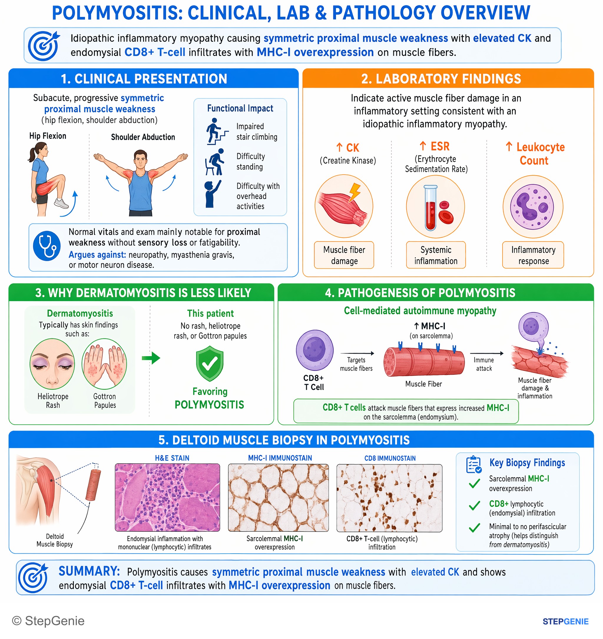
Polymyositis causes symmetric proximal muscle weakness with elevated CK and shows endomysial CD8+ T-cell infiltrates with MHC-I…
USMLE Step 1Frequently Asked Questions
What is the difference between dermatomyositis and polymyositis?
Both cause symmetric weakness of the shoulder and hip girdle muscles, usually with a raised CK. Dermatomyositis adds characteristic skin signs such as a heliotrope rash and Gottron papules, is driven by complement-mediated injury to small vessels, and shows perifascicular atrophy on biopsy. Polymyositis has no rash, is caused by CD8+ T cells invading muscle fibers that express MHC class I, and carries a lower cancer risk.
Which antibodies are associated with dermatomyositis?
Dermatomyositis-specific antibodies include anti-Mi-2 (classic rash, good treatment response), anti-MDA5 (amyopathic disease, skin ulcers and rapidly progressive interstitial lung disease), anti-TIF1-gamma and anti-NXP2 (higher cancer risk, with calcinosis for NXP2) and anti-SAE. Anti-Jo-1 and other antisynthetase antibodies define antisynthetase syndrome, which can look like dermatomyositis or polymyositis and features interstitial lung disease, mechanic's hands, arthritis and Raynaud phenomenon.
What cancers are associated with dermatomyositis?
Adult-onset myositis, especially dermatomyositis, raises cancer risk mainly in the 3 years before and after onset. The 2023 IMACS guideline lists lung, ovarian, colorectal and breast cancer and lymphoma among the most common, with nasopharyngeal carcinoma in East and Southeast Asian patients. How intensively to screen depends on risk factors such as onset after age 40, anti-TIF1-gamma or anti-NXP2 antibodies, dysphagia and skin ulceration.
How is inclusion body myositis different from polymyositis?
Inclusion body myositis usually begins after age 50, is more common in men, progresses over years and weakens the finger flexors and quadriceps, sometimes asymmetrically, while polymyositis causes symmetric proximal weakness over weeks to months. Both show CD8+ T cells invading muscle fibers, but inclusion body myositis also has rimmed vacuoles and protein aggregates. It generally does not respond to glucocorticoids or other immunosuppressants and is not linked to an increased cancer risk.
What is perifascicular atrophy?
Perifascicular atrophy is shrinkage of the muscle fibers at the edges of each fascicle and is the hallmark biopsy finding of dermatomyositis. It is thought to reflect injury to the muscle's small blood vessels, with complement deposited on capillaries and capillary loss, which leaves the border zone at the edge of each fascicle relatively ischemic. It is one of the biopsy features scored in the 2017 EULAR/ACR classification criteria.
What is antisynthetase syndrome?
Antisynthetase syndrome is an inflammatory myopathy defined by antibodies against tRNA synthetase enzymes, most often anti-Jo-1. Typical features are myositis, interstitial lung disease, nonerosive arthritis, Raynaud phenomenon, fever and mechanic's hands, which are thickened, cracked skin on the sides of the fingers. Interstitial lung disease is often the most serious feature, and the cancer risk is lower than in classic dermatomyositis.