Systemic lupus erythematosus (SLE) is one of those Step 1 “great imitators” that can look like anything—rash, arthritis, kidney failure, seizures, cytopenias—yet it’s built on a few repeatable, testable immune mechanisms. If you anchor SLE to its autoantibodies, immune complex deposition, and a handful of classic clinical patterns, you’ll stop memorizing random facts and start recognizing vignettes instantly.
Where SLE Fits in MSK (Bone & Joint Disorders)
Even though SLE is a systemic autoimmune disease, it’s extremely testable in the MSK bucket because it commonly presents with:
- Inflammatory, symmetric arthralgias/arthritis (often hands/wrists)
- Non-erosive arthritis (key distinction vs rheumatoid arthritis)
- Tendon/ligament laxity → reducible deformities (Jaccoud arthropathy)
Step framing: SLE = autoimmune + immune complex disease that frequently shows up as a joint complaint plus multi-organ clues.
Definition (What SLE Is)
Systemic lupus erythematosus is a chronic, relapsing-remitting autoimmune disease characterized by:
- Loss of self-tolerance → autoantibody production
- Type III hypersensitivity: immune complex deposition → inflammation and tissue damage
- Some manifestations are also Type II hypersensitivity (antibody-mediated cytopenias)
High-yield epidemiology:
- Classically affects women of childbearing age
- Higher prevalence/severity in African American and Hispanic populations (commonly tested association)
Pathophysiology (How It Happens)
Core immune mechanism: immune complexes (Type III)
- Autoantibodies form against nuclear antigens (DNA, histones, Sm, RNP)
- Antigen–antibody complexes circulate
- Deposition occurs in:
- Kidneys (glomeruli)
- Skin
- Joints
- Serosal surfaces (pleura/pericardium)
- Complement is activated → inflammation
→ low complement levels (consumption)
High-yield lab consequence:
- Low C3 and C4 during active disease (esp. nephritis)
Why SLE makes autoantibodies (Step 1-friendly explanation)
- Defective clearance of apoptotic debris → nuclear antigens persist
- Increased exposure of nuclear material to immune system
- B cell hyperactivity + T cell dysregulation → autoantibodies
Hypersensitivity types tested in SLE
| Manifestation | Hypersensitivity Type | Classic Example |
|---|---|---|
| Nephritis, arthritis, rash, serositis | Type III | Immune complex deposition |
| Hemolytic anemia, thrombocytopenia, leukopenia | Type II | Autoantibodies against blood cells |
Clinical Presentation (How It Shows Up on Step)
The “classic” symptom clusters
MSK
- Symmetric arthralgia/arthritis
- Non-erosive inflammatory arthritis
- Morning stiffness can occur, but X-rays lack erosions (unlike RA)
Skin/Mucosa
- Malar rash (spares nasolabial folds is a common teaching point)
- Photosensitivity
- Oral/nasal ulcers (often painless)
Kidney
- Lupus nephritis: hematuria, proteinuria, RBC casts
- May present as edema/HTN or incidental abnormal urinalysis
Serositis
- Pleuritis or pericarditis (pleuritic chest pain, friction rub)
Neuro/Psych
- Seizures, psychosis, cognitive dysfunction (varies)
Hematologic
- Autoimmune hemolytic anemia, leukopenia, thrombocytopenia
High-yield “pattern recognition” clue list
If you see young woman + joint pain + rash + proteinuria → think SLE first.
Diagnosis (What Confirms It)
Screening vs specific antibodies (know the hierarchy)
| Test | Sensitivity | Specificity | Step takeaway |
|---|---|---|---|
| ANA | High | Low | Great screening test; many false positives |
| Anti–dsDNA | Moderate | High | Correlates with lupus nephritis and disease activity |
| Anti-Smith (Sm) | Low | Very high | Most specific for SLE |
| Anti-histone | — | — | Suggests drug-induced lupus |
High-yield: ANA positive is common, but anti-dsDNA and anti-Sm are the classic Step 1 “confirmers.”
Complement and inflammatory markers
- Low C3/C4 suggests active immune complex disease (often nephritis)
- ESR may be elevated; CRP can be variable (CRP spikes more with infection—useful in differentials)
Renal evaluation (must-know for Step)
- Urinalysis: proteinuria, hematuria, RBC casts
- If nephritis suspected → kidney biopsy determines class and treatment intensity
Lupus nephritis classes (high-yield summary)
| Class | Path | Key association |
|---|---|---|
| I | Minimal mesangial | Mild/normal LM |
| II | Mesangial proliferative | Mild hematuria/proteinuria |
| III | Focal proliferative | <50% glomeruli |
| IV | Diffuse proliferative | Most common & most severe; “wire-loop” lesions |
| V | Membranous | Nephrotic syndrome pattern |
| VI | Advanced sclerosing | ESRD |
Wire-loop lesions = thickened capillary loops from subendothelial immune complex deposition (classic for Class IV).
Treatment (How You Manage It—Step 1 + Step 2 overlap)
Baseline management (most patients)
- Hydroxychloroquine for skin/joint symptoms and flare prevention
- Key adverse effect: retinopathy (requires eye exams)
- NSAIDs for mild inflammatory joint pain (watch kidneys/GI)
Moderate to severe flares / organ-threatening disease
- Glucocorticoids (systemic) for significant inflammation
- Steroid-sparing immunosuppressants depending on organ:
- Mycophenolate mofetil: common for lupus nephritis
- Cyclophosphamide: severe nephritis/CNS disease (toxicity-heavy)
- Azathioprine: maintenance in some cases
Biologics (high-yield mention)
- Belimumab (anti-BAFF) can be used in refractory cases (often tested as “newer SLE drug”)
High-Yield Associations & Classic Vignette Traps
1) Drug-induced lupus (DIL)
Clues
- Lupus-like symptoms after starting certain meds
- Usually no renal/CNS involvement (classic teaching)
- Labs: anti-histone antibodies
High-yield drugs (think “HIND”)
- Hydralazine
- Isoniazid
- Nprocainamide (often written as Procainamide)
- D-penicillamine (plus others like anti-TNF agents)
2) Antiphospholipid syndrome (APS) in SLE
Can coexist with SLE and is extremely testable.
Clues
- Arterial/venous thrombosis
- Recurrent pregnancy loss
- Livedo reticularis (sometimes)
Labs
- Anticardiolipin, anti–β2-glycoprotein I, lupus anticoagulant
- Paradox: prolonged PTT but hypercoagulable
3) Neonatal lupus and congenital heart block
Maternal antibodies can cross placenta:
- Anti-Ro (SSA) and anti-La (SSB)
Associated with: - Neonatal rash
- Congenital heart block (classic association)
4) SLE vs RA (MSK distinction you’ll be asked)
| Feature | SLE | RA |
|---|---|---|
| Joint damage | Non-erosive | Erosive |
| Deformities | Can be reducible (Jaccoud) | Fixed deformities |
| Antibodies | ANA, anti-dsDNA, anti-Sm | RF, anti-CCP |
| Extra-articular | Kidneys, CNS, serositis | Lungs (nodules, ILD), vasculitis |
“First Aid” Cross-References (What to Tie Together While Studying)
Use these as conceptual anchors (section names match the way First Aid organizes content across editions):
- Immunology: Hypersensitivity reactions
- SLE = primarily Type III, cytopenias = Type II
- Rheumatology: SLE and autoimmune connective tissue diseases
- Autoantibodies (ANA, anti-dsDNA, anti-Sm), malar rash, arthritis, nephritis
- Renal: Nephritic/nephrotic syndromes & glomerulonephritis
- Lupus nephritis classes, “wire-loop” lesions (diffuse proliferative)
- Heme/Onc: Autoimmune hemolytic anemia and thrombocytopenia
- Coombs-positive hemolysis; immune thrombocytopenia
- Reproductive/OB (often Step 2, but shows up on Step 1 questions): APS + pregnancy loss
- Hypercoagulability + prolonged PTT paradox
Rapid-Fire USMLE High-Yield Checklist
If you remember nothing else, remember this:
- ANA: sensitive screening, not specific
- Anti–dsDNA: nephritis + activity
- Anti-Smith: most specific
- Low C3/C4: active immune complex disease
- Type III hypersensitivity: nephritis, rash, arthritis, serositis
- Type II hypersensitivity: cytopenias
- Non-erosive symmetric arthritis
- Wire-loop lesions: Class IV nephritis
- Anti-histone: drug-induced lupus
- APS: thrombosis + pregnancy loss + prolonged PTT
- Anti-Ro/SSA: congenital heart block (neonatal lupus)
Visual explanations from the StepGenie QBank
Infographics StepGenie drew to explain questions in its own QBank, matched to this article's topic. Each caption is the takeaway of the question it explains.
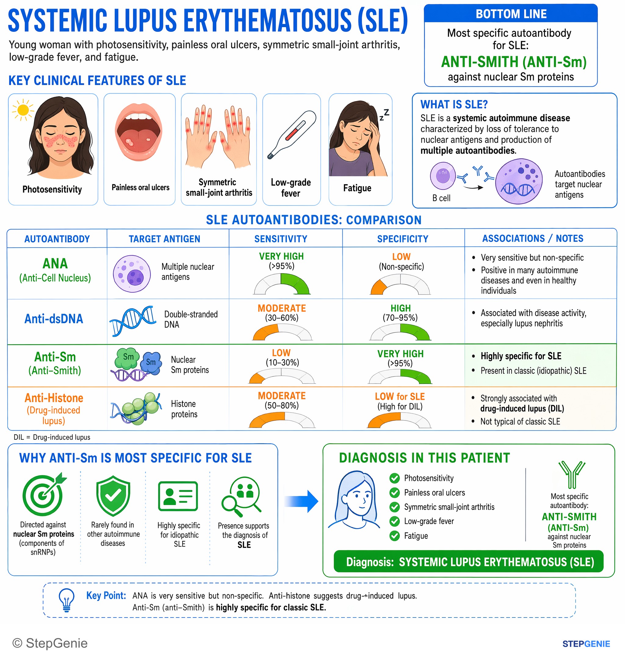
Systemic lupus erythematosus (SLE) with photosensitivity, arthritis, oral ulcers; most specific autoantibody is anti-Smith (anti-Sm)…
USMLE Step 1Frequently Asked Questions
How do you tell SLE arthritis from rheumatoid arthritis on Step 1?
SLE arthritis is classically a symmetric inflammatory arthritis that is non-erosive. The key distinction from rheumatoid arthritis is that patients can have joint pain, stiffness, and even deformities from tendon or ligament laxity, but X-rays do not show erosions. This makes a vignette with inflammatory hand or wrist symptoms plus multi-organ findings point more toward SLE than RA.
Why does SLE cause low complement levels?
SLE causes low complement because it is driven by immune complex deposition with complement activation. Autoantibodies form against nuclear antigens, the antigen-antibody complexes circulate and deposit in tissues such as the kidneys, skin, joints, and serosal surfaces, and complement is then consumed during the inflammatory response. Low C3 and C4 are especially associated with active disease, particularly lupus nephritis.
Which antibodies are most useful for diagnosing SLE?
ANA is the best screening test, while anti-dsDNA and anti-Smith are the classic confirmatory antibodies. ANA is highly sensitive but not specific, so false positives are common. Anti-dsDNA has high specificity and also correlates with lupus nephritis and disease activity. Anti-Smith is the most specific antibody for SLE, making it especially high-yield for confirming the diagnosis.
What clue should make you think of lupus nephritis?
Proteinuria, hematuria, and RBC casts should make you think of lupus nephritis. In SLE, kidney involvement may also present with edema, hypertension, or an incidental abnormal urinalysis. Anti-dsDNA antibodies and low C3 and C4 further support active immune complex disease affecting the kidneys. If nephritis is suspected, kidney biopsy is used to determine the class and guide treatment intensity.
What is the first-line treatment for most patients with SLE?
Hydroxychloroquine is baseline treatment for most patients with SLE. It is used for skin and joint symptoms and also helps prevent flares. NSAIDs can be added for mild inflammatory joint pain, while systemic glucocorticoids and immunosuppressants are used for more severe or organ-threatening disease. A key adverse effect of hydroxychloroquine is retinopathy, so patients need eye exams.