Systemic sclerosis (scleroderma) is an autoimmune disease of small-vessel damage plus fibrosis of the skin and internal organs, and it comes in two main forms. Limited cutaneous systemic sclerosis (CREST) thickens skin only on the face and distal to the elbows and knees, progresses slowly, carries anticentromere antibodies, and its signature late complication is pulmonary arterial hypertension (PAH). Diffuse cutaneous systemic sclerosis spreads proximal to the elbows and knees and onto the trunk, moves fast and carries anti-Scl-70 (anti-topoisomerase I), which flags interstitial lung disease (ILD), or anti-RNA polymerase III, which flags scleroderma renal crisis. Renal crisis is treated with an ACE inhibitor started at once (captopril is preferred), and high-dose glucocorticoids can trigger it.
Limited vs diffuse at a glance
| Feature | Limited cutaneous (CREST) | Diffuse cutaneous |
|---|---|---|
| Skin thickening | Fingers, hands, forearms, lower legs and face: distal to the elbows and knees | Also proximal to the elbows and knees: upper arms, thighs, trunk |
| Tempo | Slow, over years | Rapid; internal organs involved early |
| Signature antibody | Anticentromere | Anti-Scl-70 (topoisomerase I) or anti-RNA polymerase III |
| Lungs | PAH, usually later in the course | ILD more common |
| Kidneys | Renal crisis uncommon | Scleroderma renal crisis, mostly in the first 4–5 years |
| 10-year survival (Merck) | 92% | 65% |
Both forms share Raynaud phenomenon (the most common first symptom), esophageal dysmotility, digital ulcers and telangiectasias. Systemic sclerosis sine scleroderma has the antibodies and organ disease without skin thickening. Localized scleroderma (morphea, linear scleroderma) is a different disease: patches or streaks of thickened skin without internal organ involvement.
Why it happens
- Vascular injury first: endothelial damage and intimal proliferation narrow small arteries, which explains Raynaud phenomenon, digital ulcers and pitting scars, PAH and renal crisis.
- Immune activation: autoantibodies and cytokines, especially TGF-β, turn fibroblasts into myofibroblasts.
- Fibrosis: myofibroblasts overproduce type I and type III collagen, and collagen-degrading enzymes fall, so skin, lungs, gut and heart stiffen.
Systemic sclerosis is about 5 times more common in women and is most common between ages 20 and 50.
CREST decoded
| Letter | Feature | Exam clue |
|---|---|---|
| C | Calcinosis cutis | Firm calcium deposits in the fingertips and skin that can ulcerate |
| R | Raynaud phenomenon | Cold- or stress-triggered pallor, then cyanosis, then redness on rewarming; in systemic sclerosis it causes painful fingertip ulcers |
| E | Esophageal dysmotility | Fibrosis weakens peristalsis and makes the lower esophageal sphincter incompetent: dysphagia, reflux, strictures; Merck puts Barrett esophagus at about one-third of patients |
| S | Sclerodactyly | Tight, shiny finger skin with tapered fingertips and restricted motion |
| T | Telangiectasias | Dilated vessels on the fingers, face, lips and tongue |
Autoantibodies and what each one predicts
| Antibody | Subset | What it warns about |
|---|---|---|
| ANA | Positive in at least 90% of patients, often with a nucleolar pattern | Sensitive screen, not specific |
| Anticentromere | Limited cutaneous (CREST) | PAH later in the course; limited disease carries a lower risk of lung fibrosis and renal crisis |
| Anti-Scl-70 (anti-topoisomerase I) | Diffuse cutaneous | ILD: needs closer lung monitoring |
| Anti-RNA polymerase III | Diffuse cutaneous, rapidly progressive skin | Renal crisis (patients check blood pressure at home) and cancer |
| Anti-U1 RNP, high titer | Mixed connective tissue disease, not systemic sclerosis | Overlap of lupus, scleroderma and polymyositis features |
Classification (2013 ACR/EULAR): skin thickening of the fingers of both hands extending proximal to the MCP joints is enough on its own (9 points). Otherwise, points are added for puffy fingers or sclerodactyly, fingertip ulcers or pitting scars, telangiectasia, abnormal nailfold capillaries, PAH or ILD, Raynaud phenomenon and SSc-specific antibodies; a total of 9 or more classifies systemic sclerosis.
Organ by organ: findings and treatment
| Organ | What happens | Screen or test | Treatment |
|---|---|---|---|
| Blood vessels | Raynaud phenomenon, fingertip ulcers | Nailfold capillaries | Avoid cold; long-acting dihydropyridine calcium channel blocker (nifedipine) first; PDE-5 inhibitors or bosentan (which may cut new ulcers) next; no beta-blockers |
| Skin | Puffy fingers, then tight skin; masklike face; small mouth | Examination | Immunosuppression (mycophenolate, methotrexate) for active, spreading skin disease |
| Esophagus and gut | Reflux, dysphagia, strictures; small bowel hypomotility with bacterial overgrowth and malabsorption | Endoscopy | High-dose PPI, small frequent meals, head of bed raised; dilation for strictures |
| Lungs: ILD | Dry cough, dyspnea, restrictive PFTs with low DLCO; more common in diffuse disease and with anti-Scl-70 | Screen with PFTs plus high-resolution CT (2023 ACR); once ILD is found, PFTs every 3–6 months for the first year, then less often if stable | Mycophenolate (as effective as cyclophosphamide and better tolerated); nintedanib or tocilizumab, both FDA-approved to slow lung function decline in SSc-ILD; rituximab; transplant referral if it progresses |
| Lungs: PAH | Exertional dyspnea and a low DLCO; about 15% of patients, usually later and mostly in limited disease | Echocardiography, confirmed by right heart catheterization | Endothelin receptor antagonists, PDE-5 inhibitors (up-front ambrisentan plus tadalafil is FDA-approved for PAH) and prostacyclins such as epoprostenol |
| Heart | Myocardial fibrosis, conduction disease, arrhythmias, pericardial effusion | ECG, echocardiography | Treat the complication |
| Kidneys | Scleroderma renal crisis | Blood pressure and creatinine checks in early diffuse disease | ACE inhibitor (below) |
Lung disease (ILD and PAH) is now the most common cause of death in systemic sclerosis. The 2023 American College of Rheumatology ILD guideline conditionally recommends mycophenolate, azathioprine, rituximab, cyclophosphamide, tocilizumab or nintedanib as first-line options for SSc-ILD and strongly recommends against glucocorticoids as first-line ILD treatment. Autologous stem cell transplant is an option in selected patients with early diffuse disease: survival after the first year is better than with IV cyclophosphamide, but first-year mortality is higher.
Scleroderma renal crisis: the must-know emergency
- Who: about 10% of patients with systemic sclerosis, mostly those with early, rapidly progressive diffuse disease and anti-RNA polymerase III; about 75% of cases occur within 4 years of diagnosis. Glucocorticoids are a risk factor, so high doses are avoided.
- Why: narrowed renal arteries suddenly clamp down ("Raynaud phenomenon of the kidney"), the ischemic cortex releases renin, and angiotensin II drives malignant hypertension that injures the vessels further.
- What you see: abrupt severe hypertension, a rapidly rising creatinine and thrombotic microangiopathy (microangiopathic hemolytic anemia with schistocytes and low platelets). About 10% of cases are normotensive, and those carry a worse prognosis.
- Treatment: start an ACE inhibitor immediately; captopril is preferred because its short half-life allows fast titration. Keep it going even as creatinine rises: the 2017 EULAR recommendations call for high-dose ACE inhibition continued long term while kidney function may still improve. Temporary or long-term dialysis may be needed. Starting an ACE inhibitor before a crisis, as prevention, is not supported by evidence. The class effects are reviewed in the ACE inhibitors vs ARBs mnemonic.
Practice question
Vignette. A 44-year-old woman has 8 months of skin thickening that has spread from her fingers to her forearms, upper arms and chest. Raynaud phenomenon began a year ago. She is anti-RNA polymerase III positive. Two weeks ago, prednisone 40 mg daily was started for joint pain. She now has a severe headache and blurred vision. Blood pressure is 210/120 mm Hg. Creatinine is 2.9 mg/dL (0.8 mg/dL a month ago), hemoglobin is 9.1 g/dL with schistocytes on the smear, and platelets are 88,000/µL. What is the most appropriate next step in management?
- A. Start captopril
- B. Give pulse IV methylprednisolone
- C. Start plasma exchange
- D. Avoid ACE inhibitors because of the rising creatinine and start amlodipine
- E. Obtain a renal biopsy before starting treatment
Answer: A. Early diffuse skin disease, anti-RNA polymerase III, recent glucocorticoids, malignant hypertension, acute kidney injury and thrombotic microangiopathy add up to scleroderma renal crisis, and an ACE inhibitor started at once is what changes survival.
Why not the others?
- B. Glucocorticoids are a risk factor for renal crisis and do not treat it; this prednisone course probably helped trigger it.
- C. Schistocytes with low platelets suggest TTP, but here the microangiopathy follows from malignant hypertension in diffuse scleroderma, and the treatment is ACE inhibition.
- D. A rising creatinine is not a reason to withhold an ACE inhibitor in renal crisis. The crisis is driven by renin and angiotensin II, and prompt ACE inhibition is what improves survival; a calcium channel blocker alone does not block that loop.
- E. The diagnosis is clinical, and delaying the ACE inhibitor costs kidney function.
Exam traps
- Scleroderma esophagus vs achalasia: scleroderma weakens the lower esophageal sphincter and causes reflux; achalasia is a sphincter that fails to relax, compared in the achalasia Q-bank breakdown.
- Primary vs secondary Raynaud phenomenon: onset after 40, fingertip ulcers, abnormal nailfold capillaries or a positive ANA point to an underlying disease such as systemic sclerosis.
- Beta-blockers are contraindicated in Raynaud phenomenon.
- Proximal weakness with a high CK in a scleroderma patient suggests an overlap myositis; the myositis patterns are in the dermatomyositis vs polymyositis guide.
- Anti-U1 RNP means mixed connective tissue disease, where serious kidney disease is uncommon.
- Glucocorticoids can precipitate renal crisis and are not first-line for SSc-ILD.
Key takeaways
- Limited (CREST) = distal skin and face, slow, anticentromere, PAH later.
- Diffuse = proximal and trunk skin, fast, anti-Scl-70 → ILD, anti-RNA polymerase III → renal crisis (and cancer).
- Mechanism: vasculopathy plus fibrosis (TGF-β, myofibroblasts, type I and III collagen).
- Renal crisis: malignant hypertension, AKI and thrombotic microangiopathy in early diffuse disease; ACE inhibitor at once (captopril), continued despite a rising creatinine; avoid high-dose glucocorticoids.
- SSc-ILD: mycophenolate is a first-line option, nintedanib and tocilizumab are approved to slow lung function decline, and glucocorticoids are not first-line.
- Raynaud phenomenon: dihydropyridine calcium channel blocker first; avoid beta-blockers.
Sources
- Systemic Sclerosis — Merck Manual Professional Edition (2024)
- Systemic Sclerosis (Scleroderma) — Medscape Reference (2023)
- Systemic Sclerosis (Scleroderma) Treatment and Management — Medscape Reference
- About Scleroderma — Johns Hopkins Scleroderma Center
- 2023 ACR Guideline for the Treatment of Interstitial Lung Disease in People with Systemic Autoimmune Rheumatic Disease (summary) — American College of Rheumatology (2023)
- 2023 ACR Guideline for the Screening and Monitoring of Interstitial Lung Disease in People with Systemic Autoimmune Rheumatic Disease (summary) — American College of Rheumatology (2023)
- Kowal-Bielecka O, et al. Update of EULAR recommendations for the treatment of systemic sclerosis — Annals of the Rheumatic Diseases (2017)
- 2013 ACR/EULAR Classification Criteria for Systemic Sclerosis — American College of Rheumatology
- Ofev (nintedanib) prescribing information — DailyMed (2025)
- Actemra (tocilizumab) prescribing information — DailyMed (2025)
- Raynaud Syndrome — Merck Manual Professional Edition (2025)
- Scleroderma — National Institute of Arthritis and Musculoskeletal and Skin Diseases
Visual explanations from the StepGenie QBank
Infographics StepGenie drew to explain questions in its own QBank, matched to this article's topic. Each caption is the takeaway of the question it explains.
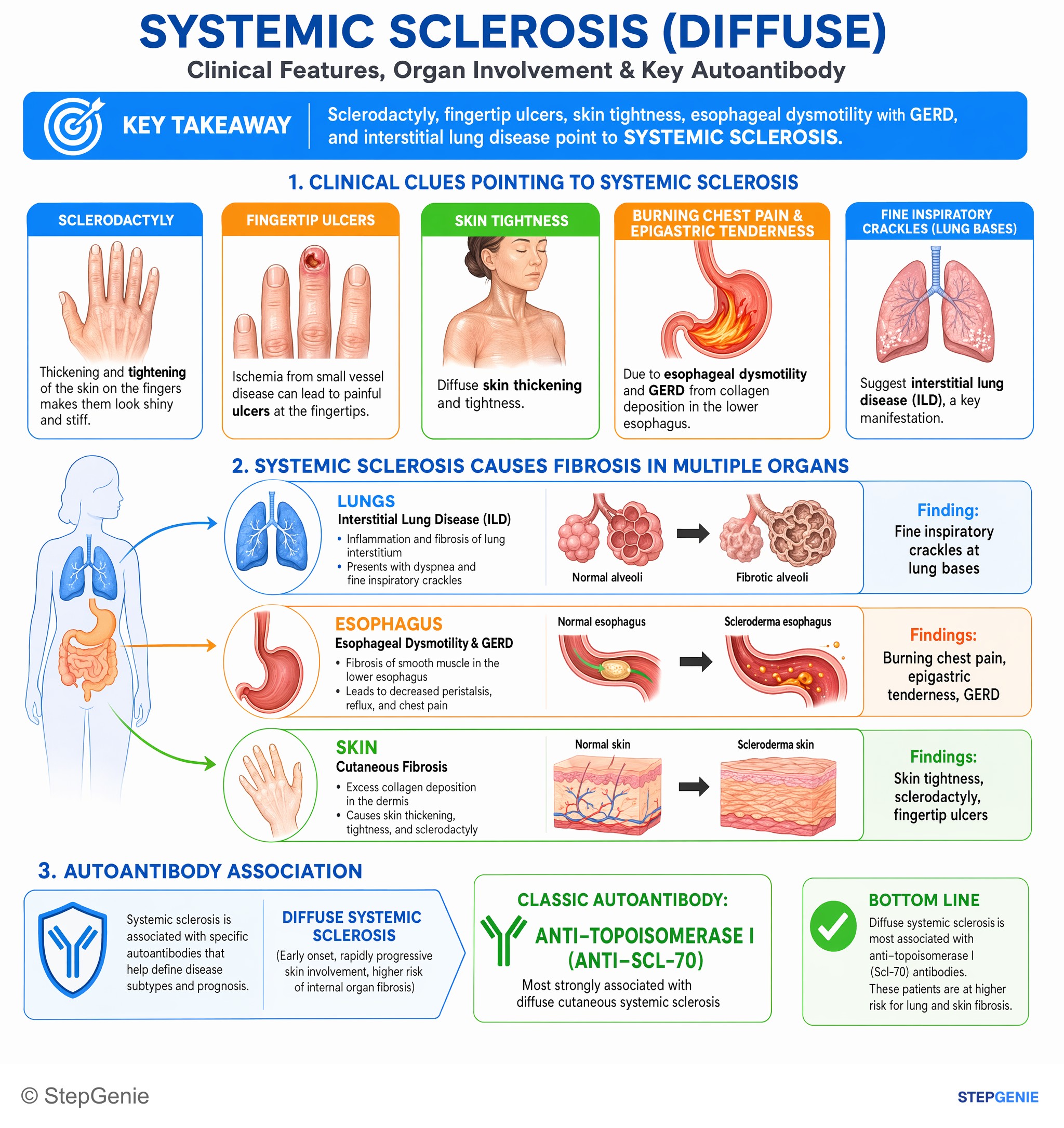
Diffuse systemic sclerosis: anti-topoisomerase I (Scl-70) → fibrosis of skin, esophagus (reflux, chest pain), and lungs (interstitial lung…
USMLE Step 1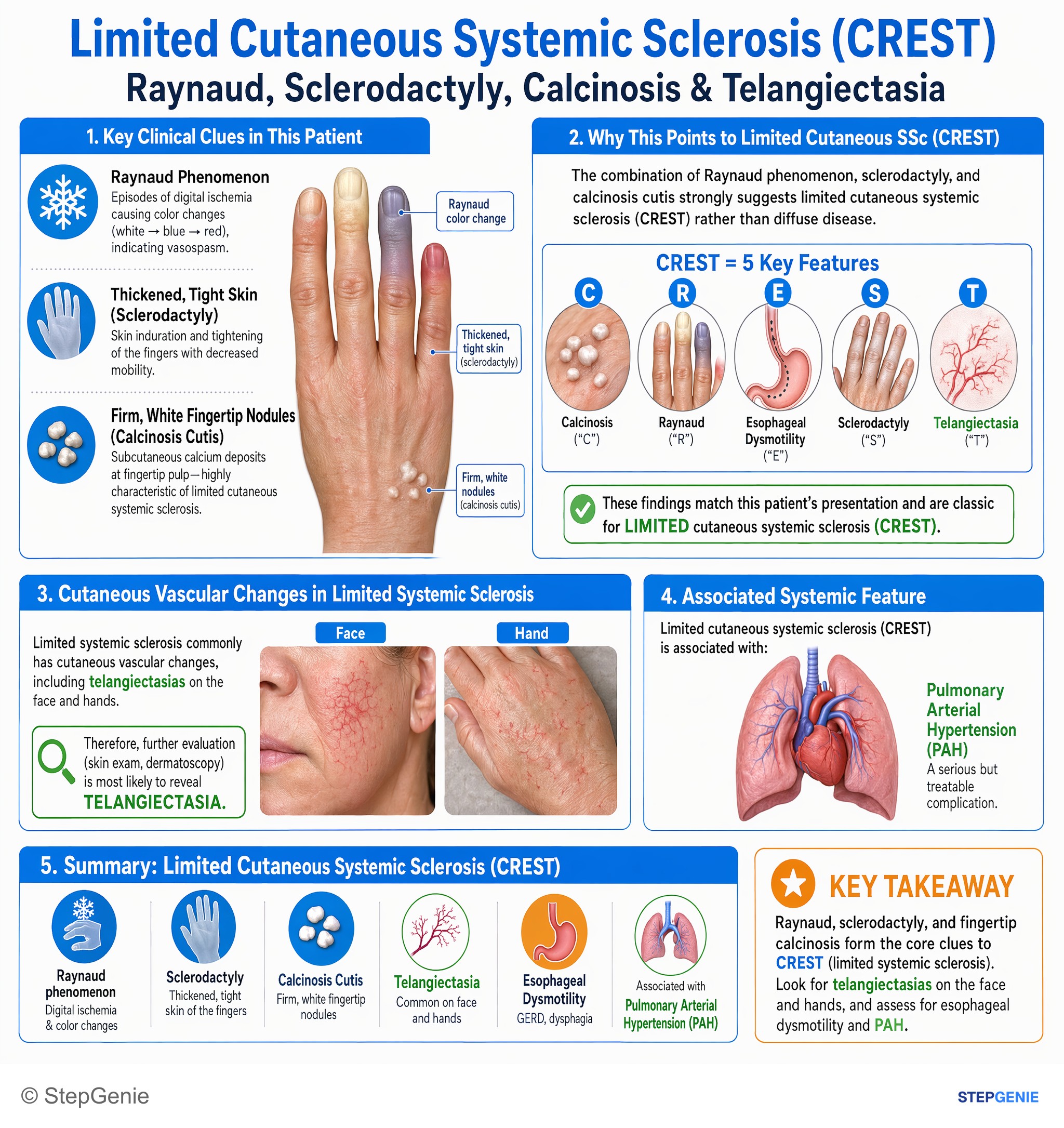
Limited cutaneous systemic sclerosis (CREST) causes Raynaud, sclerodactyly, fingertip nodules/calcinosis, and is associated with…
USMLE Step 2Frequently Asked Questions
What is the difference between limited and diffuse scleroderma?
Limited cutaneous systemic sclerosis thickens the skin of the face and of the limbs distal to the elbows and knees, progresses slowly, is associated with anticentromere antibodies and tends to cause pulmonary arterial hypertension later in the disease. Diffuse cutaneous systemic sclerosis also involves the upper arms, thighs and trunk, progresses rapidly, involves internal organs early and is associated with anti-Scl-70 (interstitial lung disease) and anti-RNA polymerase III (renal crisis). Merck cites 10-year survival of 92% for limited and 65% for diffuse disease.
What does CREST stand for in scleroderma?
CREST stands for calcinosis cutis, Raynaud phenomenon, esophageal dysmotility, sclerodactyly and telangiectasias. It is the older name for limited cutaneous systemic sclerosis, which is associated with anticentromere antibodies. Its most serious late complication is pulmonary arterial hypertension.
Which antibodies go with limited vs diffuse systemic sclerosis?
Anticentromere antibodies are associated with limited cutaneous disease (CREST). Anti-Scl-70, also called anti-topoisomerase I, is associated with diffuse disease and interstitial lung disease, while anti-RNA polymerase III is associated with diffuse disease, scleroderma renal crisis and cancer. ANA is positive in at least 90% of patients, often with a nucleolar pattern.
How is scleroderma renal crisis treated?
Scleroderma renal crisis is treated with an ACE inhibitor started immediately, and captopril is preferred because its short half-life allows rapid titration. The ACE inhibitor is continued even if creatinine keeps rising, because kidney function can still improve over time, and some patients need temporary or long-term dialysis. Glucocorticoids are a risk factor for renal crisis, so high doses are avoided in at-risk patients.
What is the treatment for scleroderma interstitial lung disease?
Mycophenolate is a first-line option; Merck notes it is as effective as cyclophosphamide and better tolerated. Nintedanib and tocilizumab are FDA-approved to slow the rate of decline in lung function in systemic sclerosis-associated ILD, and rituximab, azathioprine and cyclophosphamide are other first-line options in the 2023 American College of Rheumatology guideline. That guideline strongly recommends against glucocorticoids as first-line ILD treatment.
Why does scleroderma cause reflux and dysphagia?
Fibrosis of the esophageal wall weakens peristalsis and makes the lower esophageal sphincter incompetent, so acid refluxes and is cleared poorly. Patients develop heartburn, dysphagia and strictures, and Merck notes that about one-third develop Barrett esophagus. This is the opposite of achalasia, in which the lower esophageal sphincter fails to relax.