Osteoarthritis (OA) and rheumatoid arthritis (RA) are two of the most tested joint diseases on USMLE—and the questions love to blur them together with vague “joint pain” stems. The trick is to anchor on 3 fast discriminators: what hurts when, what the joint looks like, and what the labs/imaging show.
Tip #1: Pattern + Timing = Your fastest giveaway
The one-liner
- OA: “Wear-and-tear pain that worsens with use (better with rest).”
- RA: “Inflammatory pain with morning stiffness that improves with use.”
High-yield details
Osteoarthritis
- Pain worse at the end of the day
- Morning stiffness < 30 minutes
- Mechanical symptoms: crepitus, decreased ROM
Rheumatoid arthritis
- Pain worse in the morning
- Morning stiffness > 1 hour
- Improves with activity (“gels” then loosens up)
- Systemic inflammation: fatigue, low-grade fever can show up in vignettes
Tip #2: Joint distribution + “signature” deformities
The one-liner
- OA hits the “use joints” (hands but specific spots; plus weight-bearing joints).
- RA hits the “immune joints”—classically MCP/PIP with symmetry.
Quick distribution table (USMLE-style)
| Feature | Osteoarthritis (OA) | Rheumatoid Arthritis (RA) |
|---|---|---|
| Hand joints | DIP + PIP (± 1st CMC) | MCP + PIP, spares DIP (classic) |
| Classic nodules | Heberden (DIP) + Bouchard (PIP) | Rheumatoid nodules (extensor surfaces) |
| Common big joints | Knees, hips, spine | Knees can be involved, but hands/wrists are a big clue |
| Symmetry | Can be asymmetric | Symmetric polyarthritis |
Visual/mnemonic device
“OA = Old Axe”
Picture an old axe chopping wood:
- It wears down from overuse
- It hits the DIP “tips” of the fingers (where you grip) → Heberden nodes
- It hits weight-bearing joints (knees/hips)
“RA = Really Angry immune system”
Imagine the immune system “angrily shaking hands”:
- It targets MCP/PIP (the knuckles you shake with)
- Symmetric involvement (both hands)
Tip #3: Imaging + labs—know the “buzzwords”
The one-liner
- OA = cartilage loss → joint space narrowing + osteophytes.
- RA = synovial inflammation → erosions + ulnar deviation (and serologies).
Imaging (most tested)
OA X-ray
- Joint space narrowing (often asymmetric)
- Osteophytes
- Subchondral sclerosis
- Subchondral cysts
RA X-ray
- Periarticular osteopenia
- Marginal erosions
- Joint space narrowing (from inflammatory destruction)
- Deformities in advanced disease: ulnar deviation, swan-neck/boutonnière
Labs (high-yield)
RA
- RF (IgM against Fc portion of IgG): not super specific
- Anti-CCP: more specific (classic USMLE point)
- Elevated ESR/CRP
- Can have anemia of chronic disease
OA
- Typically normal inflammatory markers
- Diagnosis is clinical + radiographic, not serologic
Rapid-fire “if you see this, think that” (USMLE vignettes)
- DIP bony enlargements + crepitus + worse with activity → OA
- Symmetric MCP/PIP pain + morning stiffness > 1 hour → RA
- Anti-CCP positive → RA
- Osteophytes on X-ray → OA
- Marginal erosions + periarticular osteopenia → RA
- Improves with NSAIDs but still inflammatory + extra-articular clues (nodules, pleuritis, pericarditis) → RA
10-second recap (memorize this)
- OA: Old cartilage → DIP/PIP, worse with use, osteophytes, stiffness < 30 min
- RA: Angry synovium → MCP/PIP, morning stiffness > 1 hr, anti-CCP, erosions
Visual explanations from the StepGenie QBank
Infographics StepGenie drew to explain questions in its own QBank, matched to this article's topic. Each caption is the takeaway of the question it explains.
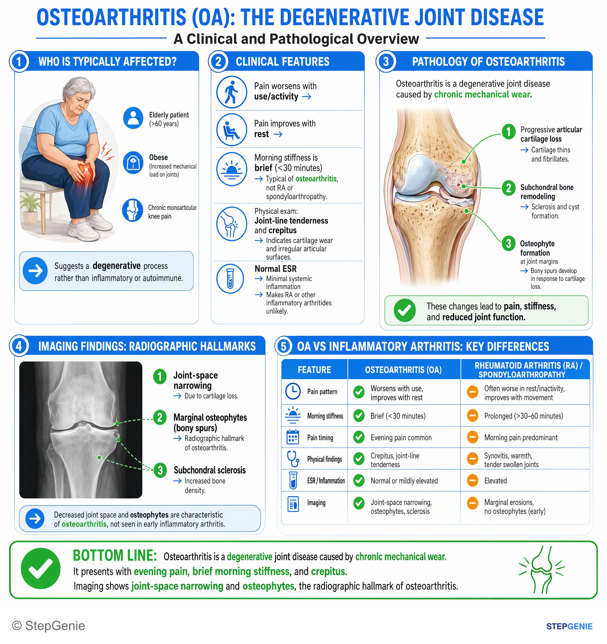
Osteoarthritis from chronic mechanical wear shows evening pain, brief morning stiffness, crepitus and imaging with joint-space narrowing…
USMLE Step 1Frequently Asked Questions
How do you quickly tell osteoarthritis from rheumatoid arthritis based on pain and stiffness?
The fastest distinction is that osteoarthritis causes mechanical pain that worsens with use, while rheumatoid arthritis causes inflammatory pain with prolonged morning stiffness that improves with activity. Osteoarthritis is typically worse at the end of the day and has morning stiffness lasting less than 30 minutes. Rheumatoid arthritis is worse in the morning, has stiffness lasting more than 1 hour, and often “gels” before loosening up with movement.
Which hand joints are usually affected in osteoarthritis vs rheumatoid arthritis?
Osteoarthritis classically affects the DIP and PIP joints, often with involvement of the first CMC, whereas rheumatoid arthritis classically affects the MCP and PIP joints and usually spares the DIP joints. Rheumatoid arthritis also tends to present as a symmetric polyarthritis, especially involving the hands and wrists. This joint distribution is one of the highest-yield ways to separate the two on exams.
What X-ray findings point to osteoarthritis instead of rheumatoid arthritis?
Osteoarthritis is suggested by joint space narrowing with osteophytes, often with subchondral sclerosis and subchondral cysts. The narrowing can be asymmetric because osteoarthritis reflects cartilage loss and mechanical wear. In contrast, rheumatoid arthritis is associated with periarticular osteopenia, marginal erosions, and inflammatory joint destruction, with advanced deformities such as ulnar deviation rather than classic osteophyte formation.
Which lab test is more specific for rheumatoid arthritis?
Anti-CCP is the more specific lab test for rheumatoid arthritis. Rheumatoid arthritis can also have a positive rheumatoid factor, elevated ESR or CRP, and anemia of chronic disease, but rheumatoid factor is not very specific. Osteoarthritis, by contrast, usually has normal inflammatory markers and is diagnosed clinically and radiographically rather than with serologic testing.
What classic deformities or nodules help distinguish osteoarthritis from rheumatoid arthritis?
Osteoarthritis is associated with Heberden nodes at the DIP joints and Bouchard nodes at the PIP joints, reflecting bony enlargement in those locations. Rheumatoid arthritis is associated with rheumatoid nodules on extensor surfaces and, in advanced disease, deformities such as ulnar deviation, swan-neck deformity, and boutonnière deformity. These characteristic findings help distinguish a degenerative process from an inflammatory one.