High-yield rheumatology images for the USMLE
StepGenie explanation infographics. Free to view, no account needed.
What do StepGenie's rheumatology images cover?
Each rheumatology and musculoskeletal image is a StepGenie infographic drawn for one of its own practice questions. Most sort an arthritis or a vasculitis by the pattern it produces — which joints, which vessel size, which antibody — rather than by its name.
Bone, joint and soft tissue injuries and infections carry the largest share of this set, followed by autoimmune disorders and vasculitides, arthritis and the spondyloarthropathies, metabolic bone disease, upper and lower limb nerve lesions, and muscular disorders.
Vasculitis is the topic these images help with most. Sorting by vessel size before anything else reduces a long and unfriendly list to three short ones, and a panel that draws large, medium and small vessel disease on the same vessel makes that sorting automatic. The arthritis panels do the equivalent with joint distribution and with the symmetry and morning stiffness pattern, which is what actually separates rheumatoid from osteoarthritis in a vignette.
The orthopedic and sports images are more concrete: they show the mechanism of injury and the structure it damages. Those pair well with the anatomy gallery, where the same nerve lesions appear from the anatomical side.
Autoantibody panels are the subset to check last. They are only useful once the clinical pattern has narrowed the differential, and reading them first tends to produce confident wrong answers on questions where two diseases share an antibody.
Topics in this set
Labels are the question-bank topics these images are filed under. Step 1 and Step 2 file some of the same material under different names, so a few labels here merge both.
Rheumatology explanation images
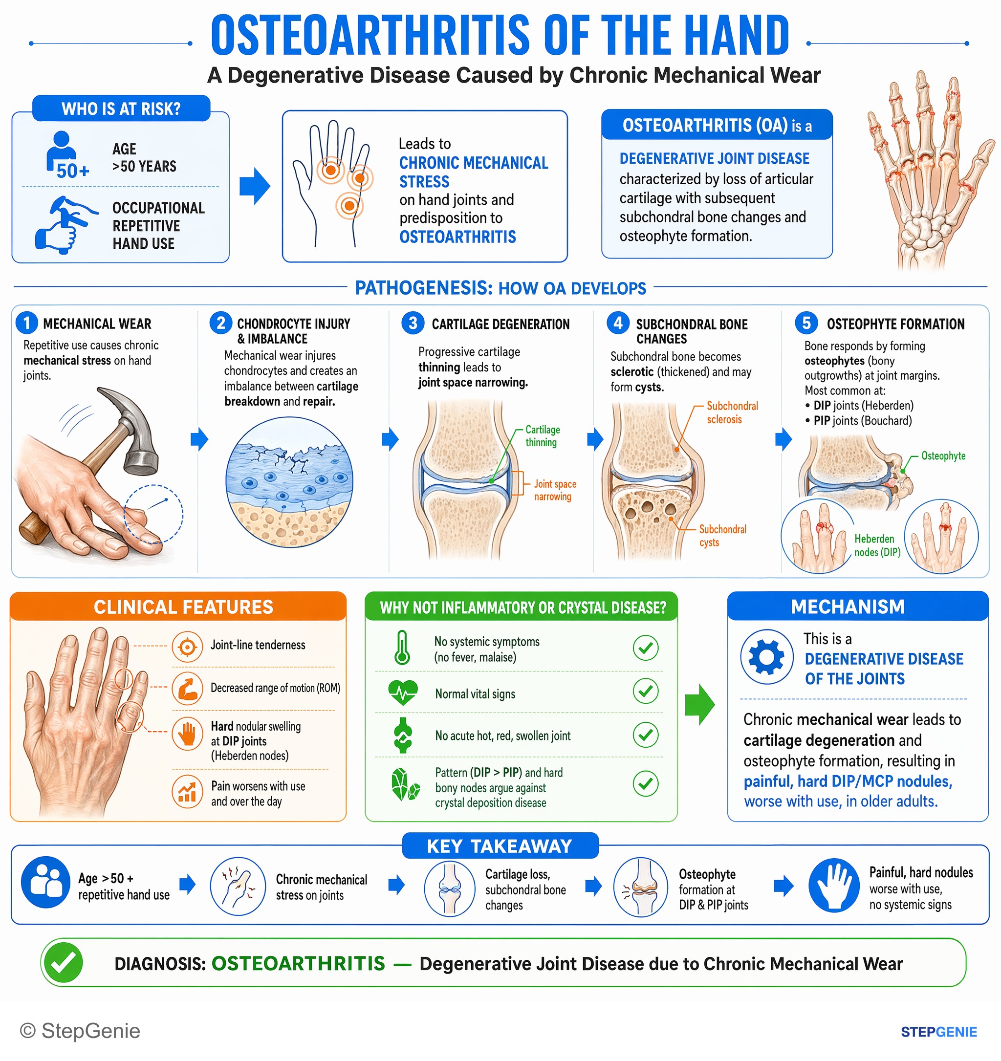
Osteoarthritis from chronic mechanical wear → cartilage degeneration and osteophyte formation → painful, hard DIP/MCP nodules, worse with…
USMLE Step 1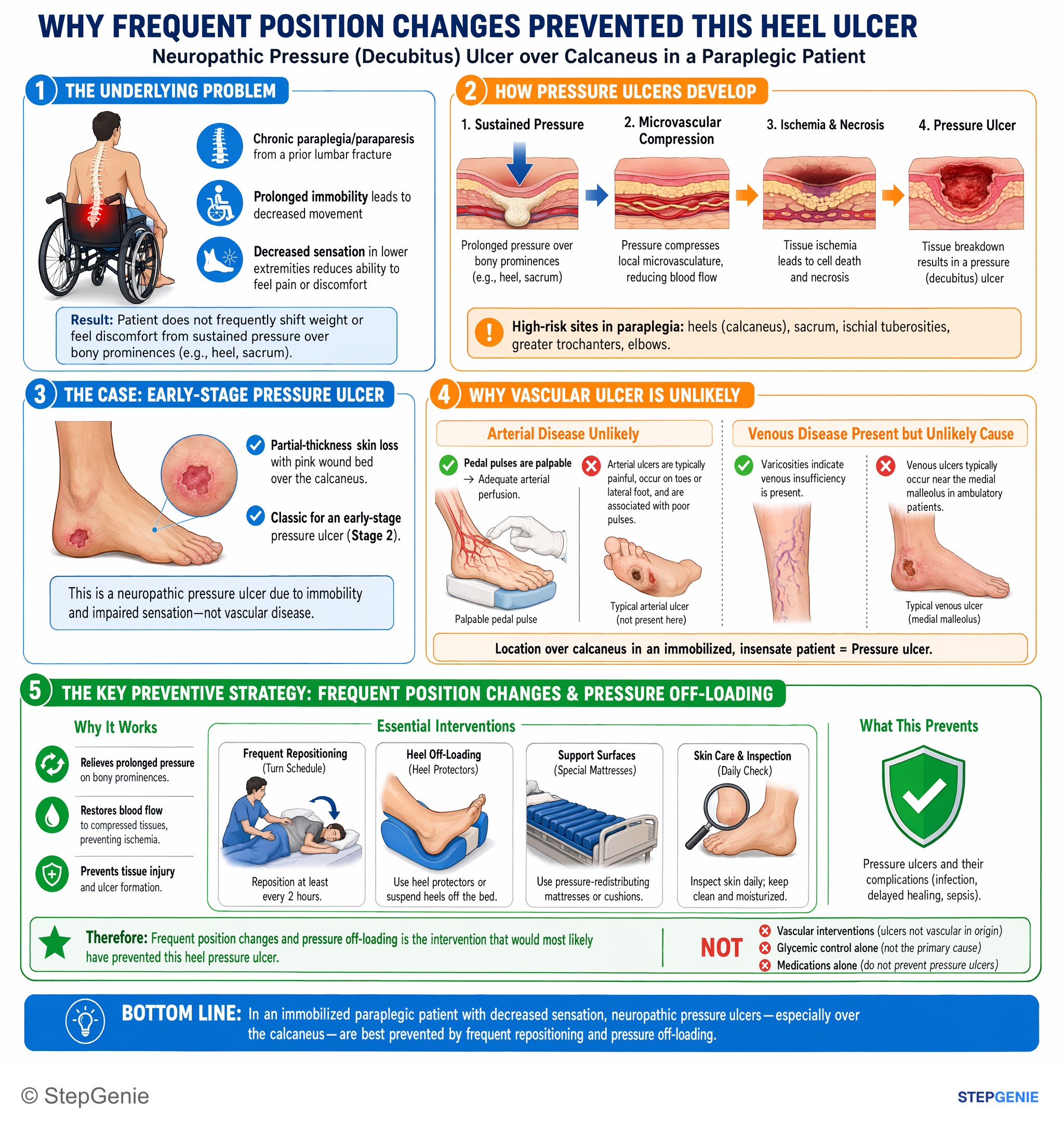
Neuropathic pressure ulcer over calcaneus in paraplegic patient is best prevented by frequent repositioning/pressure off-loading, not…
USMLE Step 2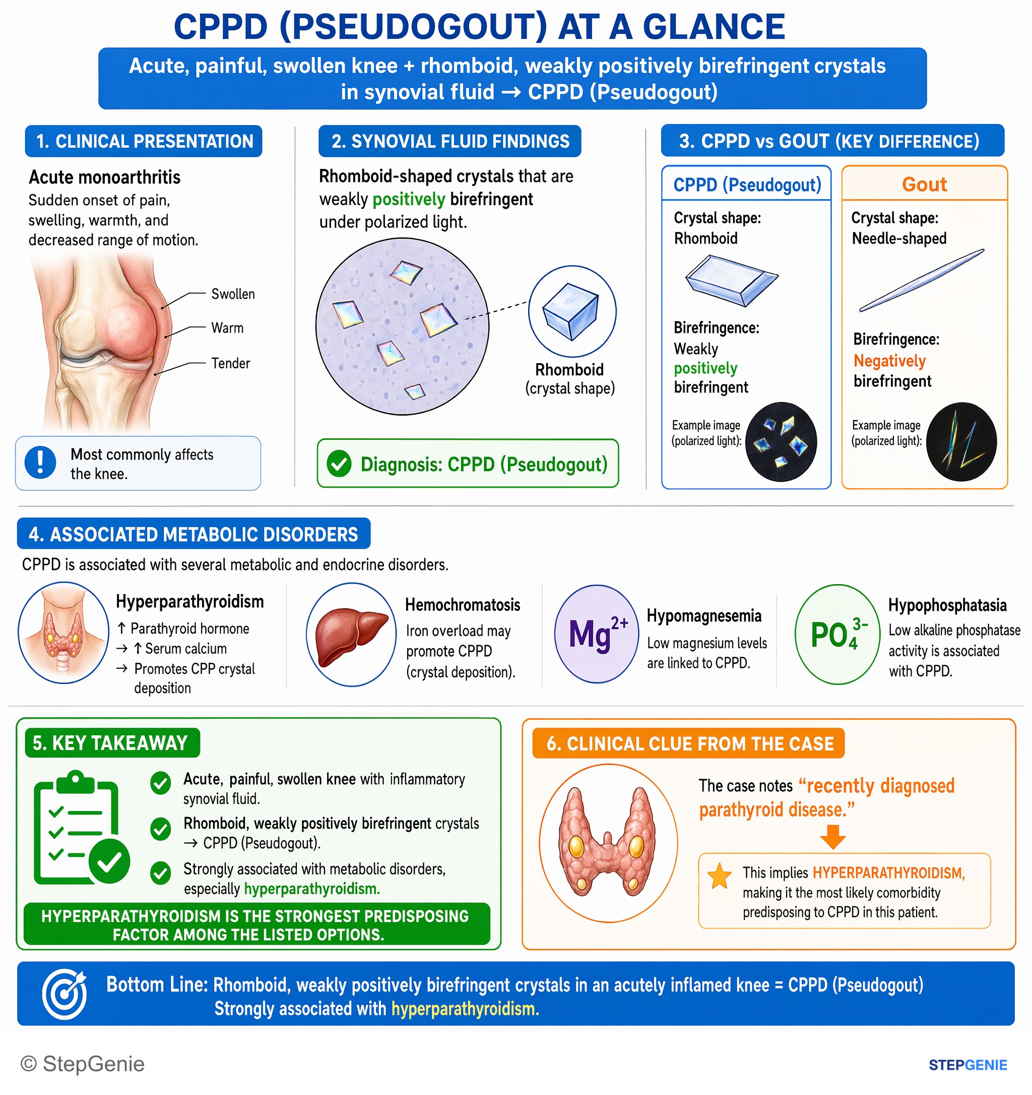
Calcium pyrophosphate deposition disease (pseudogout) is strongly associated with hyperparathyroidism, causing rhomboid, weakly positively…
USMLE Step 1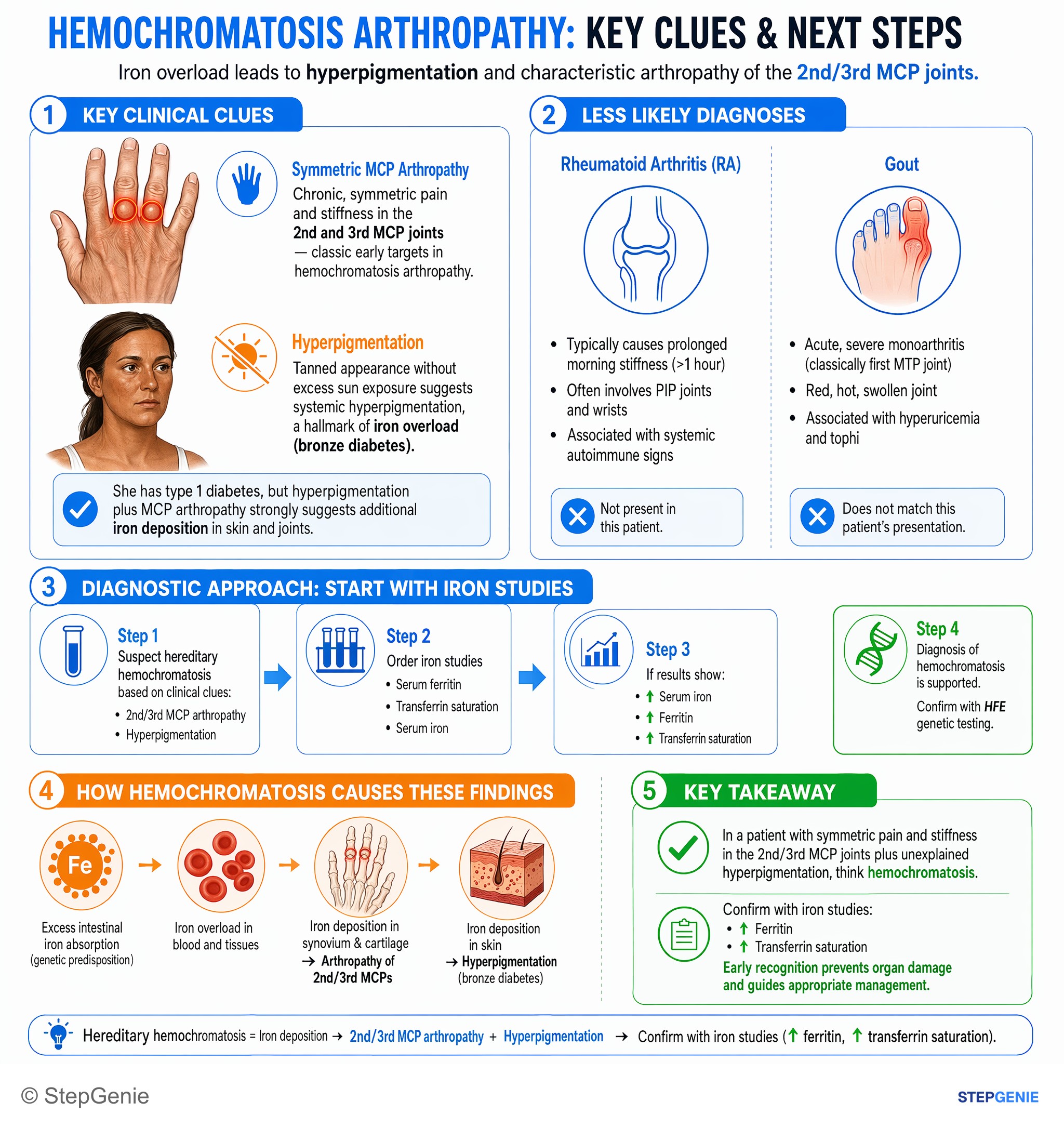
Hereditary hemochromatosis causes iron deposition → arthropathy of 2nd/3rd MCPs + hyperpigmentation; confirm with iron studies (↑ferritin…
USMLE Step 2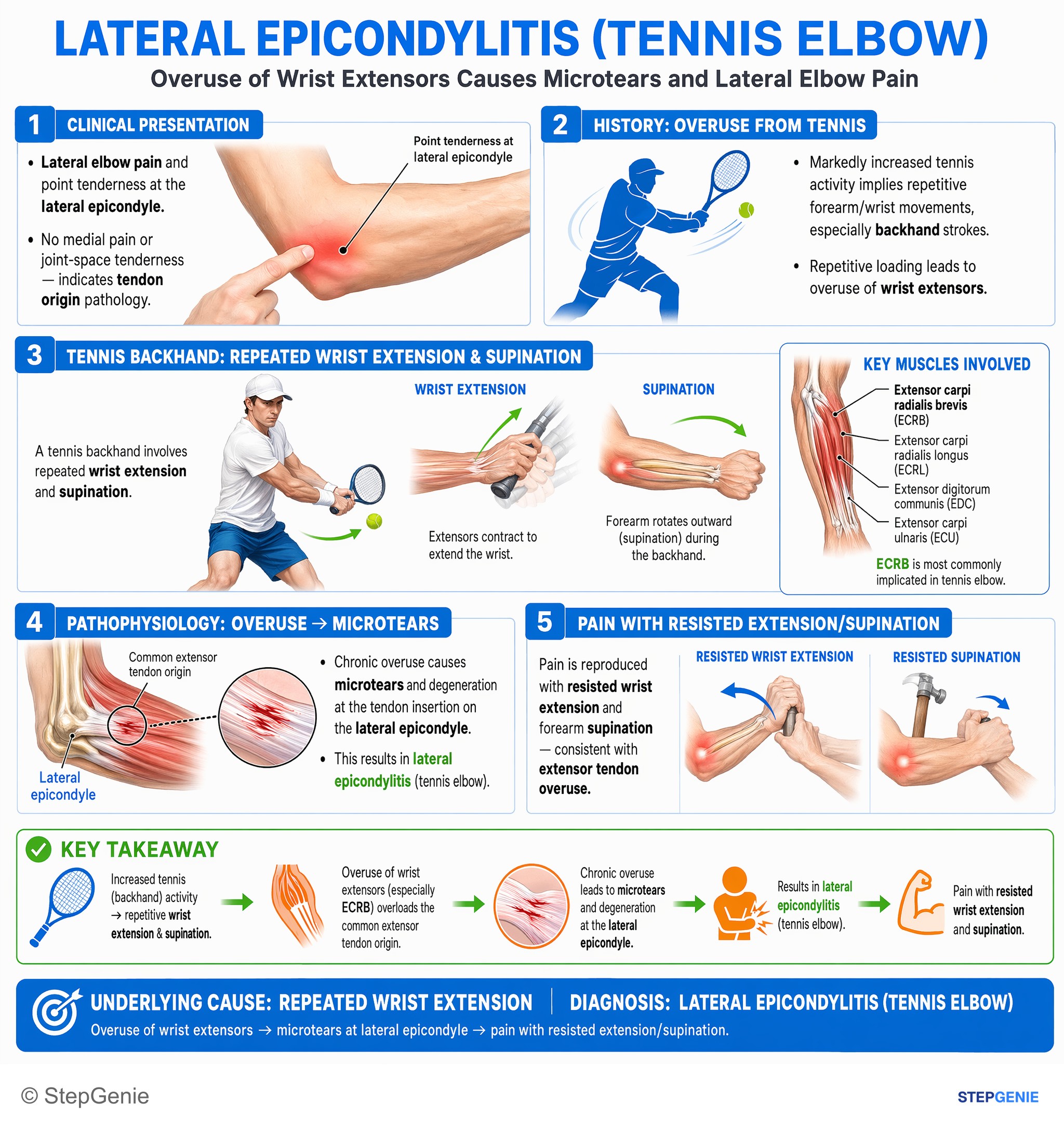
Lateral epicondylitis (tennis elbow) from overuse of wrist extensors → microtears at lateral epicondyle → pain with resisted…
USMLE Step 1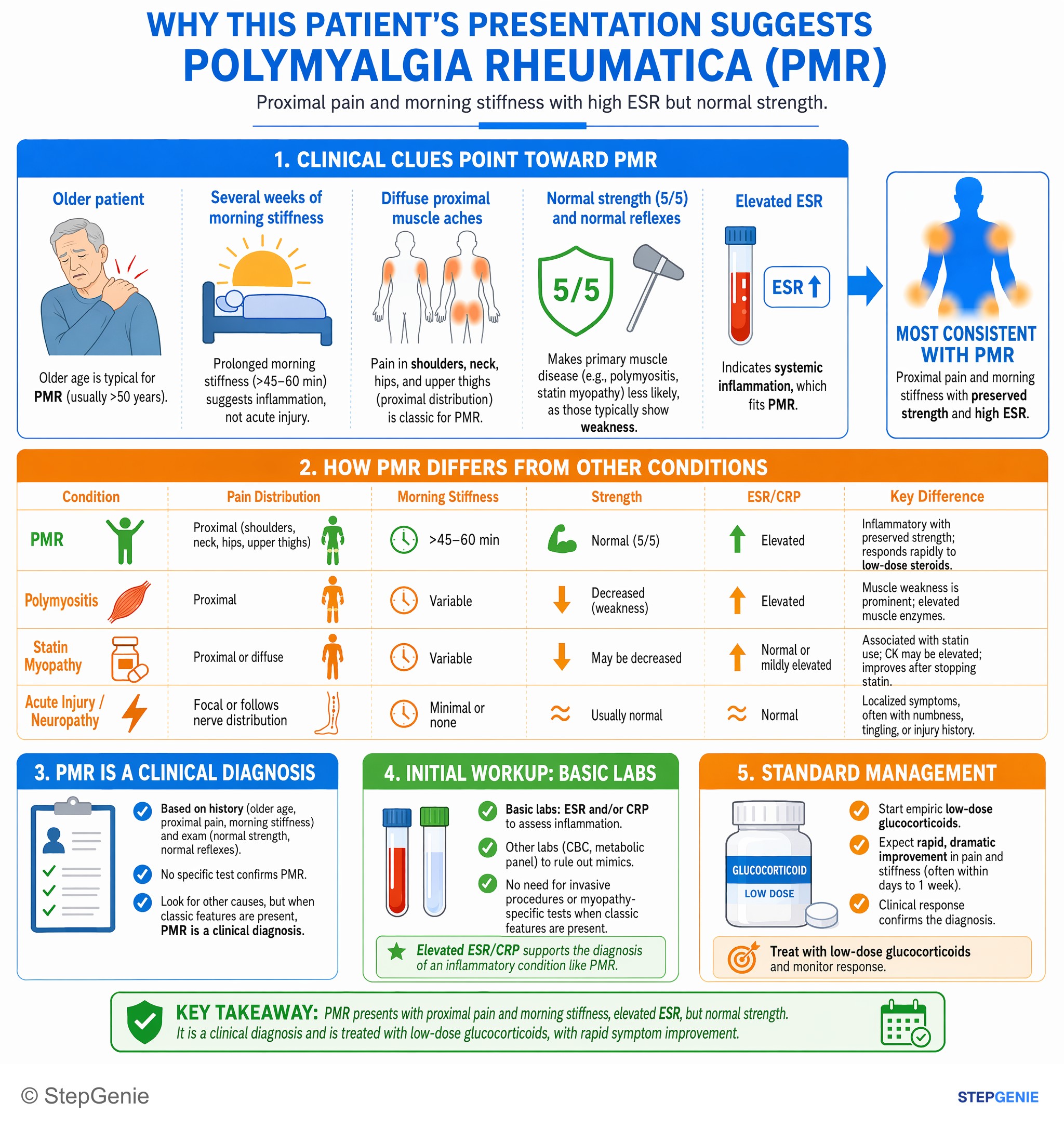
Polymyalgia rheumatica causes proximal pain and morning stiffness with high ESR but normal strength; treat empirically with low-dose…
USMLE Step 2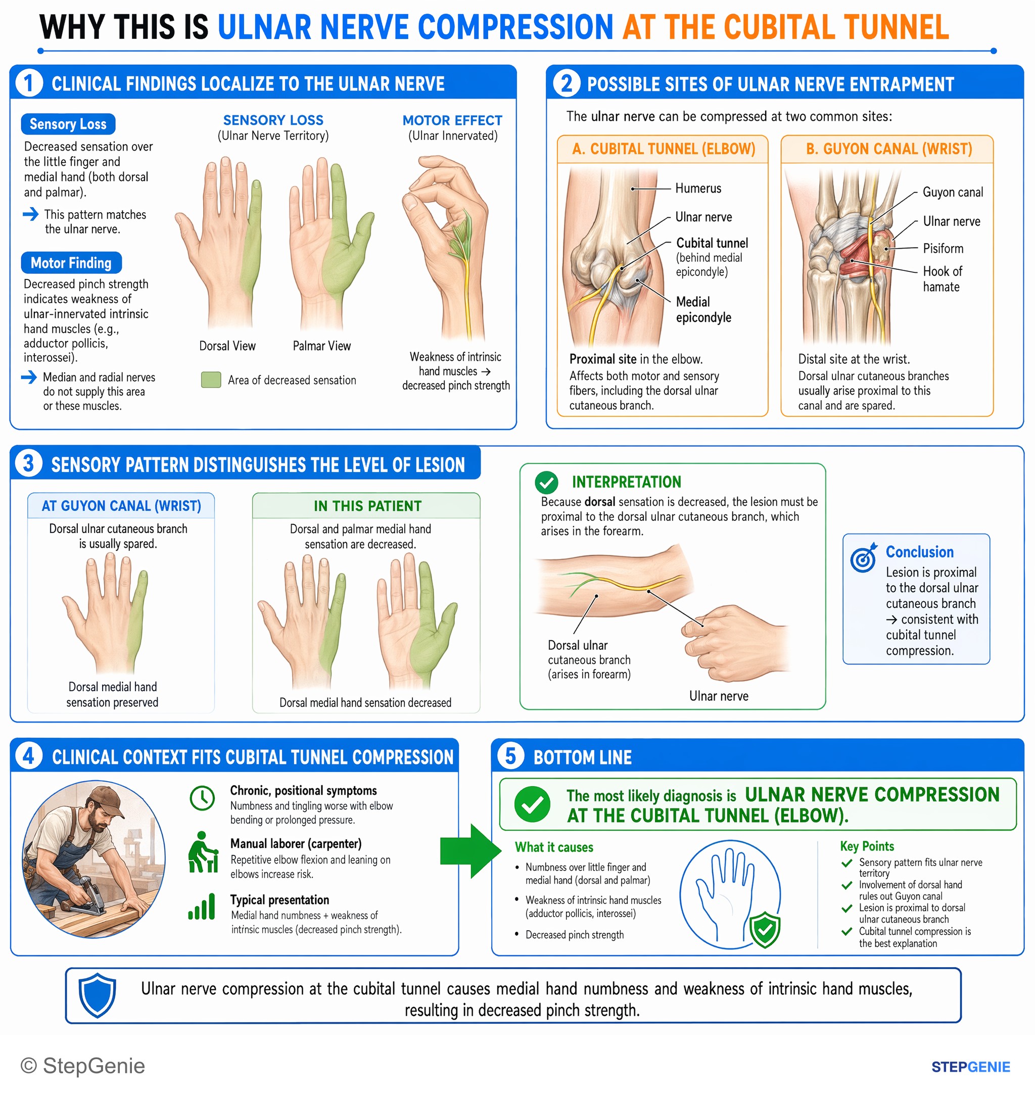
Ulnar nerve compression at the cubital tunnel causes medial hand numbness and weakness of intrinsic hand muscles with decreased pinch…
USMLE Step 1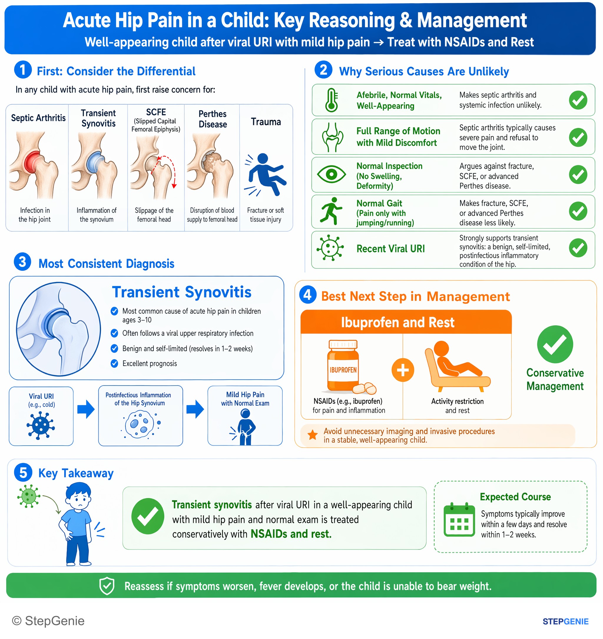
Transient synovitis after viral URI in a well-appearing child with mild hip pain and normal exam is treated conservatively with NSAIDs and…
USMLE Step 2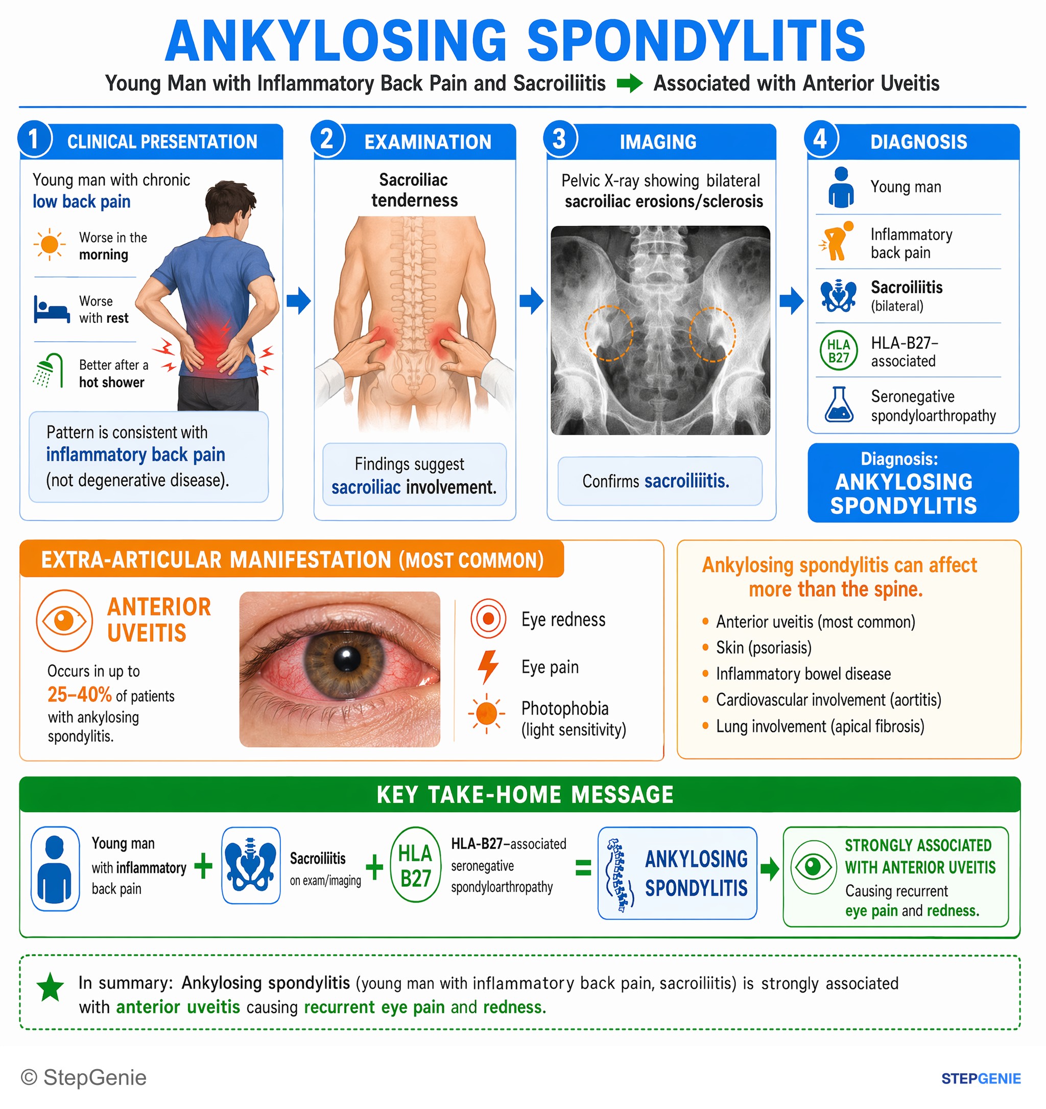
Ankylosing spondylitis (young man with inflammatory back pain, sacroiliitis) is strongly associated with anterior uveitis causing…
USMLE Step 1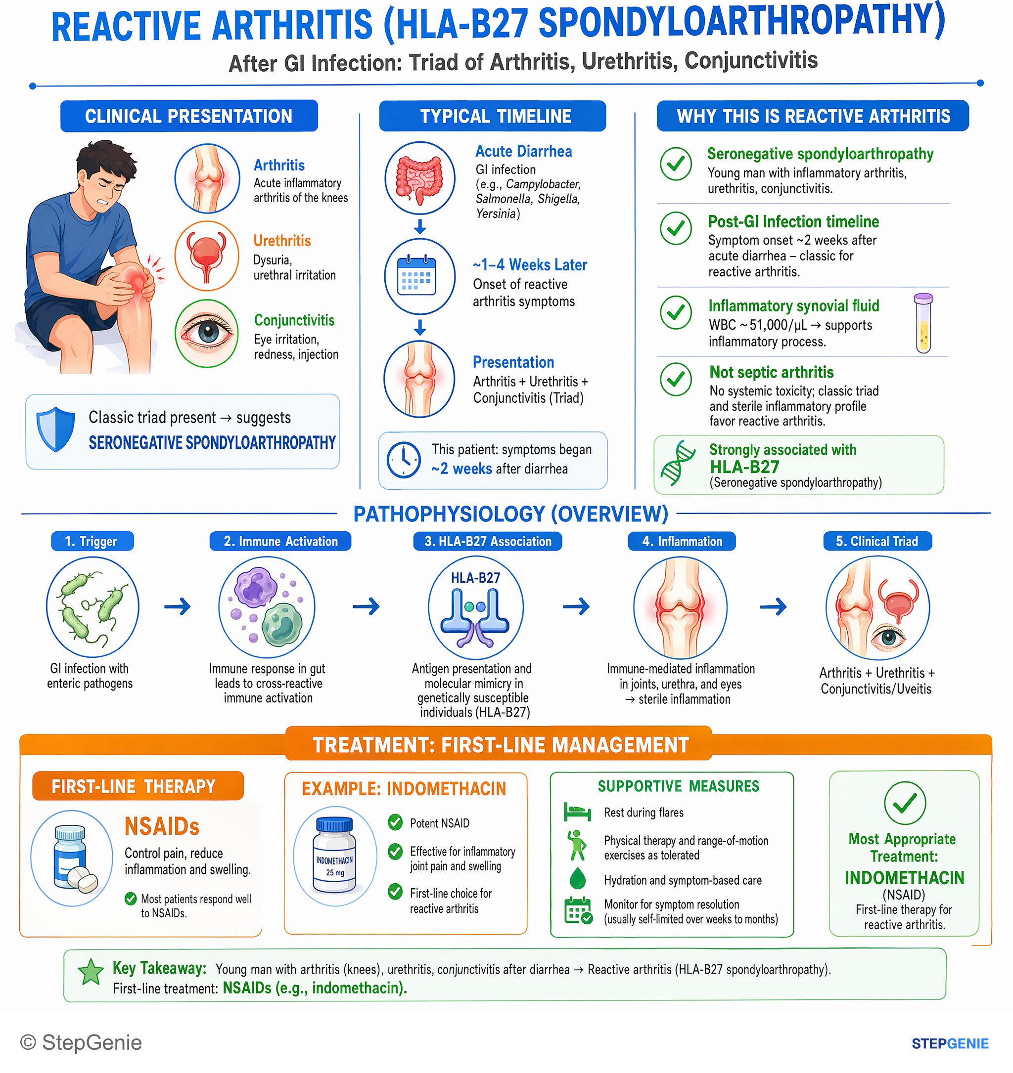
Reactive arthritis (HLA-B27 spondyloarthropathy) after diarrhea: triad of arthritis, urethritis, conjunctivitis. First-line treatment is…
USMLE Step 2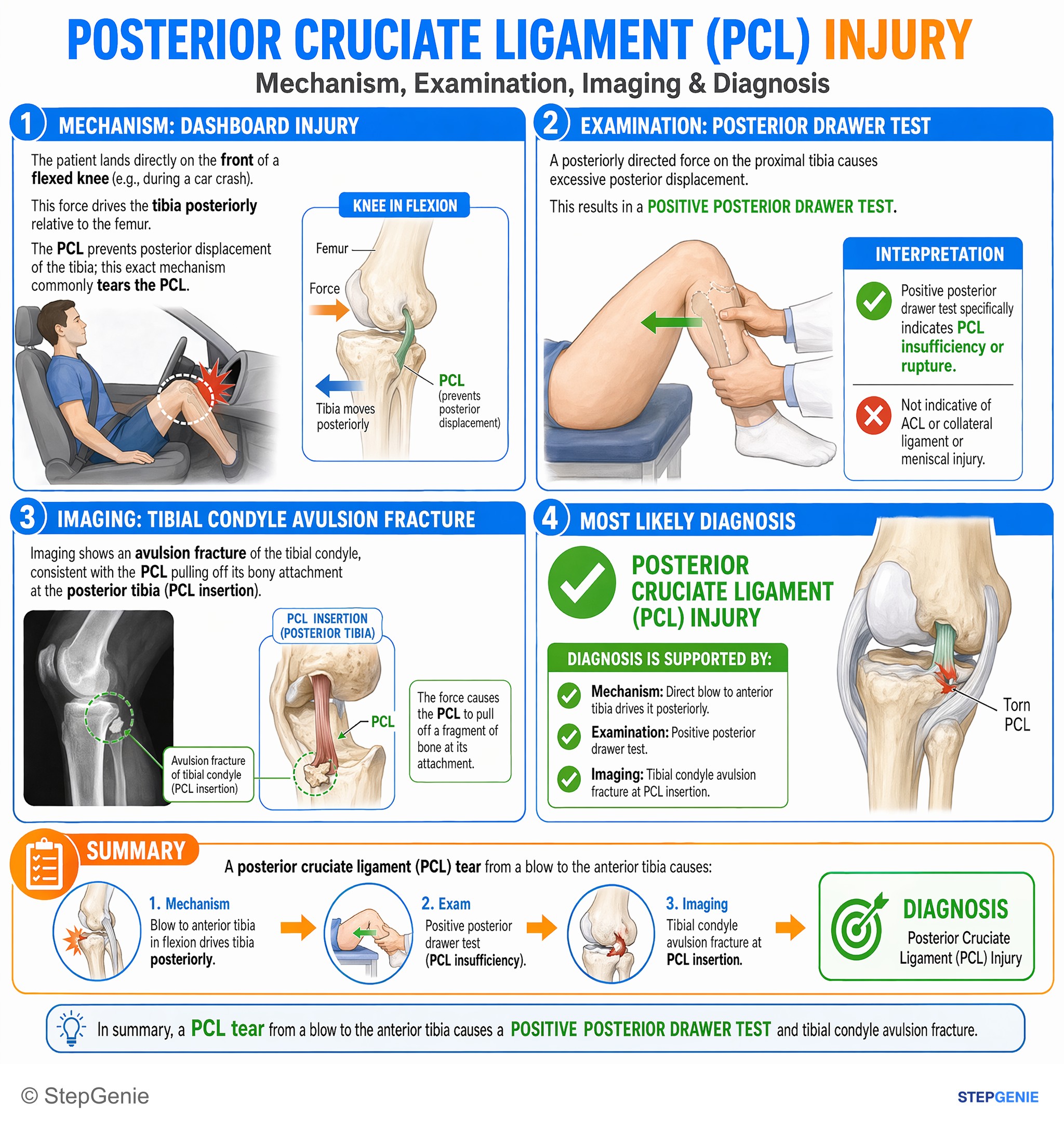
Posterior cruciate ligament (PCL) tear from a blow to the anterior tibia causes a positive posterior drawer test and tibial condyle…
USMLE Step 1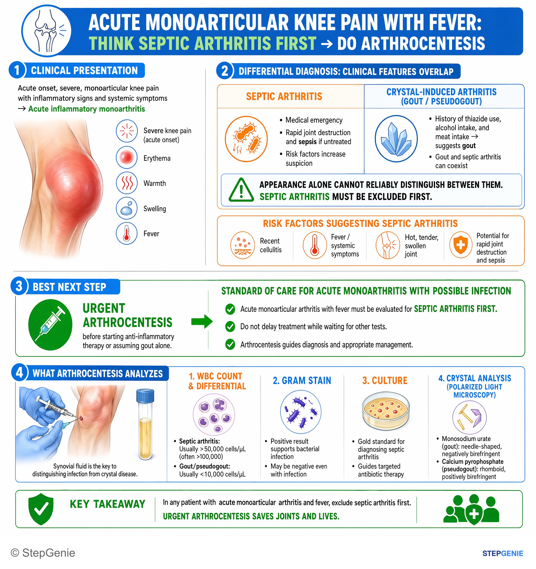
Acute monoarticular arthritis with fever must be evaluated for septic arthritis first; perform arthrocentesis for cell count, Gram stain…
USMLE Step 2288 publishable rheumatology infographics in the library — 136 from Step 1 questions and 152 from Step 2 CK, counted on October 29, 2026. This page shows 48 of them, alternating the two steps. An image is listed when its question is active, has a full structured explanation, passed verification and carries no unresolved report.
This page caps out at 48 images. 240 more rheumatology infographics sit in the in-app image library, where they are searchable and filterable by subject, system and topic.
Open the image libraryPractice the questions behind these images
Each image explains one question. The rheumatology topic page has the written explanations and the question sets, and the preview lets you answer three questions without an account.
Other image sets
Study rheumatology with the image attached
In the app, each of these sits at the end of the question it explains, alongside the step-by-step reasoning and a panel for every option you did not pick. Starting is free.
Start practicing free