High-yield dermatology images for the USMLE
StepGenie explanation infographics. Free to view, no account needed.
What do StepGenie's dermatology images cover?
Each dermatology image is an infographic StepGenie drew for one of its own practice questions. It shows the layer of skin involved and the change happening there, which is what turns a described rash into a diagnosis rather than a guess.
Inflammatory dermatoses and bullous disease carry the largest block here, then skin and soft tissue infections, skin tumors and tumor-like lesions, cutaneous neoplasia, and drug reactions.
Dermatology is hard to revise from text because the vignette describes a picture and expects you to reconstruct it. These images work the opposite way round: they draw the lesion at the level it occurs. A bullous panel that shows where the split sits — within the epidermis for pemphigus vulgaris, below it for bullous pemphigoid — explains the clinical difference in blister fragility without asking you to memorize the pairing separately.
The drug reaction images are a small group, and the one to read closely before Step 2 CK: those questions turn on recognizing the severe reactions early. The panels put the timing since exposure next to the mucosal and systemic features, which is the combination the exam uses to separate a benign rash from one that requires the drug to be stopped immediately.
Skin cancer panels are organized by the layer and the cell of origin rather than by appearance, which is what keeps basal cell carcinoma, squamous cell carcinoma and melanoma separable when the vignette only describes a lesion in words.
Topics in this set
Labels are the question-bank topics these images are filed under. Step 1 and Step 2 file some of the same material under different names, so a few labels here merge both.
Dermatology explanation images
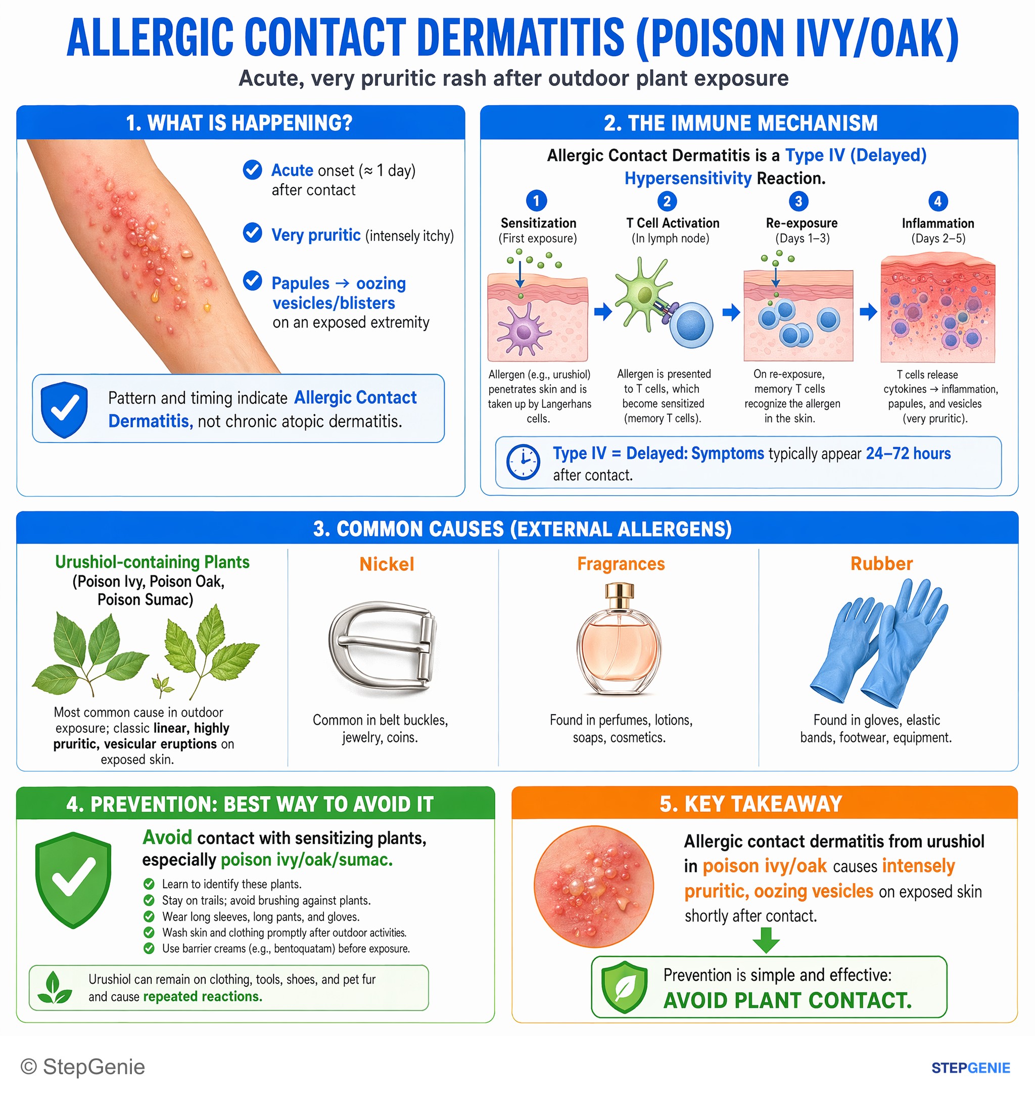
Allergic contact dermatitis from urushiol in poison ivy/oak causes intensely pruritic, oozing vesicles; prevention is by avoiding plant…
USMLE Step 1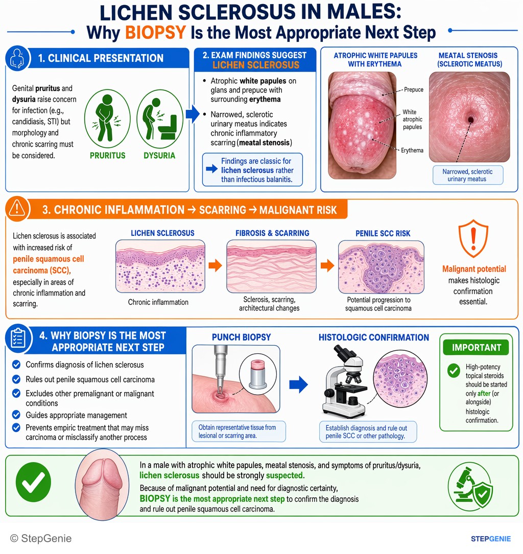
Lichen sclerosus in a male (atrophic white papules, meatal stenosis) requires biopsy to confirm and rule out penile SCC.
USMLE Step 2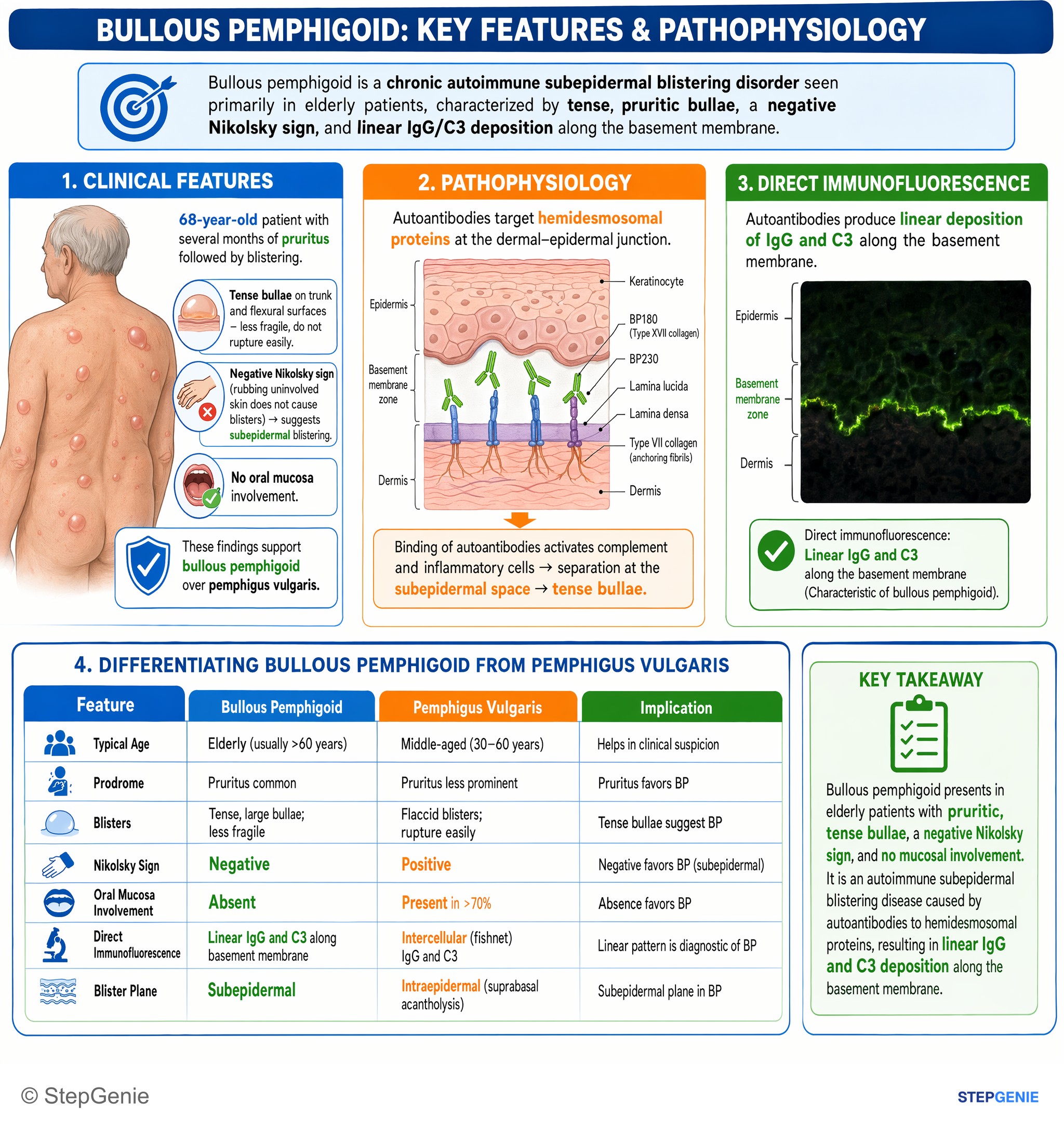
Bullous pemphigoid: tense, pruritic bullae in elderly; negative Nikolsky sign; due to autoantibodies to hemidesmosomes with linear IgG/C3…
USMLE Step 1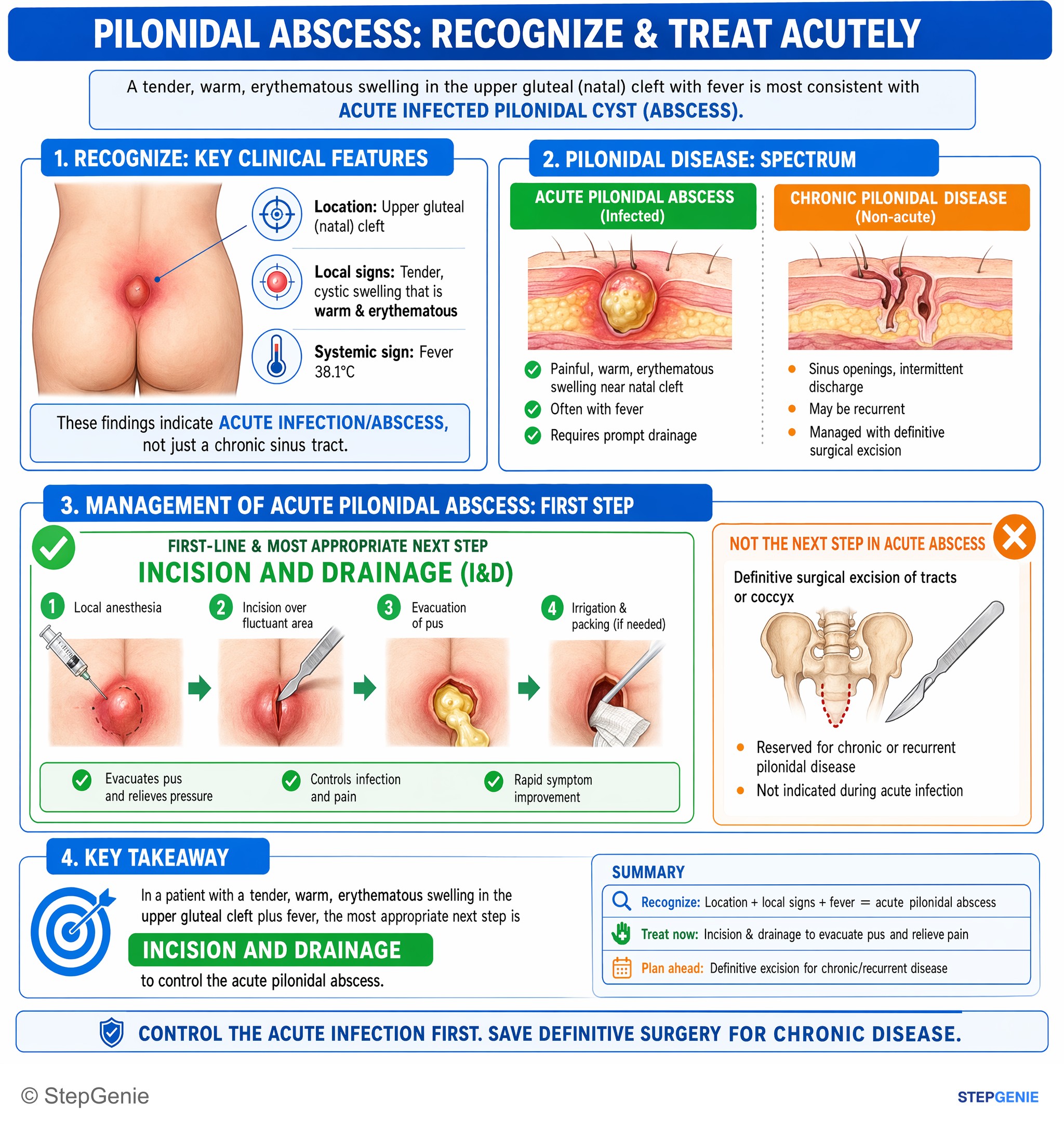
Pilonidal abscess presents as a painful, warm, erythematous cyst near the natal cleft; acute management is incision and drainage with…
USMLE Step 2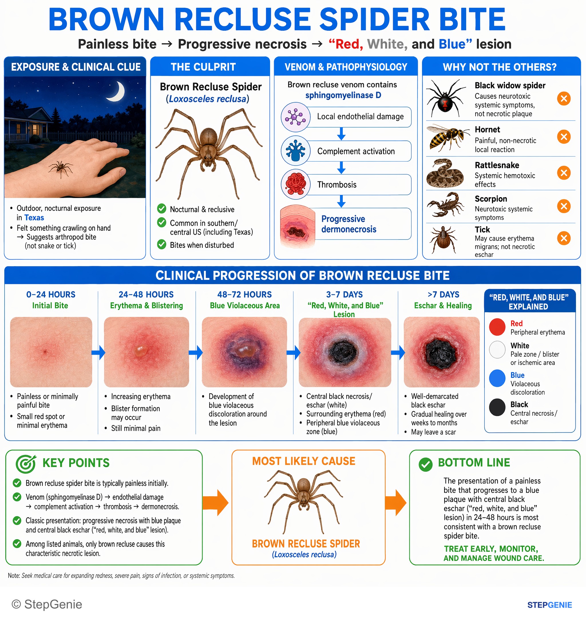
Brown recluse spider bite causes painless bite, progressive local necrosis with blue plaque and central black eschar (“red, white, and…
USMLE Step 1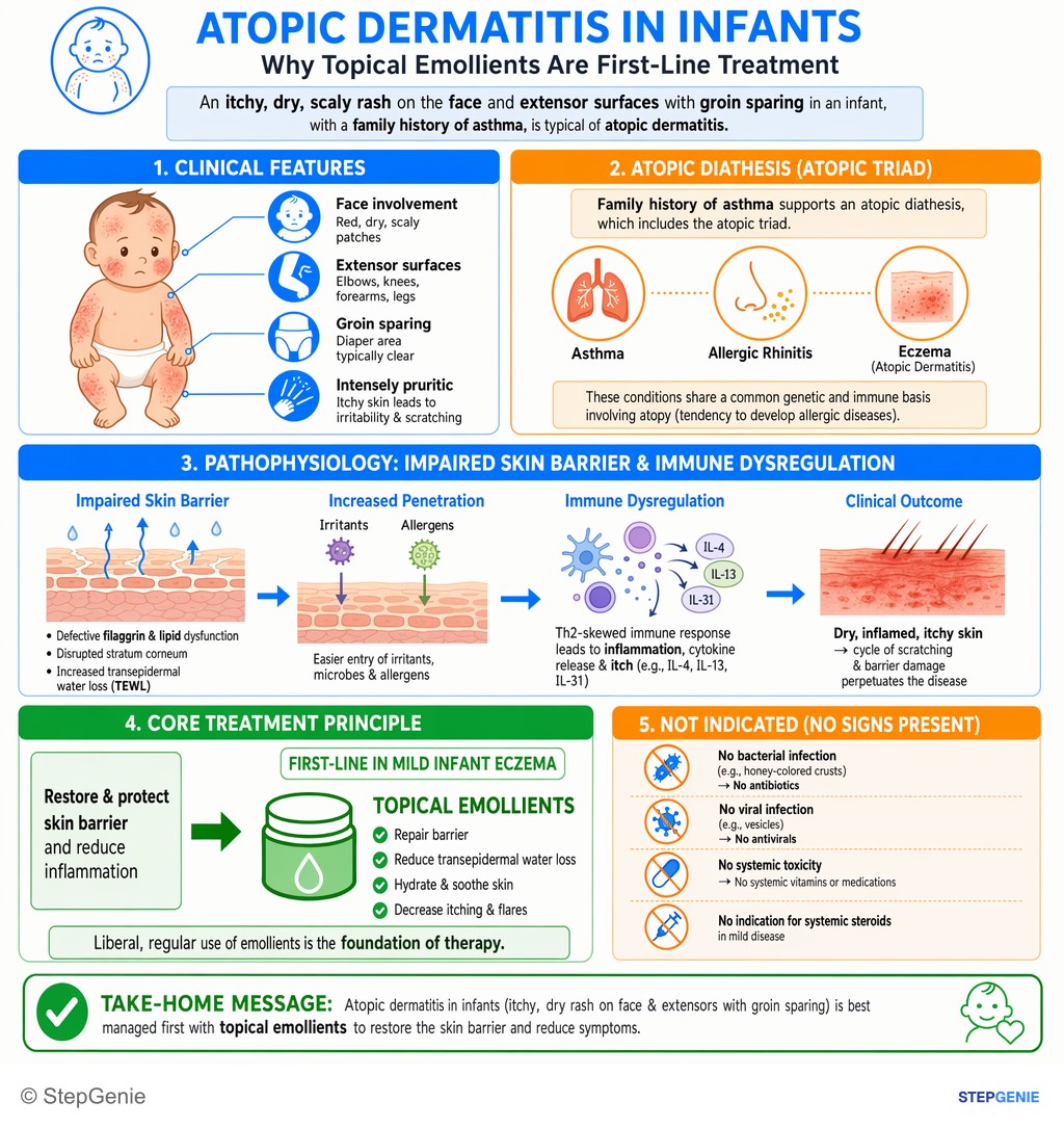
Atopic dermatitis in an infant (itchy, dry rash on face/extensors) is first-line treated with topical emollients to restore skin barrier.
USMLE Step 2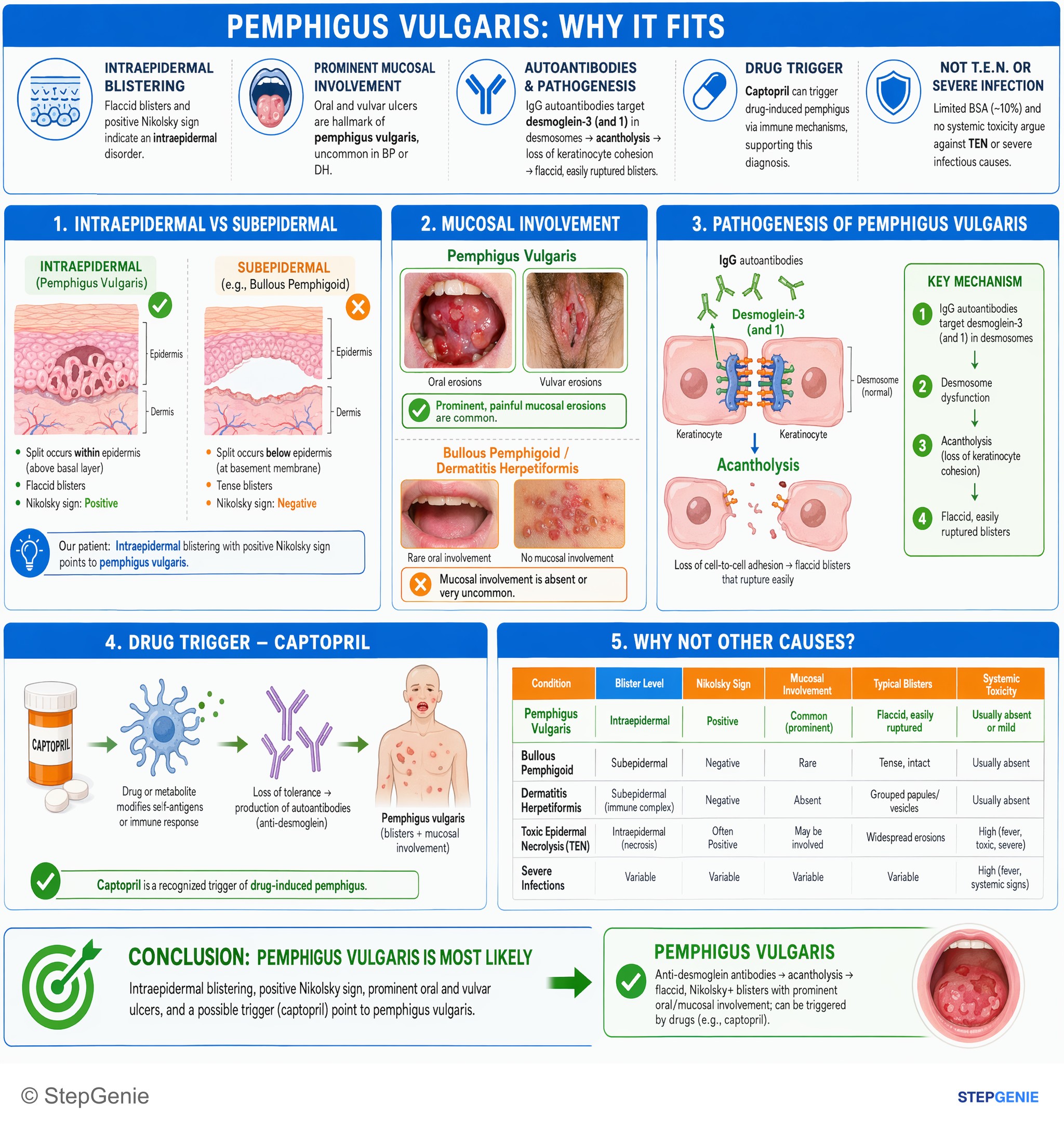
Pemphigus vulgaris: anti-desmoglein antibodies → flaccid, Nikolsky+ blisters with prominent oral/mucosal involvement; can be triggered by…
USMLE Step 1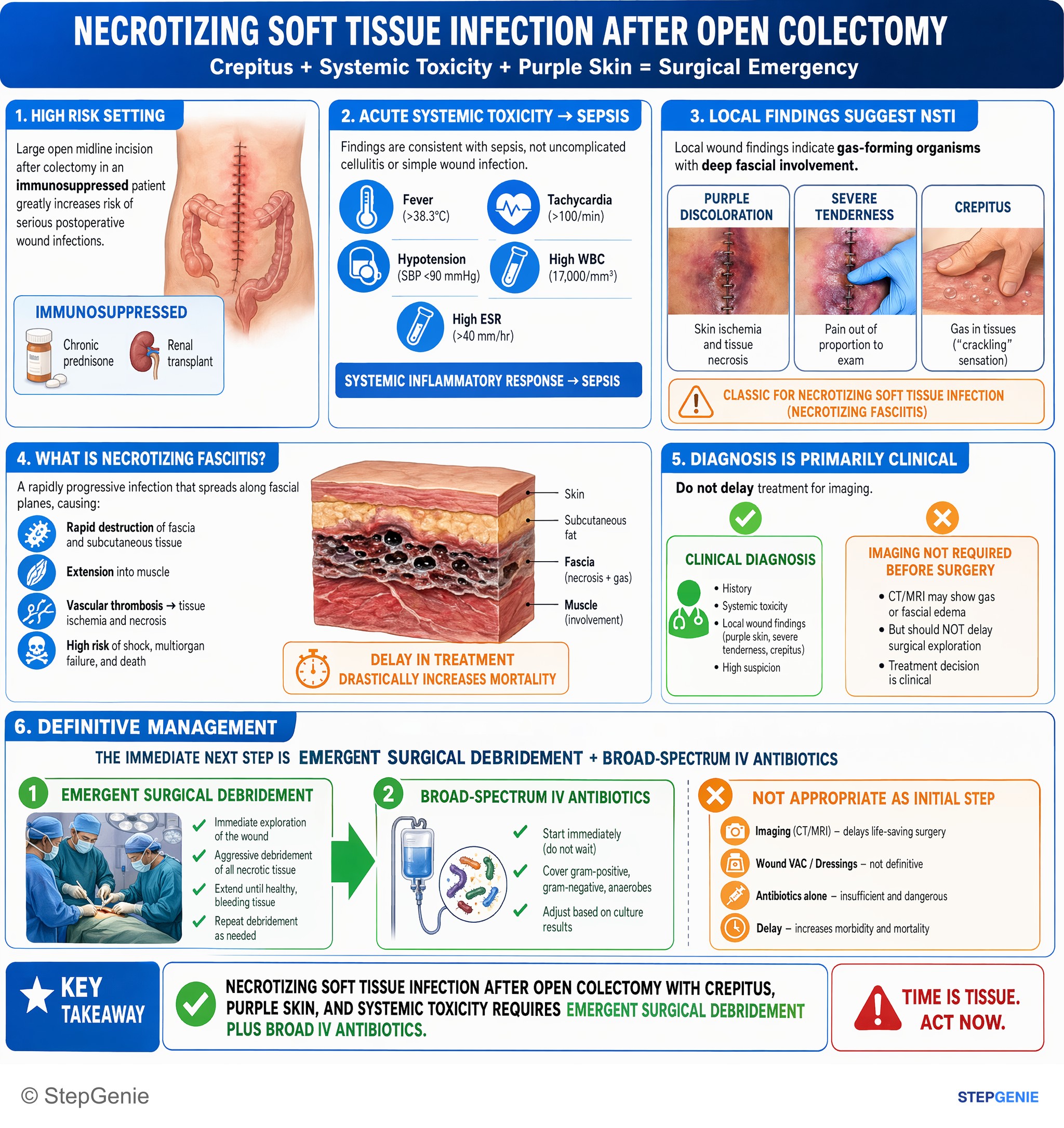
Necrotizing soft tissue infection after open colectomy with crepitus, systemic toxicity, and purple skin → emergent surgical debridement…
USMLE Step 2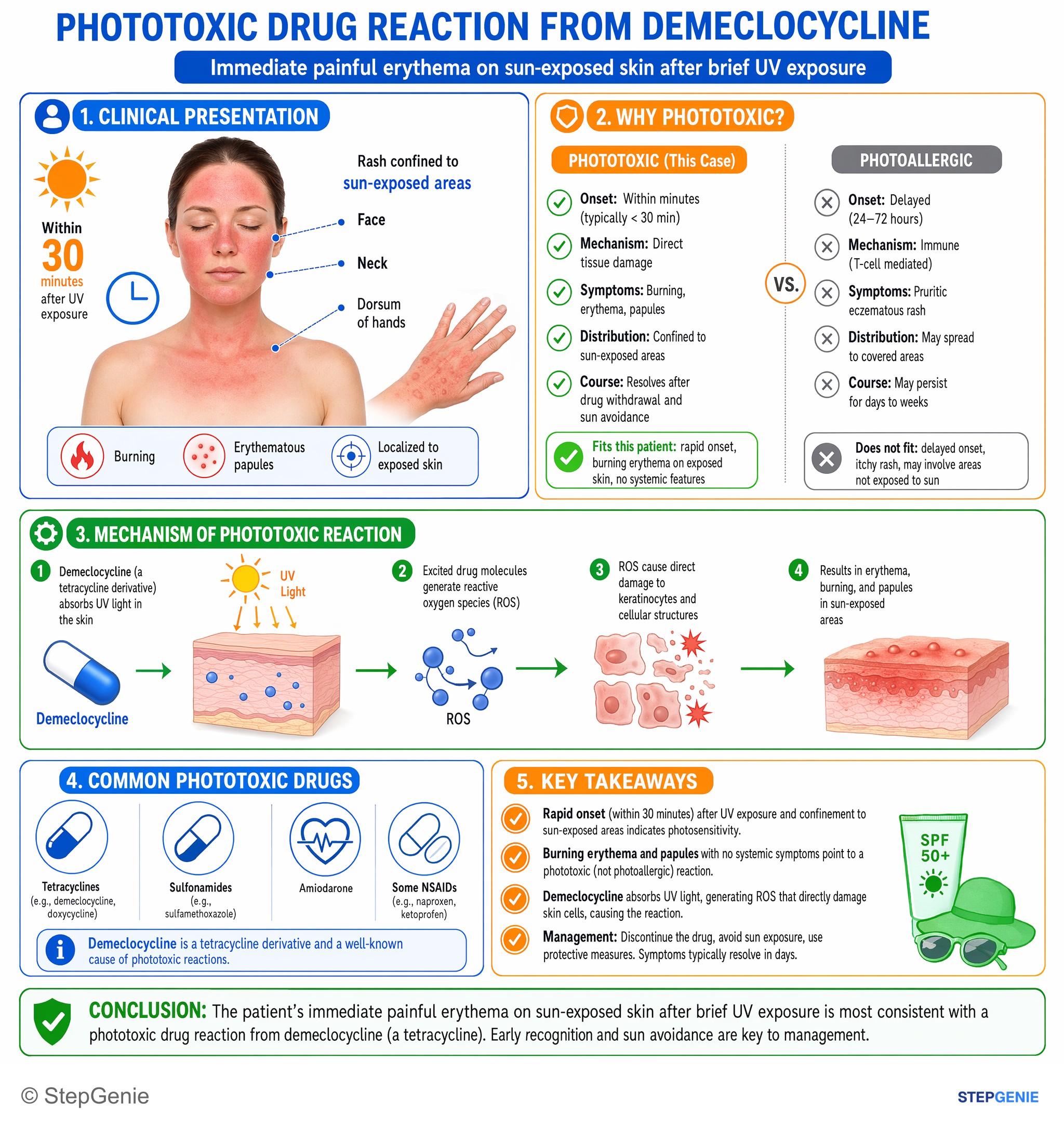
Phototoxic drug reaction from demeclocycline (tetracycline) causes immediate painful erythema on sun-exposed skin after brief UV exposure.
USMLE Step 1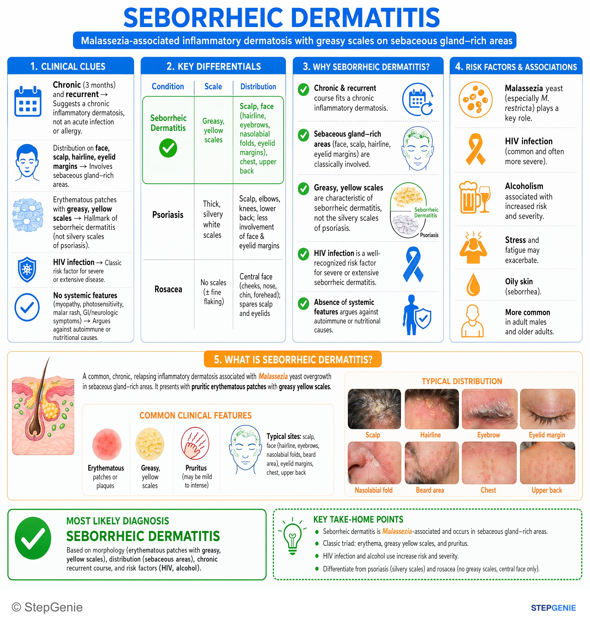
Seborrheic dermatitis: Malassezia-associated, HIV/alcohol-linked, pruritic erythematous patches with greasy yellow scales on scalp, face…
USMLE Step 2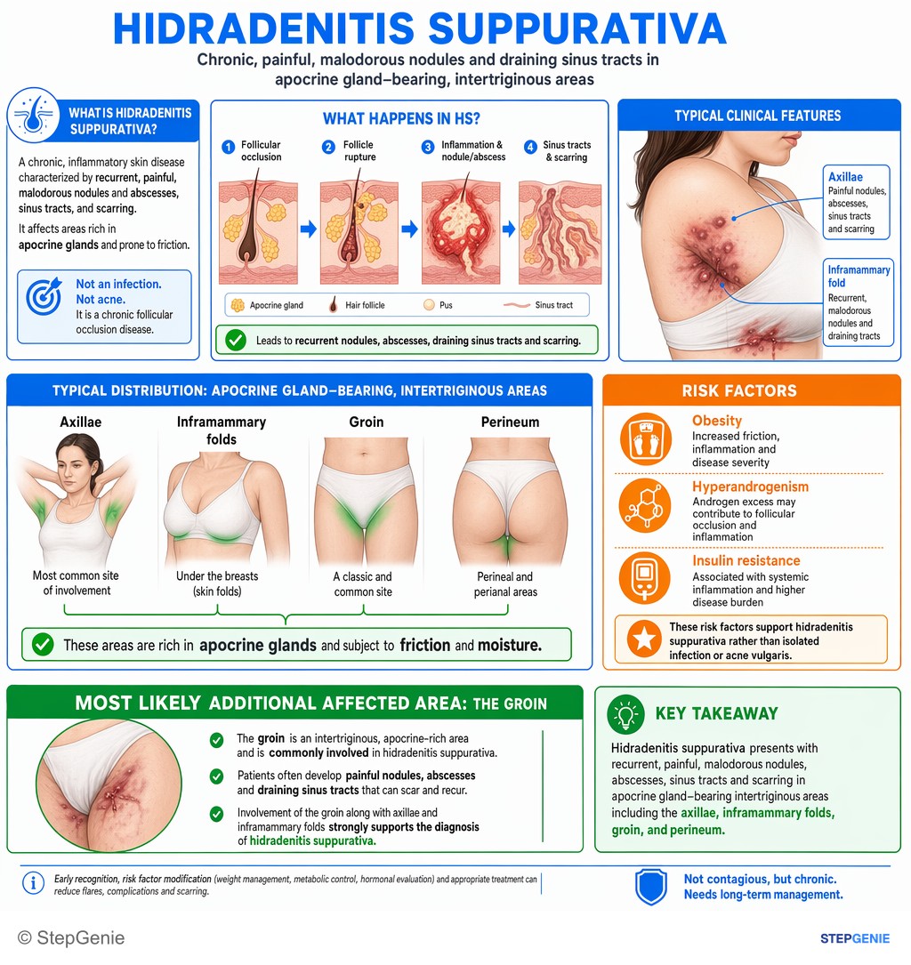
Hidradenitis suppurativa: chronic, painful, malodorous nodules and draining sinus tracts in apocrine gland–bearing, intertriginous areas…
USMLE Step 1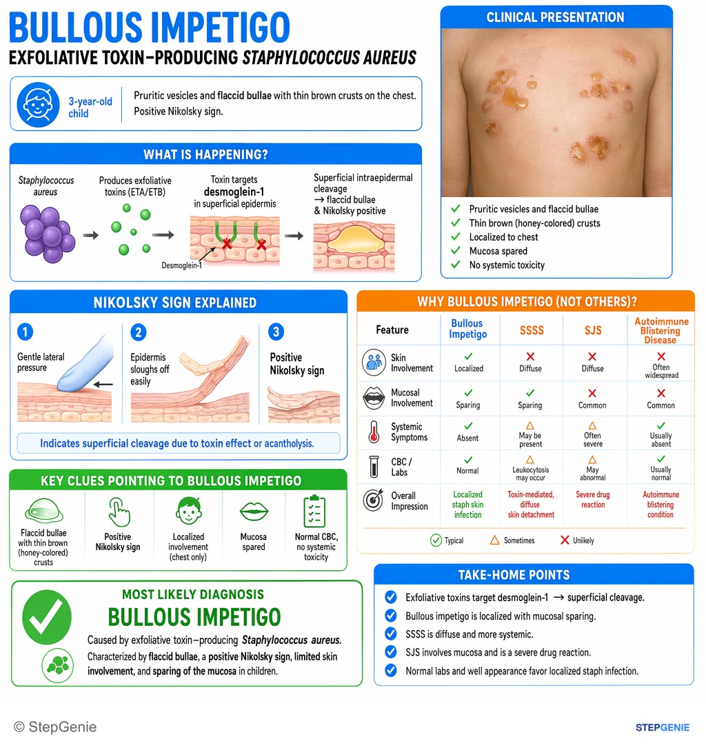
Bullous impetigo from exfoliative toxin–producing Staph aureus causes flaccid bullae, positive Nikolsky sign, limited skin involvement…
USMLE Step 2123 publishable dermatology infographics in the library — 63 from Step 1 questions and 60 from Step 2 CK, counted on October 29, 2026. This page shows 48 of them, alternating the two steps. An image is listed when its question is active, has a full structured explanation, passed verification and carries no unresolved report.
This page caps out at 48 images. 75 more dermatology infographics sit in the in-app image library, where they are searchable and filterable by subject, system and topic.
Open the image libraryPractice the questions behind these images
Each image explains one question. The dermatology topic page has the written explanations and the question sets, and the preview lets you answer three questions without an account.
Other image sets
Study dermatology with the image attached
In the app, each of these sits at the end of the question it explains, alongside the step-by-step reasoning and a panel for every option you did not pick. Starting is free.
Start practicing free