High-yield pulmonology images for the USMLE
StepGenie explanation infographics. Free to view, no account needed.
What do StepGenie's pulmonology images cover?
Each pulmonology image is a StepGenie infographic drawn for one of its own practice questions. It shows the physiology the question turns on — the pressure, the shunt, the compliance change — rather than restating the vignette, and the caption names the conclusion.
Pulmonary and critical care images cluster around pneumonia and other pulmonary infections, then critical care and trauma medicine, obstructive and restrictive disease, pulmonary vascular disease, and interstitial lung disease. Smaller groups cover normal pulmonary structure and function, lung cancer, acid-base problems and congenital anomalies.
This is a system where a picture does real work, because most of the reasoning is about numbers moving together. A ventilation-perfusion panel puts the shunt and the dead-space case side by side and shows why only one improves with supplemental oxygen. An obstructive-versus-restrictive panel puts the flow-volume loops next to the FEV1/FVC ratio so the ratio stops being a rule you memorize and becomes something you can read off a curve.
The critical care images are worth a separate pass. They tend to fix the order of actions — what you check, what you give, what you stop — which is the axis Step 2 CK questions are usually built on.
Acid-base images are shared with the nephrology gallery, which is where the compensation rules are drawn in more detail. Read the two pages together if respiratory and metabolic disorders keep blurring into each other.
Topics in this set
Labels are the question-bank topics these images are filed under. Step 1 and Step 2 file some of the same material under different names, so a few labels here merge both.
Pulmonology explanation images
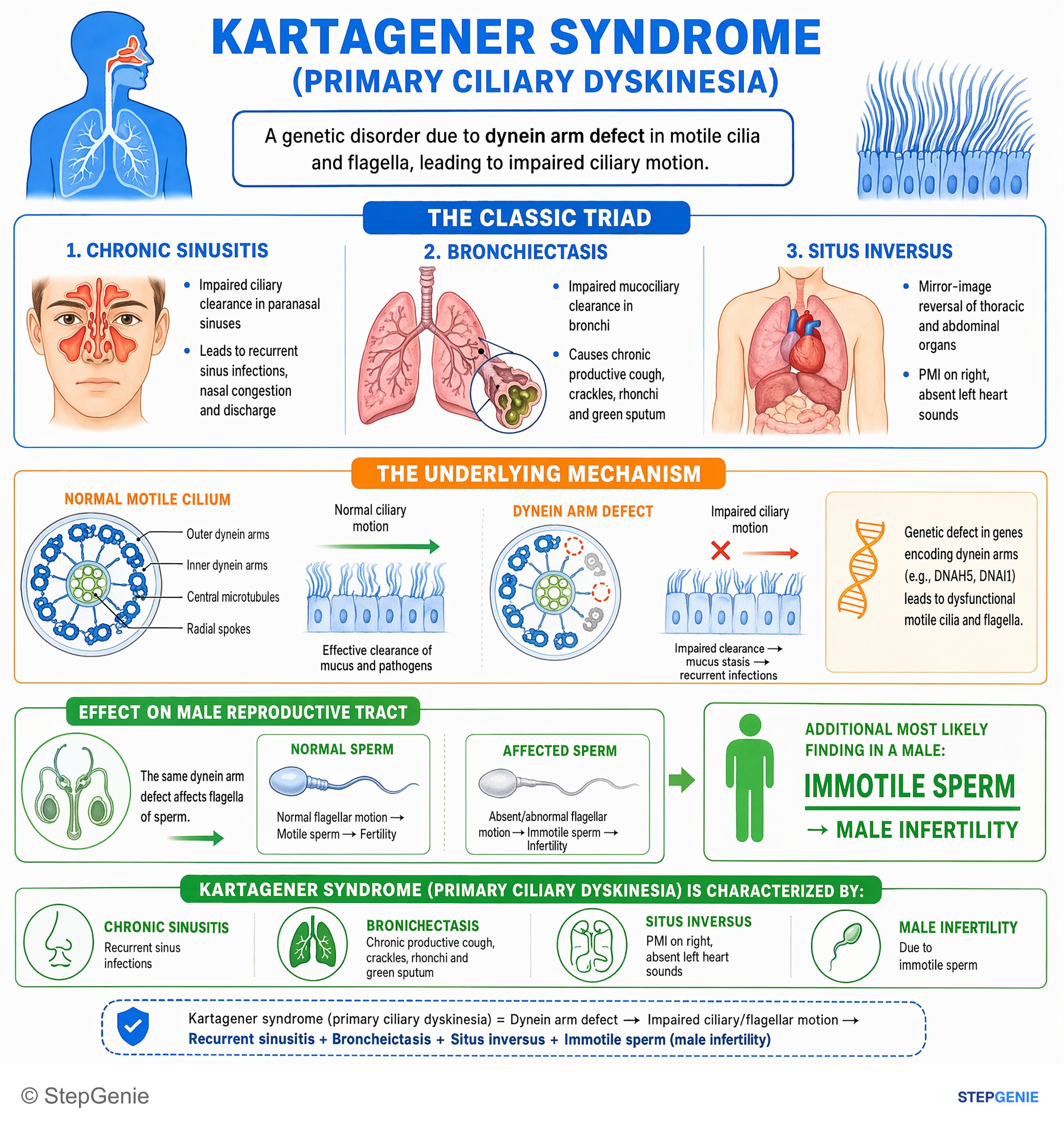
Kartagener syndrome (primary ciliary dyskinesia): dynein arm defect → recurrent sinusitis, bronchiectasis, situs inversus, and male…
USMLE Step 1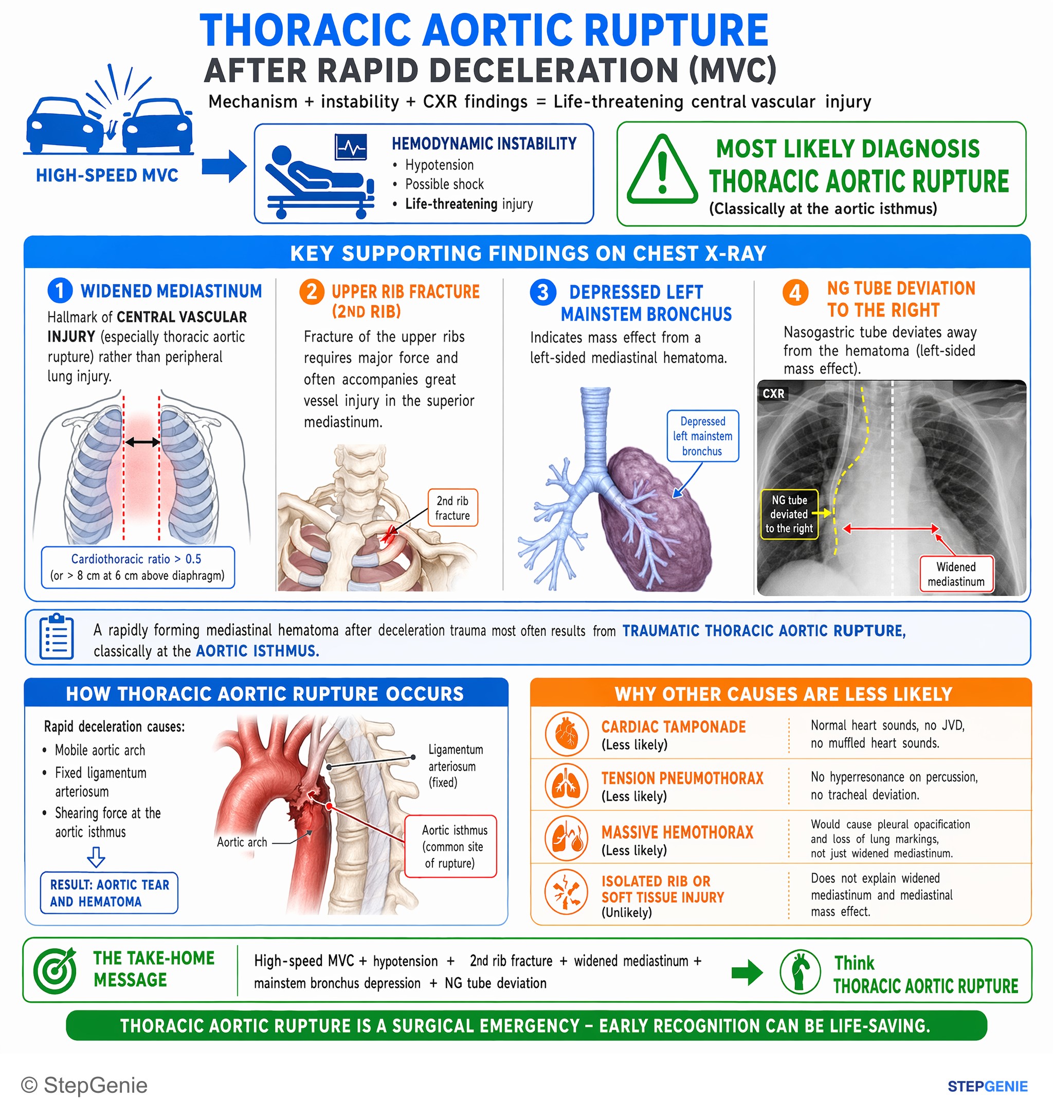
Thoracic aortic rupture after rapid deceleration: hypotension, chest trauma, rib fracture, widened mediastinum, mainstem bronchus…
USMLE Step 2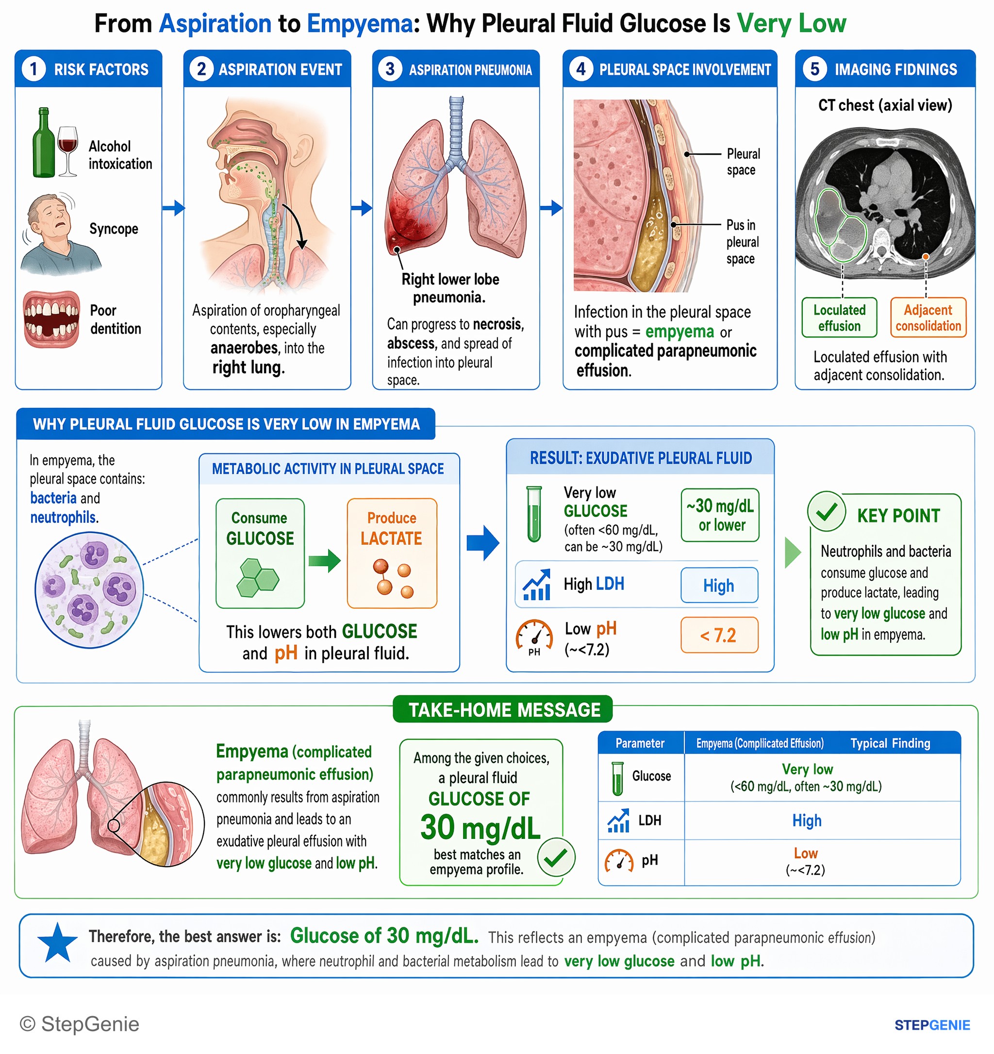
Empyema (complicated parapneumonic effusion) from aspiration pneumonia causes an exudative pleural effusion with very low glucose and low…
USMLE Step 1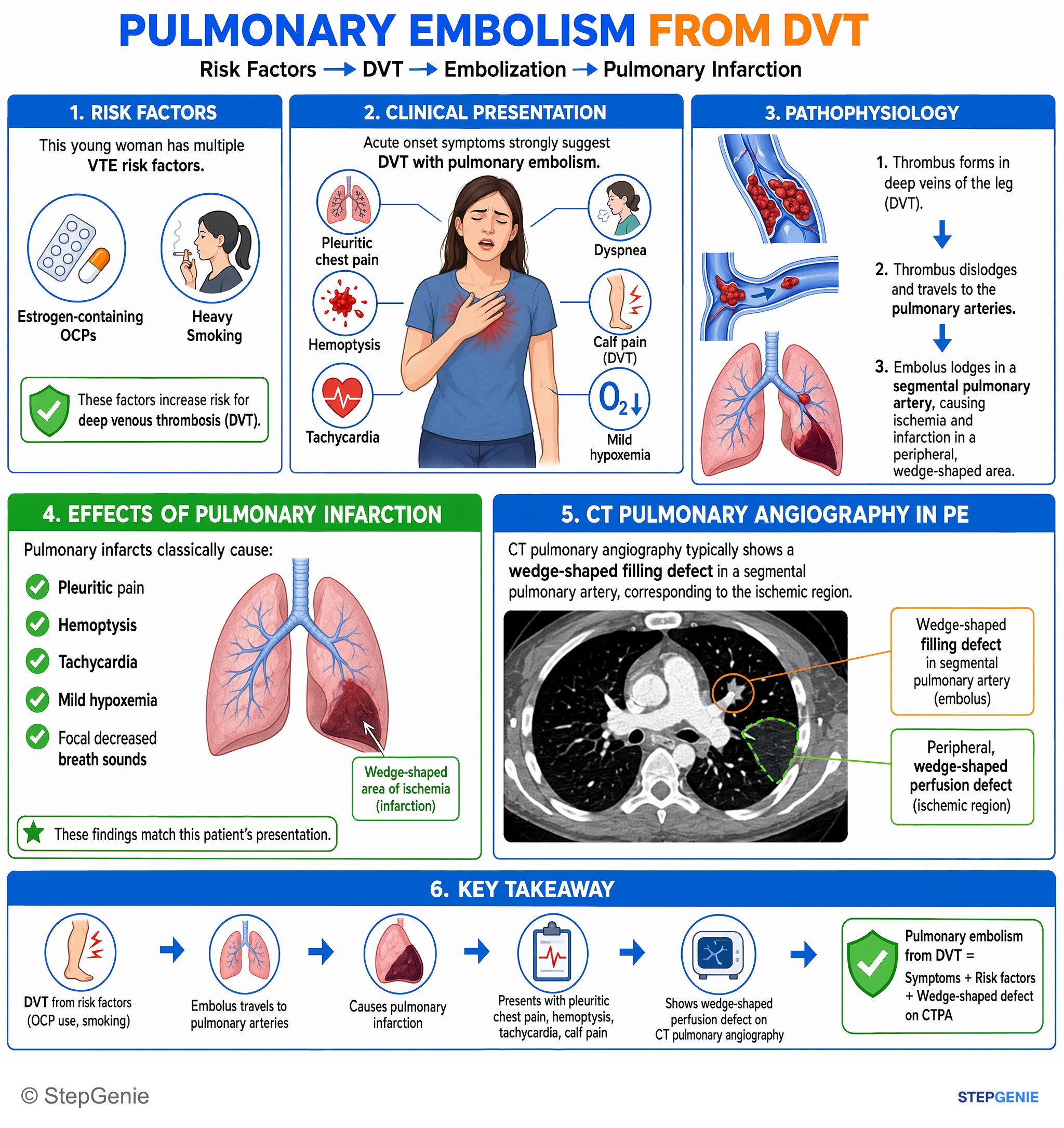
Pulmonary embolism from DVT: pleuritic chest pain, hemoptysis, tachycardia, OCP use, calf pain; shows wedge-shaped perfusion defect on CT…
USMLE Step 2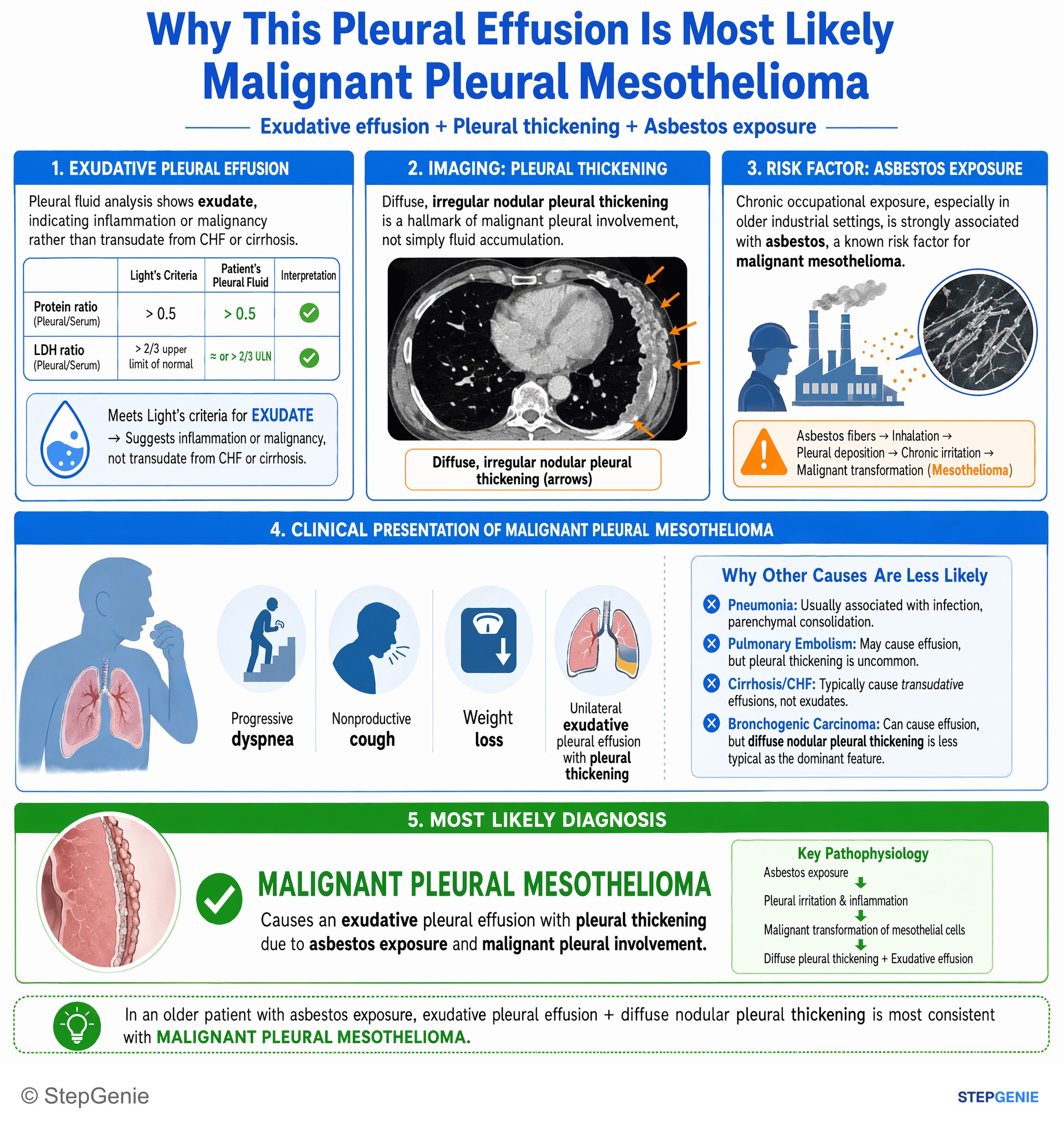
Malignant pleural mesothelioma causes an exudative pleural effusion with pleural thickening due to asbestos exposure and malignant pleural…
USMLE Step 1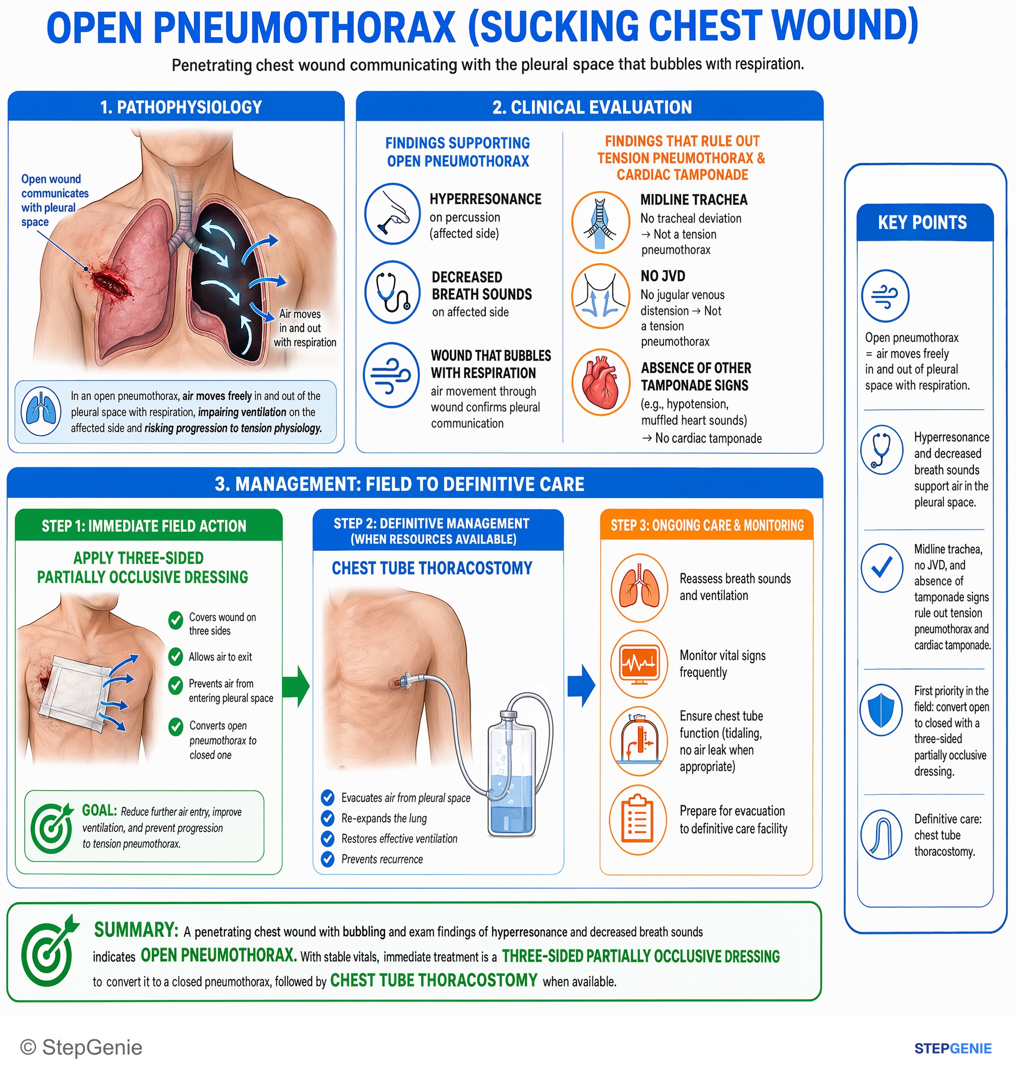
Open pneumothorax from penetrating chest wound with stable vitals → treat immediately with three‑sided partially occlusive dressing, then…
USMLE Step 2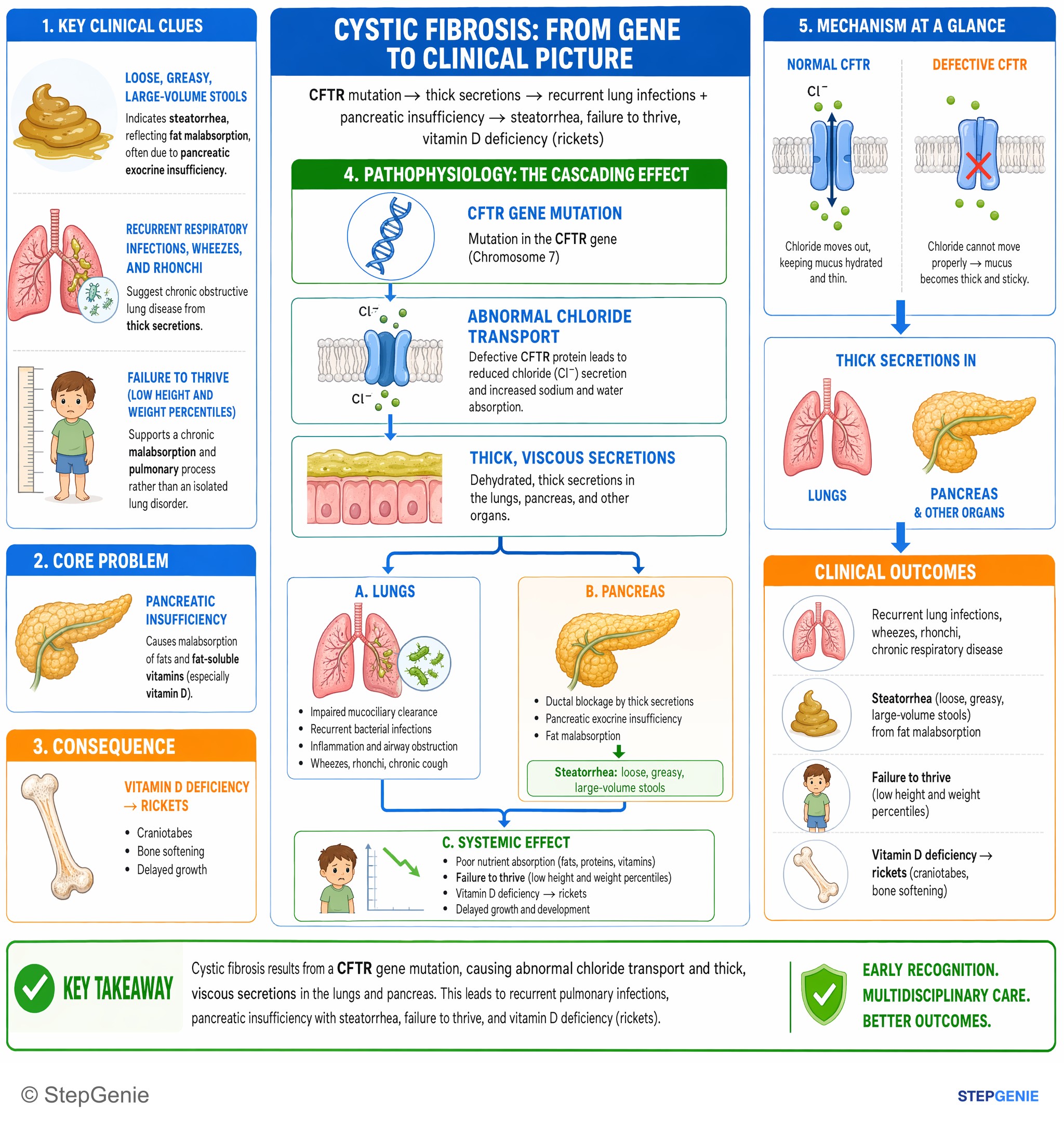
Cystic fibrosis from CFTR mutation → thick secretions → recurrent lung infections + pancreatic insufficiency → steatorrhea, failure to…
USMLE Step 1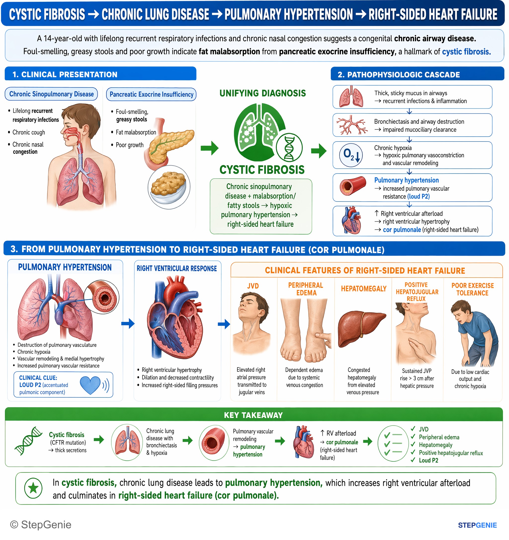
Cystic fibrosis → chronic sinopulmonary disease + malabsorption/fatty stools → hypoxic pulmonary hypertension → right-sided heart failure…
USMLE Step 2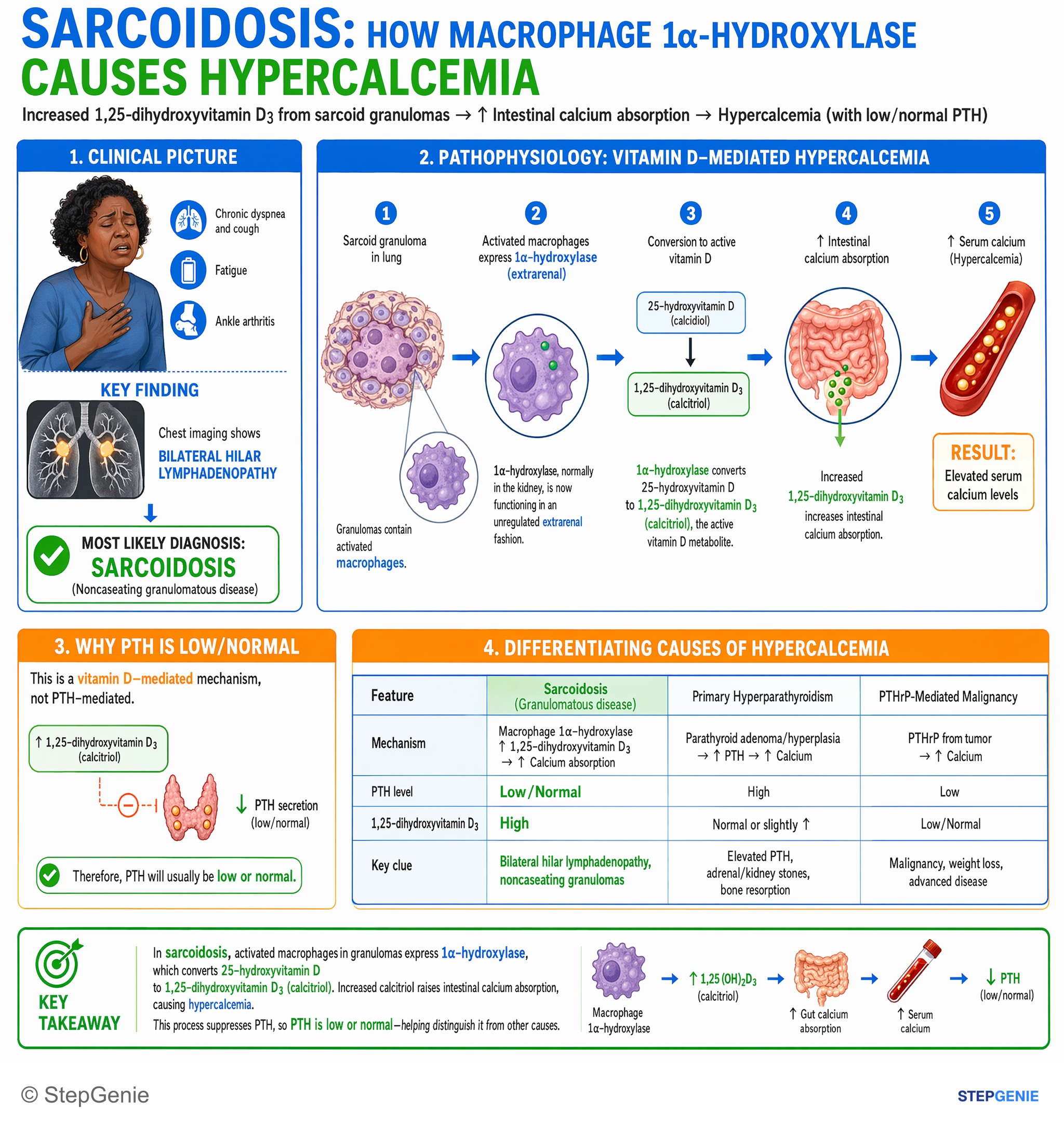
Sarcoidosis causes hypercalcemia via macrophage 1α-hydroxylase, increasing 1,25-dihydroxyvitamin D3 and gut calcium absorption without…
USMLE Step 1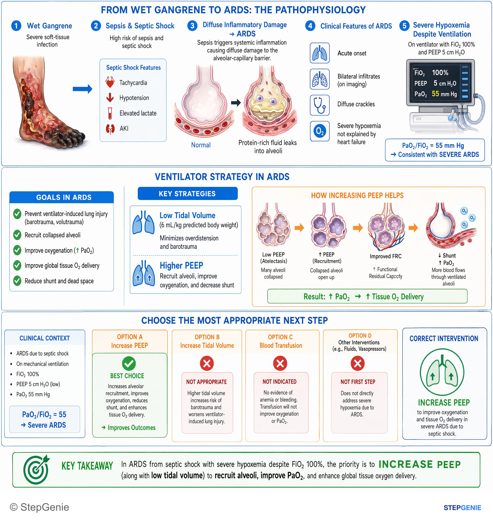
ARDS due to septic shock on mechanical ventilation with severe hypoxemia despite FiO2 100% → increase PEEP to improve oxygenation and…
USMLE Step 2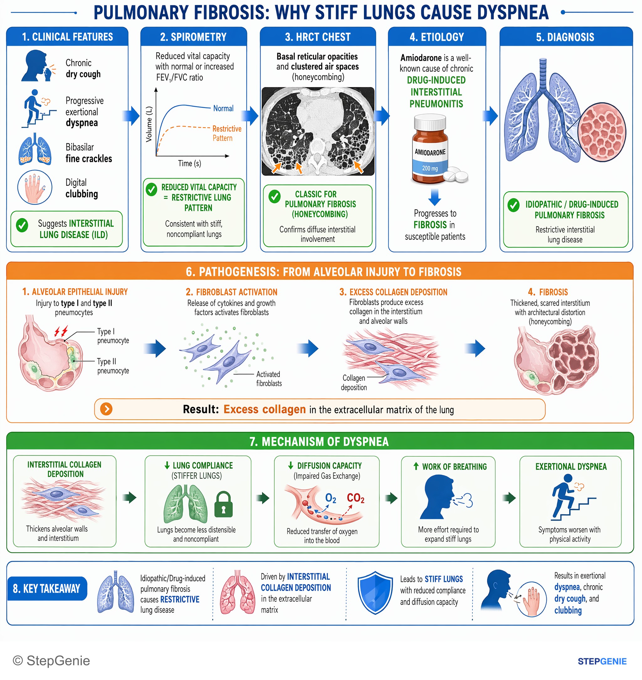
Idiopathic/Drug-induced pulmonary fibrosis causes restrictive lung disease from interstitial collagen deposition, leading to stiff lungs…
USMLE Step 1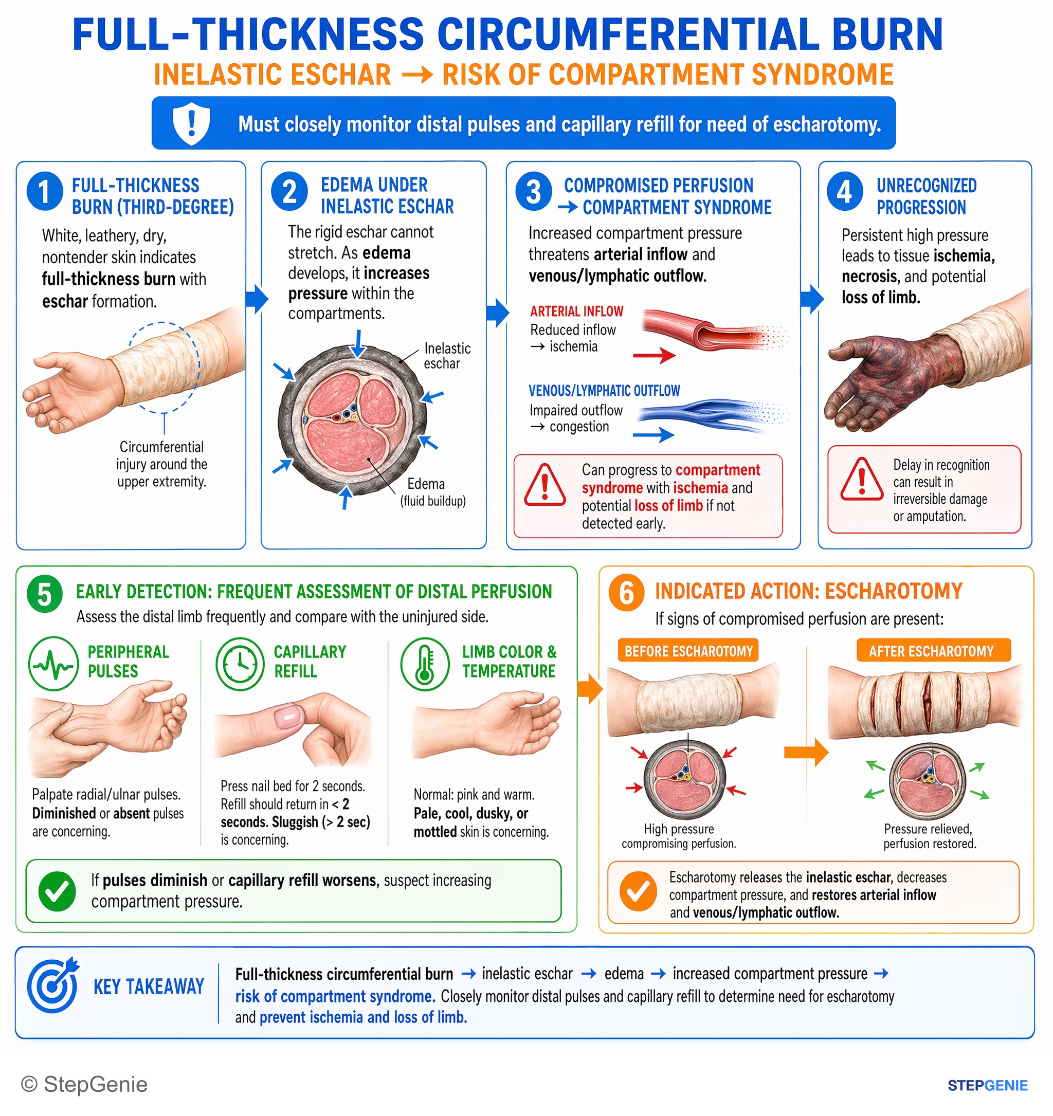
Full-thickness circumferential burn creates inelastic eschar → risk of compartment syndrome; must closely monitor distal pulses/cap refill…
USMLE Step 2254 publishable pulmonology infographics in the library — 132 from Step 1 questions and 122 from Step 2 CK, counted on October 29, 2026. This page shows 48 of them, alternating the two steps. An image is listed when its question is active, has a full structured explanation, passed verification and carries no unresolved report.
This page caps out at 48 images. 206 more pulmonology infographics sit in the in-app image library, where they are searchable and filterable by subject, system and topic.
Open the image libraryPractice the questions behind these images
Each image explains one question. The pulmonology topic page has the written explanations and the question sets, and the preview lets you answer three questions without an account.
Other image sets
Study pulmonology with the image attached
In the app, each of these sits at the end of the question it explains, alongside the step-by-step reasoning and a panel for every option you did not pick. Starting is free.
Start practicing free