You’re cruising through a neuro Q-bank when a deceptively simple vignette shows up: fluctuating weakness, normal sensation, and ocular symptoms. Easy myasthenia gravis (MG), right? Maybe—but the real test is whether you can prove MG and eliminate every distractor with confidence. That’s where points are won on Step 1 and Step 2.
Clinical Vignette (Q-Bank Style)
A 28-year-old woman comes to clinic for intermittent double vision and drooping eyelids for 3 months. Symptoms are worse at the end of the day and improve after rest. She reports difficulty chewing tough foods at dinner but feels better in the morning. No numbness or tingling. On exam, she has fatigable ptosis and mild weakness with repeated arm abduction. Reflexes are normal.
Question: What is the underlying pathophysiology?
✅ Correct Answer
Autoantibodies against postsynaptic nicotinic acetylcholine receptors at the neuromuscular junction (most commonly anti-AChR; sometimes anti-MuSK).
Why This Is Myasthenia Gravis
The “Step-ready” triad
- Fluctuating weakness (worse with use, better with rest)
- Ocular involvement (ptosis, diplopia) is common and often the first clue
- Bulbar symptoms (dysphagia, chewing fatigue, dysarthria) are classic
Key negatives that support MG
- No sensory symptoms (NMJ disorder → motor only)
- Normal reflexes (reflex arcs intact; contrast with neuropathies)
- Fatigability on exam (repeat movements worsen weakness)
Mechanism: What the Antibodies Actually Do
MG is a type II hypersensitivity condition:
- Anti-AChR antibodies (most common):
- Block receptor function
- Cross-link receptors → internalization/downregulation
- Activate complement → damage postsynaptic membrane
- Net result: decreased end-plate potentials, so repeated stimulation fails to trigger muscle action potentials → fatigable weakness
High-yield association: thymus
- Thymic hyperplasia in many patients
- Thymoma in a subset (screen with chest imaging)
- Think: “MG = check the thymus”
What You’d Expect on Testing (and What’s Worth Memorizing)
Diagnostic highlights
- Serology: anti-AChR antibodies (or anti-MuSK if AChR-negative)
- Electrophysiology: decremental response on repetitive nerve stimulation
- Bedside clue: ice pack test improves ptosis (cold inhibits AChE → more ACh in synapse)
Treatment framework (Step 1 + Step 2 relevant)
- Symptomatic: pyridostigmine (AChE inhibitor)
- Immunomodulating: steroids, azathioprine, mycophenolate, etc.
- Thymectomy: especially with thymoma; also considered in generalized MG
- Myasthenic crisis (respiratory failure):
- Immediate support: airway/ventilation
- Rapid immunotherapy: IVIG or plasmapheresis
- (Pyridostigmine is not your “save-the-day” drug in acute respiratory failure)
Distractor Breakdown: Why Every Wrong Answer Is Wrong
Here’s the real Q-bank skill: precision. Many answer choices sound neuro-ish but describe different levels of the motor pathway.
Distractor 1: “Autoantibodies against presynaptic voltage-gated calcium channels”
This is Lambert-Eaton myasthenic syndrome (LEMS), not MG.
How LEMS differs (testable contrasts):
- Weakness often proximal (hips/shoulders)
- Autonomic symptoms (dry mouth, constipation, impotence)
- Improves with use (facilitation), not fatigability
- Hyporeflexia is common
- Often paraneoplastic: small cell lung cancer
Buzz phrase: “Gets stronger with repetition” → think LEMS.
Distractor 2: “Demyelination in the peripheral nervous system”
This points to Guillain-Barré syndrome (GBS) (AIDP variant).
Why it doesn’t fit:
- GBS causes ascending weakness over days to weeks
- Areflexia is typical
- Often post-infectious (Campylobacter jejuni is classic)
- Can have sensory symptoms and neuropathic pain
- Not classically fluctuating or ocular-predominant
High-yield add-on: CSF in GBS shows albuminocytologic dissociation (high protein, normal WBC).
Distractor 3: “Destruction of motor neurons in the anterior horn”
This describes amyotrophic lateral sclerosis (ALS).
Why it doesn’t fit:
- ALS has mixed UMN + LMN signs
- UMN: hyperreflexia, spasticity, Babinski
- LMN: atrophy, fasciculations, weakness
- No sensory loss (true), but symptoms are progressive, not fluctuating
- Ocular muscles are typically spared until late
USMLE pearl: ALS = degeneration of corticospinal tract + anterior horn cells.
Distractor 4: “Antibodies against acetylcholine receptors in the autonomic ganglia”
This is not classic MG and usually points toward autoimmune autonomic ganglionopathy.
Why it doesn’t fit:
- Prominent autonomic failure: orthostatic hypotension, anhidrosis, GI dysmotility, urinary retention
- Not primarily ocular/bulbar fatigable weakness
Step takeaway: If the stem screams autonomic, don’t force MG.
Distractor 5: “Mutation causing defective dystrophin”
That’s Duchenne muscular dystrophy (DMD) (X-linked recessive).
Why it doesn’t fit:
- Onset in early childhood
- Gowers sign, waddling gait
- Calf pseudohypertrophy
- Markedly elevated CK
- Cardiomyopathy is common
- Weakness is chronic/progressive—not fluctuating and not ocular-predominant
Distractor 6: “Peripheral nerve infarction due to vasculitis”
This suggests mononeuritis multiplex.
Why it doesn’t fit:
- Asymmetric, patchy deficits
- Often includes sensory loss and pain
- Associated with systemic disease (PAN, RA, hepatitis B/C, etc.)
- Doesn’t produce classic fatigable ptosis/diplopia pattern
Quick Comparison Table (High-Yield)
| Disorder | Lesion Level | Weakness Pattern | Reflexes | Sensory | Key Clues |
|---|---|---|---|---|---|
| Myasthenia gravis | Postsynaptic NMJ (AChR/MuSK) | Worse with use | Normal | Normal | Ptosis/diplopia, bulbar fatigue, thymic disease |
| LEMS | Presynaptic NMJ (VG Ca channels) | Improves with use | ↓ | Normal | Autonomic sx, SCLC association |
| GBS (AIDP) | Peripheral nerve demyelination | Ascending, subacute | Absent/↓ | Often present | Post-infectious, albuminocytologic dissociation |
| ALS | UMN + LMN | Progressive | ↑ (UMN) + LMN signs | Normal | Fasciculations + spasticity, no ocular early |
| DMD | Muscle fiber membrane (dystrophin) | Progressive proximal | Variable | Normal | Childhood onset, high CK, cardiomyopathy |
High-Yield MG Pearls You’ll Use Again
- MG = fatigable weakness with ocular/bulbar prominence.
- Normal pupils (pupillary involvement suggests autonomic issues—think botulism).
- Anti-AChR most common; anti-MuSK often more bulbar/respiratory involvement.
- Screen for thymoma.
- Myasthenic crisis: treat with IVIG/plasmapheresis and secure the airway.
Q-Bank Mindset: How to Eliminate Distractors Fast
When you see weakness, ask two questions:
- Where is the lesion? (muscle vs NMJ vs nerve vs spinal cord vs brain)
- What pattern is it?
- Fluctuating + ocular → MG
- Improves with use + autonomic → LEMS
- Ascending + areflexia → GBS
- UMN + LMN signs → ALS
- Childhood + high CK → DMD
Nail those patterns, and suddenly every answer choice becomes a clue—not a trap.
Visual explanations from the StepGenie QBank
Infographics StepGenie drew to explain questions in its own QBank, matched to this article's topic. Each caption is the takeaway of the question it explains.
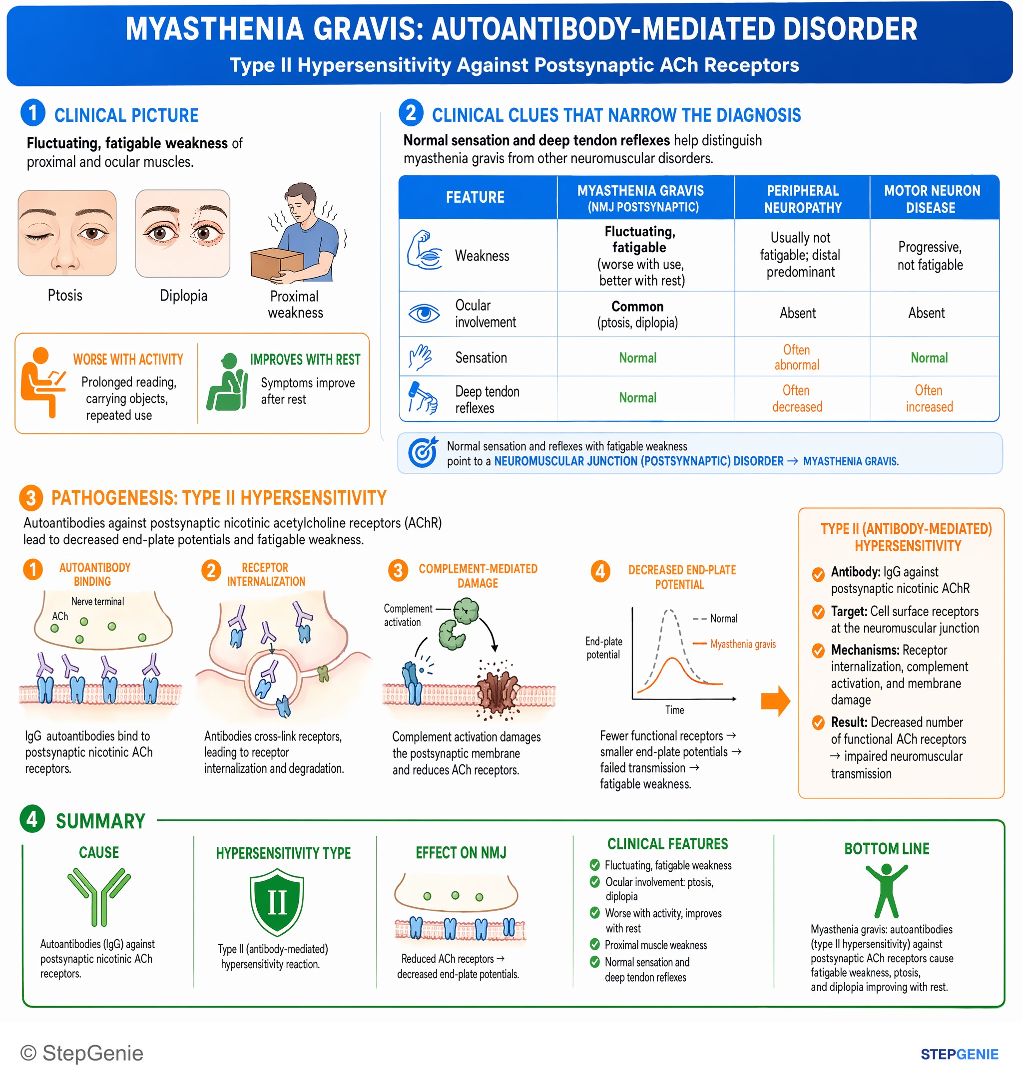
Myasthenia gravis: autoantibodies (type II hypersensitivity) against postsynaptic ACh receptors cause fatigable weakness, ptosis, and…
USMLE Step 1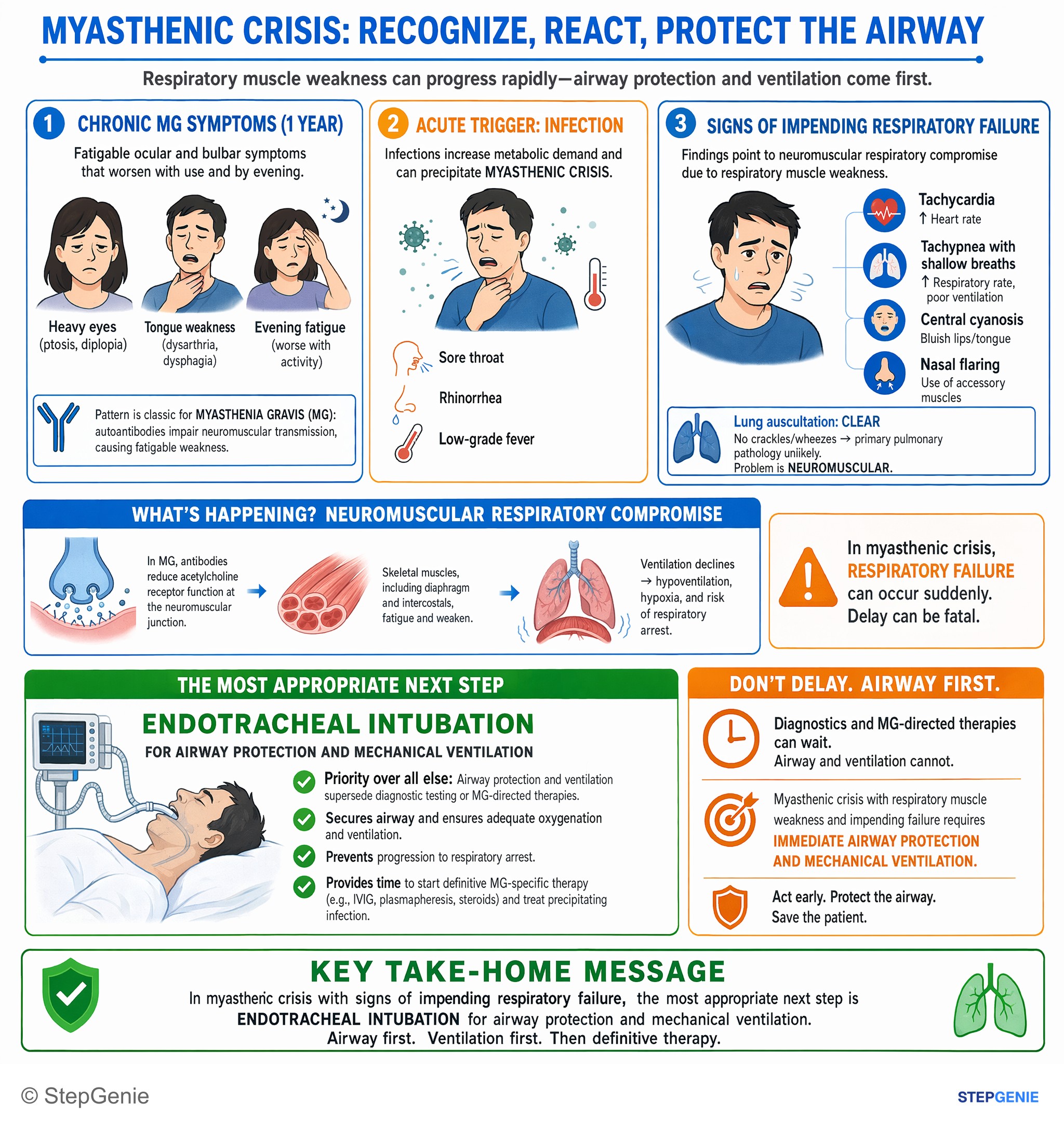
Myasthenic crisis with respiratory muscle weakness and impending failure requires immediate airway protection and mechanical ventilation…
USMLE Step 2Frequently Asked Questions
Why does myasthenia gravis cause weakness that gets worse with use and better with rest?
Myasthenia gravis causes fatigable weakness because autoantibodies impair postsynaptic nicotinic acetylcholine receptors at the neuromuscular junction. These antibodies block receptor function, promote receptor internalization, and activate complement that damages the postsynaptic membrane. The result is decreased end-plate potentials, so repeated stimulation eventually fails to trigger muscle action potentials, making weakness worsen with use and improve after rest.
How do you tell myasthenia gravis from Lambert-Eaton myasthenic syndrome on a question stem?
Myasthenia gravis is suggested by ocular and bulbar weakness that worsens with use, normal reflexes, and no sensory symptoms, whereas Lambert-Eaton usually improves with use. Lambert-Eaton also commonly has autonomic symptoms such as dry mouth, constipation, or impotence, hyporeflexia, and is often associated with small cell lung cancer. A buzz phrase like getting stronger with repetition points away from myasthenia gravis.
What exam findings make myasthenia gravis more likely?
The most supportive findings are fluctuating motor weakness with fatigability, ocular symptoms, normal sensation, and normal reflexes. Classic clues include ptosis, diplopia, chewing fatigue, and weakness that becomes more obvious with repeated movements. Because myasthenia gravis is a neuromuscular junction disorder, it produces motor symptoms without sensory loss, and the reflex arcs remain intact.
What tests are worth knowing for myasthenia gravis?
The key tests are anti-AChR antibody serology, anti-MuSK testing if AChR antibodies are negative, and electrophysiology showing a decremental response on repetitive nerve stimulation. A useful bedside clue is the ice pack test, which improves ptosis because cold inhibits acetylcholinesterase and leaves more acetylcholine in the synapse. It is also worth checking the thymus because thymic abnormalities are associated with myasthenia gravis.
What is the treatment approach for myasthenia gravis, including myasthenic crisis?
Treatment includes symptomatic therapy with pyridostigmine, immunomodulating therapy such as steroids, azathioprine, or mycophenolate, and thymectomy especially when thymoma is present. In myasthenic crisis, the immediate priorities are airway and ventilatory support plus rapid immunotherapy with IVIG or plasmapheresis. Pyridostigmine is not the rescue treatment for acute respiratory failure.