ALS is a classic “boards love it” neurodegenerative diagnosis because it’s pure motor, hits both UMN and LMN, and spares sensation—a combo that instantly narrows your differential. This post is built as quick-hit, shareable tables + a mnemonic you can picture in your head.
ALS in one line (the Step-ready definition)
Amyotrophic lateral sclerosis (ALS) = progressive degeneration of upper and lower motor neurons → mixed UMN + LMN signs with preserved sensation and cognition (usually), often fatal from respiratory failure.
The visual/mnemonic device: “ALS = Anterior horn + Lateral corticospinal”
Picture the spinal cord cross-section:
- A = Anterior horn cell loss → LMN signs
- L = Lateral corticospinal tract degeneration → UMN signs
- S = Sensation spared (dorsal columns/spinothalamic usually intact)
One-liner: ALS damages motor neuron cell bodies (anterior horn) and descending UMN tracts (lateral columns), so you get both UMN + LMN findings without sensory loss.
Quick-hit comparison table: ALS vs look-alikes (high-yield)
| Disorder | UMN signs? | LMN signs? | Sensory loss? | Key distinguishing clues | Classic test/association |
|---|---|---|---|---|---|
| ALS | ✅ | ✅ | ❌ (typically) | Progressive weakness, fasciculations, atrophy, hyperreflexia, spasticity; bulbar symptoms (dysarthria/dysphagia) | EMG: denervation (fibrillations), fasciculations; can be familial (SOD1, C9orf72) |
| Cervical myelopathy / cord compression | ✅ (below lesion) | ✅ (at lesion level) | ✅ often | Neck pain, radicular symptoms, sensory level, bowel/bladder issues possible | MRI cervical spine |
| Multiple sclerosis | ✅ | ❌ (usually) | ✅ often | CNS demyelination; optic neuritis, INO, relapsing-remitting | MRI plaques; CSF oligoclonal bands |
| Guillain-Barré (AIDP) | ❌ | ✅ | ± (often paresthesias) | Ascending weakness, areflexia, autonomic instability; often post-infectious | CSF: albuminocytologic dissociation |
| Myasthenia gravis | ❌ | ❌ (no true LMN) | ❌ | Fluctuating weakness, worse with use, ptosis/diplopia; normal reflexes | AChR/MuSK Ab; improves with rest/edrophonium historically |
| Parkinson disease | ❌ | ❌ | ❌ | Bradykinesia, resting tremor, rigidity; not true weakness early | Lewy bodies (α-synuclein) |
| Primary lateral sclerosis (PLS) | ✅ | ❌ | ❌ | Pure UMN syndrome (spasticity, hyperreflexia) | Diagnosis of exclusion; slower course |
| Spinal muscular atrophy | ❌ | ✅ | ❌ | Childhood onset; tongue fasciculations; genetic | SMN1 mutation |
USMLE move: If you see UMN + LMN + no sensory deficits, ALS rockets to the top—but always consider cord compression and rule it out (especially if pain, sensory level, or sphincter issues).
Core clinical features (what shows up in vignettes)
UMN signs (corticospinal tract)
- Hyperreflexia
- Spasticity
- Clonus
- Babinski sign
LMN signs (anterior horn cells)
- Muscle atrophy
- Fasciculations
- Weakness
- Hyporeflexia (can be regionally present even when other areas are hyperreflexic)
Bulbar involvement (high-yield)
- Dysarthria, dysphagia
- Emotional lability (pseudobulbar affect can occur)
- Tongue fasciculations/atrophy may appear
Sensory and other systems
- Sensation is typically preserved
- Extraocular movements usually spared until very late
- Bowel/bladder usually spared early (if prominent, think spinal cord pathology)
ALS vs MS in one sentence (common confusion)
- ALS = motor neuron degeneration (UMN + LMN), no sensory loss.
- MS = CNS demyelination (UMN + sensory/visual), lesions separated in time/space.
Diagnosis: what to know for USMLE
Best next step logic
- Clinical suspicion (mixed UMN/LMN signs) → EMG/NCS to support LMN involvement and exclude mimics
- MRI brain/spine is often used to rule out structural lesions (cord compression, tumor, syrinx)
EMG pearls
- Shows active denervation: fibrillations, positive sharp waves
- Fasciculation potentials are common
- Helps show widespread LMN involvement across regions
Pathology & genetics (high-yield snippets)
- Degeneration of motor neurons in:
- Primary motor cortex
- Brainstem motor nuclei
- Anterior horn of spinal cord
- Some familial cases:
- SOD1 mutation (free radical handling)
- C9orf72 repeat expansion (also linked with frontotemporal dementia)
Treatment & prognosis (Step 2–leaning but testable)
Disease-modifying (modest benefit)
- Riluzole (decreases glutamatergic transmission; can modestly prolong survival)
- Edaravone (free radical scavenger; may slow functional decline in some patients)
Supportive care that matters
- Noninvasive ventilation (BiPAP) improves survival and quality of life
- PEG feeding for dysphagia/weight loss
- Multidisciplinary ALS clinic care is associated with better outcomes
Cause of death (very commonly tested)
- Respiratory failure from diaphragmatic/respiratory muscle weakness
Rapid-fire “If you see this → think ALS” checklist
- Progressive weakness + fasciculations
- Hyperreflexia + muscle atrophy in the same patient
- No sensory loss
- Bulbar symptoms (dysarthria/dysphagia)
- Normal eye movements early
Mini-mnemonic recap (shareable)
“AL(S)” = Anterior horn (LMN) + Lateral corticospinal (UMN) + Sensation spared.
Visual explanations from the StepGenie QBank
Infographics StepGenie drew to explain questions in its own QBank, matched to this article's topic. Each caption is the takeaway of the question it explains.
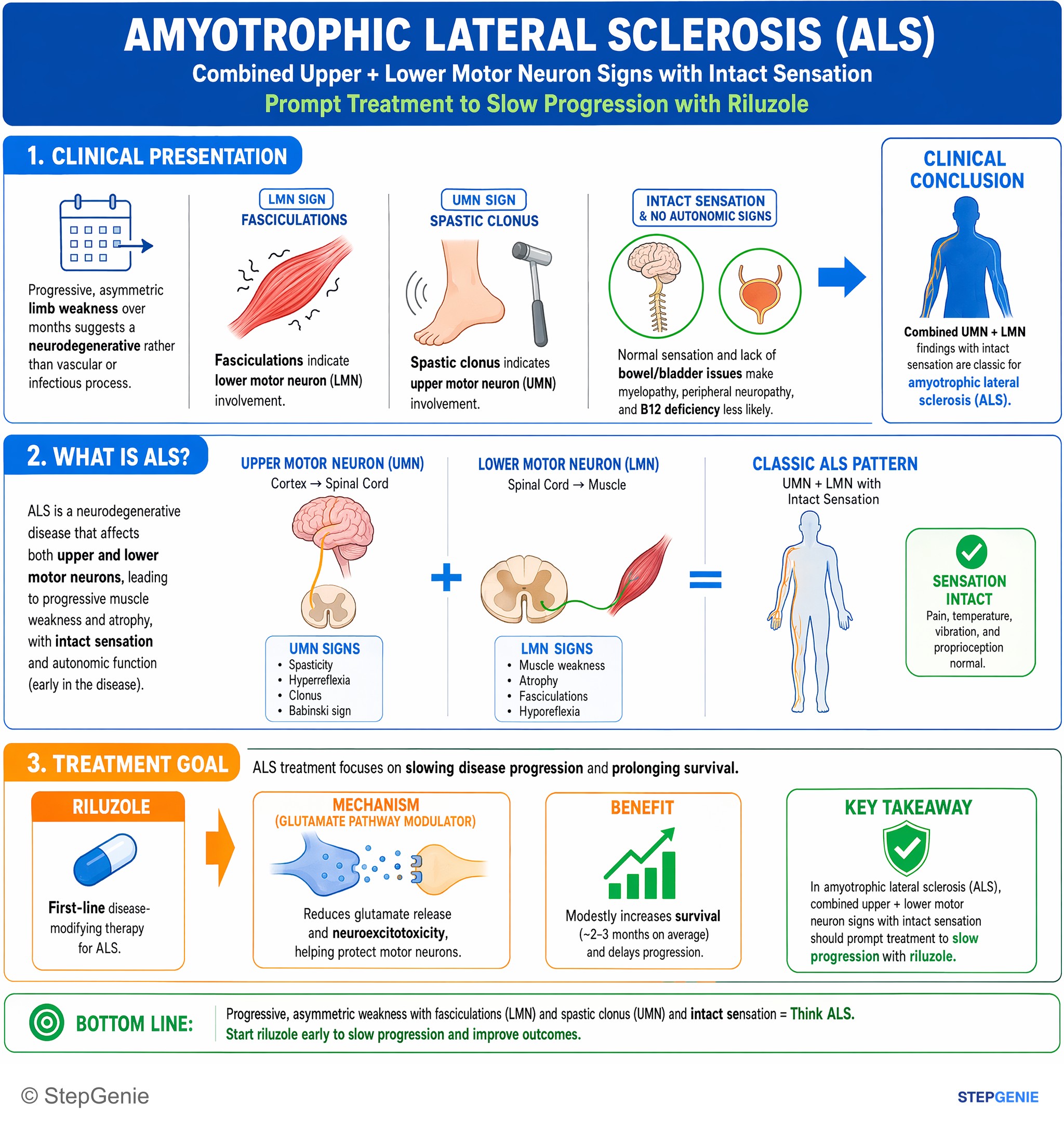
Amyotrophic lateral sclerosis (ALS): combined upper + lower motor neuron signs with intact sensation; treat to slow progression with…
USMLE Step 2Frequently Asked Questions
How do you recognize ALS on a vignette?
ALS is suggested by mixed upper and lower motor neuron findings with preserved sensation. Classic clues are progressive weakness plus fasciculations, muscle atrophy, hyperreflexia, and spasticity in the same patient. Bulbar symptoms such as dysarthria and dysphagia also fit, and extraocular movements are usually spared until very late. The combination of UMN signs, LMN signs, and no sensory loss is the key pattern.
Why does ALS cause both UMN and LMN signs without sensory loss?
ALS causes both UMN and LMN signs because it degenerates the lateral corticospinal tracts and the anterior horn cells. Lateral corticospinal tract damage produces upper motor neuron findings like hyperreflexia and spasticity, while anterior horn cell loss causes lower motor neuron findings such as atrophy and fasciculations. Sensation is typically spared because the dorsal columns and spinothalamic pathways are usually intact.
How do you tell ALS from cervical cord compression?
ALS is more likely when there are mixed UMN and LMN signs without sensory deficits, whereas cervical cord compression often has sensory and structural clues. Neck pain, radicular symptoms, a sensory level, and bowel or bladder issues point toward cervical myelopathy rather than ALS. MRI of the cervical spine is the classic study to evaluate cord compression, while ALS workup often includes EMG and MRI to exclude structural lesions.
What tests support the diagnosis of ALS?
The main supportive test is EMG with nerve conduction studies, used after clinical suspicion based on mixed UMN and LMN findings. EMG can show active denervation with fibrillations, positive sharp waves, and common fasciculation potentials, helping demonstrate widespread lower motor neuron involvement across regions. MRI of the brain and spine is often obtained to rule out structural mimics such as cord compression, tumor, or syrinx.
What is the treatment for ALS and what usually causes death?
ALS treatment includes disease-modifying therapy and supportive care, but respiratory failure is a common cause of death. Riluzole can modestly prolong survival by decreasing glutamatergic transmission, and edaravone may slow functional decline in some patients. Noninvasive ventilation with BiPAP improves survival and quality of life, and PEG feeding is used for dysphagia or weight loss caused by bulbar involvement.