The cerebellum does not start movements: it compares the movement you intended with the movement actually happening and corrects the error, so damage causes incoordination (ataxia), not weakness. Cerebellar signs are ipsilateral: midline lesions (vermis, flocculonodular lobe) cause truncal and gait ataxia and nystagmus, while lateral hemisphere lesions cause limb dysmetria, intention tremor and dysdiadochokinesia on the same side, and patients tend to fall toward the lesion. Inside the circuit, Purkinje cells are the only output of the cerebellar cortex and they are inhibitory (GABA) onto the deep nuclei, which send the cerebellum's excitatory output mostly through the superior cerebellar peduncle.
Vermis vs hemisphere: the three functional zones
| Zone | Parts | Output nucleus and target | Job | Lesion signs |
|---|---|---|---|---|
| Vestibulocerebellum | Flocculonodular lobe | Vestibular nuclei (they act as its deep nuclei) | Balance; eye movements (VOR, holding gaze) | Nystagmus, truncal instability, wide-based stance |
| Spinocerebellum | Vermis + intermediate (paravermal) zone | Fastigial (vermis) to vestibular nuclei and reticular formation; interposed (globose + emboliform) to the contralateral red nucleus | Trunk and proximal muscles: posture, stance, gait; the intermediate zone adjusts ongoing limb movements | Truncal and gait ataxia, titubation (tremor of head or trunk) |
| Cerebrocerebellum | Lateral hemispheres | Dentate to the contralateral VL thalamus (and red nucleus), then motor and premotor cortex | Planning and timing of skilled limb movements, especially the arms and hands | Ipsilateral limb ataxia: dysmetria, intention tremor, dysdiadochokinesia; falls toward the lesion |
- Rule of thumb: midline = trunk and gait (often bilateral); lateral = limbs on the same side.
- Deep nuclei, lateral to medial: Dentate, Emboliform, Globose, Fastigial ("Don't Eat Greasy Food"). Emboliform + globose = the interposed nucleus; the medial fastigial nucleus serves the medial vermis. A lesion of the deep nuclei has the same effect as losing the whole cerebellum.
Why cerebellar signs are ipsilateral: the double cross
- Output crosses twice. Right dentate nucleus → superior cerebellar peduncle → crosses in the midbrain → left red nucleus and left VL thalamus → left motor cortex → corticospinal tract crosses back at the pyramidal decussation → right limbs. (The rubrospinal route is double-crossed too.)
- Input matches. Spinal proprioception enters on the same side; cerebral input crosses in the pons, so the left cortex teams up with the right cerebellum to run the right limbs.
- Localizing contrast: cerebral hemisphere lesion = contralateral signs; cerebellar hemisphere lesion = ipsilateral signs; brainstem lesion = crossed signs (ipsilateral cranial nerve, contralateral body).
Circuitry: mossy fibers, climbing fibers and Purkinje cells
The cortex has three layers: molecular (outer), Purkinje cell (one cell thick) and granular (inner).
| Element | Source or location | Ends on | Effect | Remember |
|---|---|---|---|---|
| Mossy fibers | Pontine nuclei, spinal cord, vestibular nuclei, reticular formation | Granule cells (and deep nuclei) | Excitatory | Every input except the olive; sensory and motor "context" |
| Granule cells → parallel fibers | Granular layer | Purkinje dendrites (each parallel fiber contacts hundreds) | Excitatory (glutamate) | More than half of all neurons in the brain |
| Climbing fibers | Inferior olive only | Purkinje cells (one climbing fiber per Purkinje cell) and deep nuclei | Very powerful excitation; calcium "complex" spikes | Error (teaching) signal for motor learning |
| Basket, stellate, Golgi cells | Interneurons | Purkinje cells (basket, stellate); granule cells (Golgi) | Inhibitory | Shape the timing of the circuit |
| Purkinje cells | Purkinje layer | Deep nuclei (and vestibular nuclei) | Inhibitory (GABA), firing tonically at about 70 spikes/s | Sole output of the cerebellar cortex |
| Deep nuclei | Cerebellar white matter | Thalamus, red nucleus, vestibular nuclei, reticular formation | Excitatory | Output of the whole cerebellum |
The logic: Purkinje cells keep a constant brake on the deep nuclei and adjust it moment to moment. Climbing-fiber bursts change the strength of the parallel fiber-Purkinje synapses, so the next attempt at the same movement lands closer to target. That is motor learning; when it fails, movements overshoot, tremble near the target and lose their rhythm.
Cerebellar peduncles: what runs where
| Peduncle | Connects to | Direction | Main contents |
|---|---|---|---|
| Inferior (restiform body) | Medulla | Mostly in | Spinal proprioception (dorsal spinocerebellar and cuneocerebellar tracts), olivocerebellar climbing fibers, vestibular input; out: fastigial fibers to the vestibular nuclei and reticular formation |
| Middle (brachium pontis) | Pons | In | Pontocerebellar fibers relaying the cerebral cortex (cortex → pontine nuclei → cross → opposite hemisphere) |
| Superior (brachium conjunctivum) | Midbrain | Mostly out | Dentate and interposed output → crosses → contralateral red nucleus and VL thalamus; in: ventral spinocerebellar tract |
Memory hook: Inferior = Input from the cord and the olive; Middle = the Mind (cortex via the pons); Superior = Sends the output.
Cerebellar signs and how to test them
| Sign | What you see | Bedside test |
|---|---|---|
| Dysmetria | Overshoots or undershoots the target | Finger-to-nose, heel-to-shin |
| Intention tremor | Low-frequency (below about 4 Hz) tremor that crescendos as the limb nears the target | Finger-to-nose |
| Dysdiadochokinesia | Slow, irregular rapid alternating movements | Pronation-supination, finger tapping |
| Gait ataxia | Wide-based, reeling gait; cannot tandem walk | Gait and tandem gait |
| Truncal ataxia, titubation | Swaying of the trunk or head when sitting or standing | Sitting and standing balance |
| Nystagmus | Fast component maximal toward the side of the lesion | Gaze testing |
| Hypotonia | Reduced resistance to passive movement | Tone |
| Dysarthria, scanning speech | Slurred; slow, hesitating at the start of words or syllables | Speech |
A quick checklist is DANISH: Dysdiadochokinesia, Ataxia, Nystagmus, Intention tremor, Scanning (slurred) speech, Hypotonia.
Cerebellar vs sensory ataxia: the Romberg trap
| Cerebellar ataxia | Sensory ataxia | |
|---|---|---|
| Lesion | Cerebellum or its pathways | Dorsal columns, dorsal roots or large-fiber nerves |
| Eyes open vs closed | Unsteady even with eyes open | Much worse with eyes closed or in the dark |
| Romberg | Not the localizing sign | Positive |
| Vibration and position sense | Normal | Reduced |
| Other clues | Nystagmus, dysarthria, intention tremor | Areflexia; B12 deficiency, tabes dorsalis |
Romberg tests proprioception, not the cerebellum. For the dorsal column side of this comparison, see posterior cord syndrome.
High-yield lesion patterns
| Cause | Where | Classic clue |
|---|---|---|
| Chronic alcohol use | Classically the anterior superior vermis | Wide-based gait with legs far worse than arms; alcoholic degeneration is the most common cause of cerebellar disorders (Merck) |
| Thiamine deficiency (Wernicke) | Mammillary bodies, medial thalamus, brainstem nuclei | Wide-based gait ataxia, nystagmus or ophthalmoplegia, confusion: give parenteral thiamine, especially before IV dextrose |
| Medulloblastoma | Child, midline posterior fossa: about 80% arise around the fourth ventricle | Truncal ataxia, morning headache and vomiting from obstructive hydrocephalus; CNS dissemination in 10-40% at diagnosis, so MRI the whole brain and spine (medulloblastoma mnemonic) |
| Pilocytic (cystic) astrocytoma | Child, cerebellum | The other classic pediatric posterior fossa tumor; low grade, cystic |
| Paraneoplastic cerebellar degeneration | Diffuse, Purkinje cell loss | Subacute ataxia, sometimes before the cancer is found, with breast, ovarian or small cell lung cancer; anti-Yo (PCA-1) antibodies mainly in women with breast or ovarian cancer |
| Drugs and toxins | Diffuse | Phenytoin, lithium, carbon monoxide, heavy metals, solvents |
| Multiple sclerosis | Cerebellar pathways | Intention tremor, scanning speech, nystagmus |
| Hereditary ataxias | Cerebellum ± cord | Friedreich ataxia (autosomal recessive GAA repeat in FXN, gait unsteadiness starting at ages 5-15); autosomal dominant spinocerebellar ataxias; ataxia-telangiectasia (see trinucleotide repeat disorders) |
| Congenital | Vermis or tonsils | Dandy-Walker (absent or small vermis with an enlarged fourth ventricle and posterior fossa); Chiari I (low-lying cerebellar tonsils pushed into the spinal canal) |
Blood supply: SCA, AICA and PICA
| Artery | Arises from | Cerebellar territory | Stroke clue |
|---|---|---|---|
| SCA | Top of the basilar, just below the PCAs | Superior surface, including the superior vermis | Ipsilateral limb and gait ataxia, dysarthria, few cranial nerve signs |
| AICA | Lower basilar | Anterior inferior surface, flocculus, middle cerebellar peduncle (plus lateral pons) | Lateral pontine syndrome: ataxia with ipsilateral facial weakness, hearing loss and vertigo |
| PICA | Vertebral artery | Inferior vermis and the underside of the hemisphere (plus lateral medulla) | Lateral medullary (Wallenberg) syndrome: vertigo, dysphagia, hoarseness, crossed pain-temperature loss, Horner syndrome, ipsilateral ataxia |
- An isolated cerebellar infarct can look "peripheral": vertigo, vomiting and inability to walk. CT misses some small posterior circulation strokes, so diffusion-weighted MRI is the test.
- Watch the first days: a large cerebellar infarct or hemorrhage can swell and compress the fourth ventricle (hydrocephalus) and brainstem, with worsening headache, vomiting and drowsiness. That is a neurosurgical emergency: for cerebellar infarcts, the 2026 AHA/ASA guideline recommends ventriculostomy for obstructive hydrocephalus and suboccipital decompressive craniectomy for brainstem compression or infarcts of 35 mL or more.
- For the full artery-by-artery map, use the stroke localization guide and the anterior vs posterior circulation stroke overview.
How USMLE tests it
- Side: a right cerebellar lesion gives right-sided limb signs and falls to the right. Added cranial nerve signs mean brainstem (PICA or AICA territory).
- Tremor type: intention tremor = cerebellum; rest tremor that improves with movement = Parkinson disease; postural or action tremor that improves with alcohol = essential tremor.
- Gait: cerebellar ataxia is wide-based; parkinsonian gait is narrow-based and shuffling.
- Circuit questions: Purkinje = GABA and the only cortical output; climbing fibers = inferior olive only; mossy fibers = everything else.
- Child with ataxia: midline mass at the fourth ventricle with hydrocephalus = medulloblastoma; cystic cerebellar mass = pilocytic astrocytoma.
Practice question
A 52-year-old man with many years of heavy alcohol use has had a progressively unsteady gait for 1 year. He walks with a wide base and cannot tandem walk. Finger-to-nose testing and rapid alternating hand movements are normal, and there is no nystagmus. Vibration and joint-position sense are intact in the feet. Standing with his feet together, he sways to the same degree with his eyes open or closed. Degeneration of which structure best explains his findings?
A. Anterior superior cerebellar vermis B. Dorsal columns of the spinal cord C. Flocculonodular lobe D. Left cerebellar hemisphere E. Substantia nigra pars compacta
Answer: A. Chronic alcohol use classically degenerates the anterior superior vermis, part of the spinocerebellum that controls the trunk and legs. The result is a wide-based gait ataxia with relatively normal arms.
- B: dorsal column disease causes sensory ataxia: reduced vibration and position sense and a positive Romberg (much worse with eyes closed). Both are absent here.
- C: flocculonodular (vestibulocerebellar) lesions cause imbalance with prominent nystagmus; he has none.
- D: a hemisphere lesion causes ipsilateral limb dysmetria, intention tremor and dysdiadochokinesia; his arm testing is normal.
- E: nigral degeneration causes parkinsonism: bradykinesia, rigidity, rest tremor and a narrow-based, shuffling gait.
Key takeaways
- The cerebellum corrects movement; lesions cause ataxia, not weakness, and the signs are ipsilateral because the output crosses twice.
- Midline (vermis, flocculonodular lobe) = truncal and gait ataxia, nystagmus; hemisphere = ipsilateral limb dysmetria, intention tremor, dysdiadochokinesia.
- Purkinje cells are GABAergic and the only output of the cerebellar cortex; the deep nuclei send the excitatory output.
- Climbing fibers come only from the inferior olive (error signal); mossy fibers bring every other input via granule cells.
- Inferior peduncle = cord and olive in; middle = pons in; superior = output.
- Alcohol hits the anterior superior vermis; medulloblastoma sits midline at the fourth ventricle; anti-Yo = paraneoplastic Purkinje cell loss.
- SCA, AICA (plus hearing loss), PICA (plus Wallenberg); image suspected cerebellar stroke with MRI DWI.
Sources
- Cerebellar Disorders — Merck Manual Professional Edition (2026)
- Cerebellum (Section 3, Chapter 5) — Neuroscience Online, McGovern Medical School at UTHealth
- Tremor — Merck Manual Professional Edition (2026)
- How To Assess Sensation — Merck Manual Professional Edition (2025)
- Nutritional Cerebellar Degeneration, With Comments on Its Relationship to Wernicke Disease and Alcoholism — Laureno, Handbook of Clinical Neurology, PubMed (2012)
- Childhood Medulloblastoma and Other Central Nervous System Embryonal Tumors Treatment (PDQ®)–Health Professional Version — National Cancer Institute (2025)
- Dandy-Walker Malformation — MedlinePlus Genetics, NLM
- Wernicke Encephalopathy — Merck Manual Professional Edition (2025)
- Lab 4: The Ventricles and Blood Supply, Vertebral-Basilar System — Neuroanatomy Online, McGovern Medical School at UTHealth
- Lateral Medullary Syndrome — National Institute of Neurological Disorders and Stroke (2026)
- Ischemic Stroke — Merck Manual Professional Edition (2026)
- 2026 Guideline for the Early Management of Patients With Acute Ischemic Stroke — Prabhakaran et al., Stroke, PubMed (2026)
Visual explanations from the StepGenie QBank
Infographics StepGenie drew to explain questions in its own QBank, matched to this article's topic. Each caption is the takeaway of the question it explains.
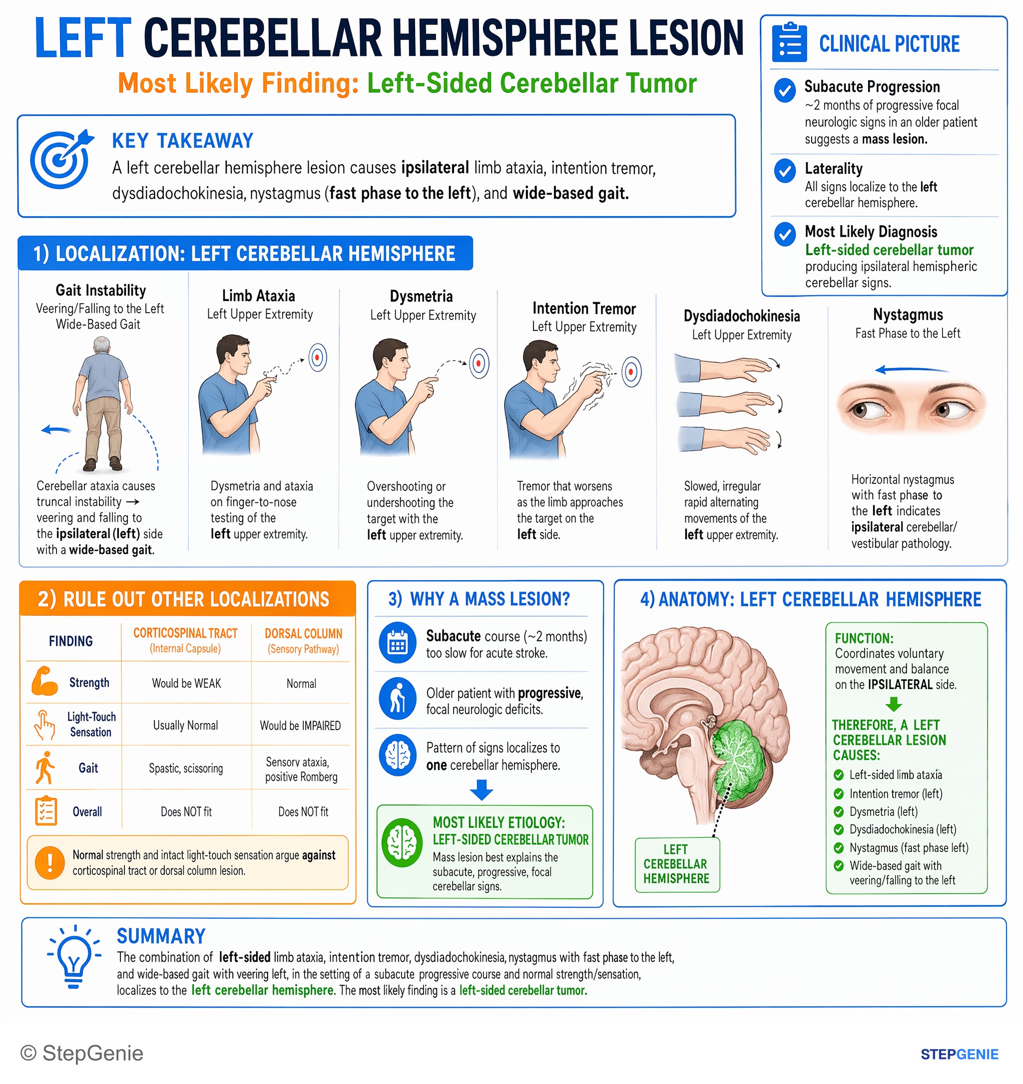
Left cerebellar hemisphere lesion causes ipsilateral limb ataxia, intention tremor, dysdiadochokinesia, nystagmus, and wide-based gait.
USMLE Step 1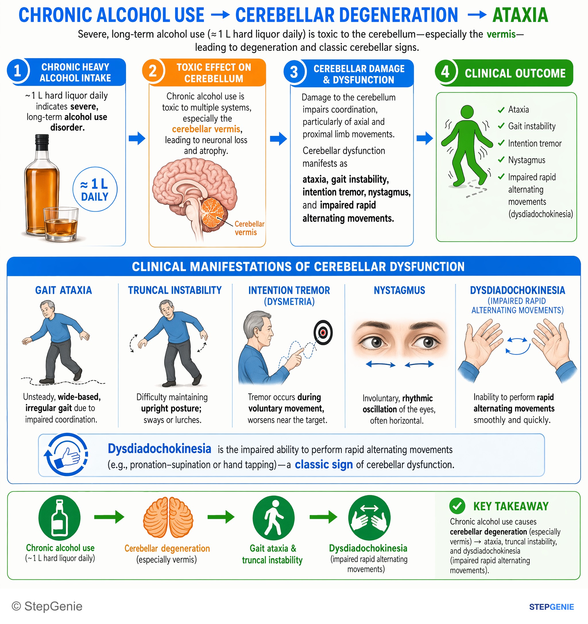
Chronic alcohol use causes cerebellar degeneration (especially vermis) → gait ataxia, truncal instability, and dysdiadochokinesia…
USMLE Step 1Frequently Asked Questions
Why do cerebellar lesions cause ipsilateral signs?
The cerebellum's output crosses twice. The dentate and interposed nuclei send fibers through the superior cerebellar peduncle, which cross in the midbrain to the opposite red nucleus and ventrolateral thalamus and then reach the opposite motor cortex. The corticospinal tract crosses back at the pyramidal decussation. The net effect is that each cerebellar hemisphere controls the same side of the body, so a right cerebellar lesion causes right-sided limb ataxia and a tendency to fall to the right.
What is the difference between a cerebellar vermis lesion and a hemisphere lesion?
The vermis is midline and controls the trunk, posture and gait, so a vermis lesion causes truncal ataxia, titubation and a wide-based, unsteady gait, often with fairly normal arm coordination. The lateral hemispheres control skilled limb movements, so a hemisphere lesion causes ipsilateral limb ataxia: dysmetria on finger-to-nose or heel-to-shin testing, intention tremor and dysdiadochokinesia. Flocculonodular lesions add nystagmus and imbalance. Chronic alcohol use and medulloblastoma are classic midline causes.
What neurotransmitter do Purkinje cells use?
Purkinje cells use GABA and are inhibitory. They are the only output of the cerebellar cortex and project to the deep cerebellar nuclei, and from the flocculonodular lobe to the vestibular nuclei. Their excitatory inputs are parallel fibers from granule cells, which relay mossy fiber input, and a single climbing fiber from the inferior olive that acts as an error signal for motor learning. The deep nuclei then send the excitatory output of the cerebellum.
What travels in each cerebellar peduncle?
The inferior cerebellar peduncle is mostly afferent: spinal proprioception (dorsal spinocerebellar and cuneocerebellar tracts), climbing fibers from the inferior olive and vestibular input, plus fastigial output to the vestibular nuclei. The middle cerebellar peduncle carries pontocerebellar fibers that relay the cerebral cortex from the opposite pontine nuclei. The superior cerebellar peduncle is mostly efferent: dentate and interposed output that crosses to the contralateral red nucleus and VL thalamus, plus the ventral spinocerebellar tract coming in.
How does chronic alcohol use affect the cerebellum?
Alcoholic cerebellar degeneration is the most common cause of cerebellar disease. It classically affects the anterior superior vermis, the midline part that controls the trunk and legs. Patients develop a wide-based, unsteady gait, usually over weeks to months, and cannot tandem walk, while arm coordination is often relatively preserved and nystagmus is uncommon. Acute intoxication causes reversible ataxia, nystagmus and slurred speech. Thiamine deficiency (Wernicke encephalopathy) can add confusion and eye movement abnormalities and needs parenteral thiamine.
Which arteries supply the cerebellum?
Three paired arteries supply the cerebellum. The superior cerebellar artery (SCA), from the top of the basilar artery, supplies the superior surface. The anterior inferior cerebellar artery (AICA), from the lower basilar, supplies the anterior inferior surface, flocculus and middle cerebellar peduncle along with the lateral pons, so AICA strokes add facial weakness and hearing loss. The posterior inferior cerebellar artery (PICA), from the vertebral artery, supplies the inferior vermis and underside of the hemisphere along with the lateral medulla.