Anterior circulation strokes (internal carotid → ACA and MCA) damage the cerebral hemispheres, so they cause one-sided cortical signs: contralateral weakness and numbness plus aphasia (dominant hemisphere), neglect (nondominant), gaze deviation or a visual field cut. Posterior circulation strokes (vertebral → basilar → PICA, AICA, SCA, PCA) hit the brainstem, cerebellum, thalamus and occipital lobes, so the clues are vertigo, ataxia, diplopia, dysarthria, dysphagia, crossed face-body findings, bilateral signs, reduced consciousness or a homonymous hemianopia. Work-up and reperfusion follow the same rules for both: noncontrast CT first to exclude hemorrhage, IV thrombolysis (alteplase or tenecteplase) within 4.5 hours, and thrombectomy for large vessel occlusion up to 24 hours in selected patients (2026 AHA/ASA guideline).
Anterior vs posterior circulation at a glance
| Anterior (carotid) | Posterior (vertebrobasilar) | |
|---|---|---|
| Arteries | ICA → ophthalmic, ACA, MCA (lenticulostriate branches) | Vertebrals → PICA; basilar → AICA, SCA, pontine perforators → PCAs |
| Supplies | Most of the frontal, parietal and temporal lobes; basal ganglia and internal capsule | Brainstem, cerebellum, thalamus, occipital and medial temporal lobes |
| Hallmark signs | Aphasia, neglect, gaze deviation, face-arm or leg weakness, monocular vision loss (ophthalmic artery) | Vertigo, nystagmus, ataxia, diplopia, dysarthria, dysphagia, crossed signs, homonymous hemianopia (PCA) |
| Side | Usually unilateral | Unilateral or bilateral; more likely to impair consciousness, especially basilar |
| Noncontrast CT | Early changes often subtle or absent | Also misses some small posterior fossa infarcts: diffusion-weighted MRI is the test |
Quick sort: a cortical sign (language, neglect, gaze deviation) = anterior. The posterior "D's" (dizziness or vertigo, diplopia, dysarthria, dysphagia, dystaxia) with crossed findings (ipsilateral face or cranial nerve, contralateral body) or bilateral limbs = posterior. Artery-by-artery map: the stroke localization guide.
Anterior circulation syndromes
The hallmark of each territory is below; each artery's full syndrome, with a worked case, is on its own linked page.
| Artery | Deficits | One-line clue |
|---|---|---|
| Ophthalmic / ICA | Transient monocular vision loss (amaurosis fugax) as a warning of carotid disease; ICA occlusion can give combined ACA + MCA deficits | "Curtain over one eye" → carotid duplex |
| ACA | Contralateral weakness and sensory loss maximal in the leg; urinary incontinence; apathy (abulia) and other frontal signs | Leg + bladder + behavior (ACA syndrome) |
| MCA | Contralateral face and arm > leg weakness and sensory loss; contralateral homonymous hemianopia; aphasia if dominant (usually left), neglect and apraxia if nondominant; eyes deviate toward the lesion | Face-arm + language or neglect (MCA syndrome) |
| Lenticulostriate (lacunar) | Pure motor hemiparesis (internal capsule), ataxic hemiparesis, dysarthria-clumsy hand; no cortical signs | Hypertension, lipohyalinosis (lacunar infarcts) |
- MCA divisions: superior division → face-arm weakness plus Broca (expressive) aphasia; inferior division → Wernicke (receptive) aphasia plus a contralateral superior quadrantanopia. Left vs right MCA signs are compared on the MCA syndrome page.
- Lacunes are not only anterior: pure sensory stroke is a thalamic lacune from the PCA's thalamic perforators, and pontine perforators of the basilar artery give the same motor syndromes.
Posterior circulation syndromes
| Artery or syndrome | What it hits | Findings |
|---|---|---|
| PCA | Occipital cortex ± thalamus, medial temporal lobe, midbrain | Contralateral homonymous hemianopia, often with macular sparing; can add memory loss, alexia without agraphia (dominant side) and thalamic or midbrain signs (PCA syndrome) |
| Basilar (ventral pons) | Corticospinal and corticobulbar tracts | Locked-in syndrome: quadriplegia, cannot speak, chew or swallow, but awake and aware, with blinking and vertical eye movements preserved; complete basilar occlusion can cause coma and death |
| Vertebral or PICA: lateral medulla (Wallenberg) | Nucleus ambiguus, spinal trigeminal nucleus, spinothalamic tract, vestibular nuclei, inferior cerebellar peduncle, descending sympathetics | Dysphagia, hoarseness, vertigo, nausea, nystagmus, hiccups; pain-temperature loss on the ipsilateral face and contralateral body; ipsilateral Horner syndrome and ataxia |
| AICA: lateral pons | Facial nucleus, cochlear and vestibular nuclei, middle cerebellar peduncle, plus the same sensory and sympathetic pathways | Ipsilateral facial paralysis and hearing loss, vertigo, ataxia, crossed pain-temperature loss |
| Anterior spinal / vertebral: medial medulla | Pyramid, medial lemniscus, CN XII | Contralateral hemiparesis and loss of vibration and position sense; tongue deviates toward the lesion |
| SCA | Superior cerebellum | Ipsilateral limb and gait ataxia, dysarthria |
For the medial vs lateral brainstem rules, see the brainstem syndromes mnemonic. Do not dismiss acute vertigo with a normal CT: vertigo plus ataxia, nystagmus or any focal sign is a posterior circulation stroke until MRI says otherwise.
Mechanisms that shape the stem
- Cardioembolic (atrial fibrillation, LV thrombus, endocarditis): sudden, maximal at onset, often MCA branches or the top of the basilar.
- Large-artery atherothrombosis: carotid bifurcation (amaurosis fugax, MCA or ACA signs) or vertebrobasilar disease; may stutter.
- Small-vessel (lacunar): lipohyalinosis of penetrating arteries from hypertension or diabetes.
- Arterial dissection: a young patient with neck pain or headache, sometimes after neck trauma; vertebral dissection is a classic posterior circulation cause.
- Watershed (hypoperfusion): after cardiac arrest or severe hypotension; bilateral ACA-MCA border zone infarcts weaken both arms, mainly proximally, while sparing the face and legs ("man in a barrel").
Imaging: what to order, in order
- Fingerstick glucose first: hypoglycemia mimics stroke.
- Noncontrast head CT: excludes hemorrhage before thrombolysis; often normal in the first hours of ischemia and misses some small posterior circulation infarcts.
- CTA (or MRA) of the head and neck in anyone who might have a large vessel occlusion, especially within 24 hours of onset, to select thrombectomy, without delaying thrombolysis.
- Perfusion CT or MRI (or an MRI DWI-FLAIR mismatch) when the onset time is unknown or the patient is beyond 4.5 hours, to select extended-window thrombolysis; before thrombectomy at 6-24 hours it helps if immediately available but is not required.
- Diffusion-weighted MRI is the most sensitive test for acute ischemia and the one that settles posterior fossa questions.
- Mechanism work-up: ECG/telemetry for atrial fibrillation, echocardiography, carotid imaging, lipids, HbA1c.
Acute treatment: windows and thresholds
Reperfusion saves the penumbra (hypoperfused but viable tissue around the core), so every clock starts at last known well.
| Decision | Rule (2026 AHA/ASA guideline unless noted) |
|---|---|
| IV thrombolysis, standard window | Within 4.5 hours of onset or last known well, for disabling deficits regardless of NIHSS score: tenecteplase 0.25 mg/kg IV bolus (max 25 mg) or alteplase 0.9 mg/kg (max 90 mg; 10% as a 1-minute bolus, the rest over 60 minutes) (Class 1) |
| FDA labels | Both Activase and TNKase are labeled for use within 3 hours of symptom onset; the 4.5-hour window comes from the guideline. TNKase gained its stroke indication in 2025 |
| Extended-window thrombolysis | Unknown onset but within 4.5 hours of symptom recognition, with an MRI DWI-FLAIR mismatch: can be beneficial. Wake-up stroke within 9 hours of the midpoint of sleep, or 4.5-9 hours from last known well, with salvageable penumbra on automated perfusion imaging: may be reasonable (both Class 2a) |
| Mild, nondisabling deficits | Dual antiplatelet therapy is preferred over thrombolysis |
| Blood pressure and thrombolysis | Lower to <185/110 mm Hg before starting; keep <180/105 for the first 24 hours. Intensive lowering to systolic <140 brings no benefit |
| Thrombectomy, 0-6 hours | ICA or M1 occlusion, NIHSS ≥6, prestroke mRS 0-1, ASPECTS 3-10 (Class 1) |
| Thrombectomy, 6-24 hours | ICA or M1 occlusion, NIHSS ≥6, prestroke mRS 0-1, ASPECTS ≥6 (Class 1); also selected large cores (ASPECTS 3-5) if age <80 with no significant mass effect (Class 1). Perfusion CT or MRI helps if immediately available (Class 2a) but is not required |
| Basilar artery occlusion | Thrombectomy within 24 hours if NIHSS ≥10, prestroke mRS 0-1 and PC-ASPECTS ≥6 (Class 1, new in 2026) |
| After successful thrombectomy | A systolic target <140 mm Hg for the first 72 hours is harmful (Class 3: Harm) |
| No reperfusion therapy | Below 220/120 mm Hg, starting BP treatment in the first 48-72 hours does not improve outcome (Class 3: No Benefit); at ≥220/120 the benefit is uncertain (Class 2b) and the guideline advises an initial reduction of about 15%; treat sooner if a comorbid condition requires it (eg, aortic dissection, acute coronary event, heart failure) |
| Glucose | Target 140-180 mg/dL; treat <60 mg/dL; tight control to 80-130 is not recommended |
- Label contraindications (Activase): current intracranial or subarachnoid hemorrhage, active internal bleeding, intracranial or intraspinal surgery or serious head trauma within 3 months, intracranial conditions that raise bleeding risk, bleeding diathesis, severe uncontrolled hypertension. The major complication is intracranial hemorrhage.
- Posterior strokes get the same treatment: thrombolysis within the window, and thrombectomy for basilar occlusion.
Secondary prevention in one breath
Noncardioembolic stroke → antiplatelet therapy (short-course aspirin plus clopidogrel after minor stroke or high-risk TIA); atrial fibrillation → anticoagulation; recent nondisabling stroke with 70-99% ipsilateral carotid stenosis → carotid endarterectomy; statin aiming for at least a 50% LDL reduction or LDL <70 mg/dL; risk-factor control.
How USMLE tests it
- "Next step" in a new deficit: glucose, then noncontrast CT. Do not give thrombolysis before hemorrhage is excluded.
- Same-side face, opposite-side body = brainstem. Aphasia or neglect points to a cerebral hemisphere (usually MCA territory), not the brainstem.
- Hearing loss + facial palsy = AICA; hoarseness + dysphagia = PICA; tongue toward the lesion = medial medulla.
- Hemianopia with macular sparing = PCA, not MCA.
- "tPA within 3-4.5 hours" still works: 3 hours on the labels, 4.5 in the guideline, and tenecteplase is now an equal first-line option.
Practice question
A 67-year-old man with hypertension and a long smoking history has had vertigo, vomiting and difficulty swallowing for 2 hours. His voice is hoarse. Examination shows left ptosis and miosis, nystagmus, left arm and leg ataxia, and decreased pain and temperature sensation over the left face and the right arm, trunk and leg. Strength and tongue movements are normal. A noncontrast head CT shows no hemorrhage. Occlusion of which artery best explains his findings?
A. Left anterior inferior cerebellar artery B. Left anterior spinal artery C. Basilar artery D. Left middle cerebral artery E. Left posterior inferior cerebellar artery
Answer: E. Hoarseness and dysphagia (nucleus ambiguus), ipsilateral facial and contralateral body pain-temperature loss (spinal trigeminal nucleus and spinothalamic tract), ipsilateral Horner syndrome, vertigo, nystagmus and ipsilateral ataxia localize to the left lateral medulla (Wallenberg syndrome). The culprit is the PICA or the intracranial vertebral artery that gives rise to it. A normal CT does not exclude a posterior fossa infarct, and at 2 hours he can still be evaluated for thrombolysis.
- A: AICA (lateral pons) causes ipsilateral facial paralysis and hearing loss; the nucleus ambiguus is not involved, so no hoarseness or dysphagia.
- B: medial medullary infarction causes contralateral hemiparesis, contralateral loss of vibration and position sense, and tongue deviation toward the lesion; his strength and tongue are normal.
- C: basilar occlusion produces bilateral signs, quadriparesis (locked-in syndrome) or coma.
- D: MCA stroke causes cortical signs (contralateral face-arm weakness, aphasia), not crossed sensory loss or Horner syndrome.
Key takeaways
- Anterior (ICA, ACA, MCA) = unilateral cortical signs: aphasia, neglect, gaze deviation, face-arm (MCA) or leg (ACA) weakness, amaurosis fugax.
- Posterior = vertigo, ataxia, diplopia, dysphagia, crossed or bilateral signs, hemianopia.
- PICA/vertebral = Wallenberg; AICA = facial palsy + hearing loss; basilar = locked-in; PCA = hemianopia with macular sparing.
- CT first to exclude blood, CTA for large vessel occlusion; DWI MRI is most sensitive, especially for posterior strokes.
- Thrombolysis within 4.5 hours (alteplase 0.9 mg/kg or tenecteplase 0.25 mg/kg); BP <185/110 before, <180/105 after.
- Thrombectomy: ICA/M1 within 6 hours (ASPECTS 3-10) and at 6-24 hours (ASPECTS ≥6, or 3-5 if younger than 80); basilar occlusion within 24 hours if NIHSS ≥10.
Sources
- 2026 Guideline for the Early Management of Patients With Acute Ischemic Stroke — Prabhakaran et al., Stroke, PubMed (2026)
- Top Things to Know: 2026 Guideline for the Early Management of Patients With Acute Ischemic Stroke — American Heart Association (2026)
- Response to Request for Modification of the 2026 AHA/ASA Acute Ischemic Stroke Guidelines: Endovascular Thrombectomy Imaging Selection in the 6- to 24-Hour Window — Stroke, PMC (2026)
- What Are Major Pharmacotherapy Updates From the 2026 AHA/ASA Stroke Guidelines? — UIC Drug Information Group (2026)
- Overview of Stroke — Merck Manual Professional Edition (2026)
- Ischemic Stroke — Merck Manual Professional Edition (2026)
- TNKASE (tenecteplase) label — DailyMed, NLM (2025)
- ACTIVASE (alteplase) label — DailyMed, NLM (2026)
- Lateral Medullary Syndrome — National Institute of Neurological Disorders and Stroke (2026)
- Locked-In Syndrome — National Institute of Neurological Disorders and Stroke (2026)
- Lab 4: The Ventricles and Blood Supply, Vertebral-Basilar System — Neuroanatomy Online, McGovern Medical School at UTHealth
- Visual Processing: Cortical Pathways (Section 2, Chapter 15) — Neuroscience Online, McGovern Medical School at UTHealth
Visual explanations from the StepGenie QBank
Infographics StepGenie drew to explain questions in its own QBank, matched to this article's topic. Each caption is the takeaway of the question it explains.
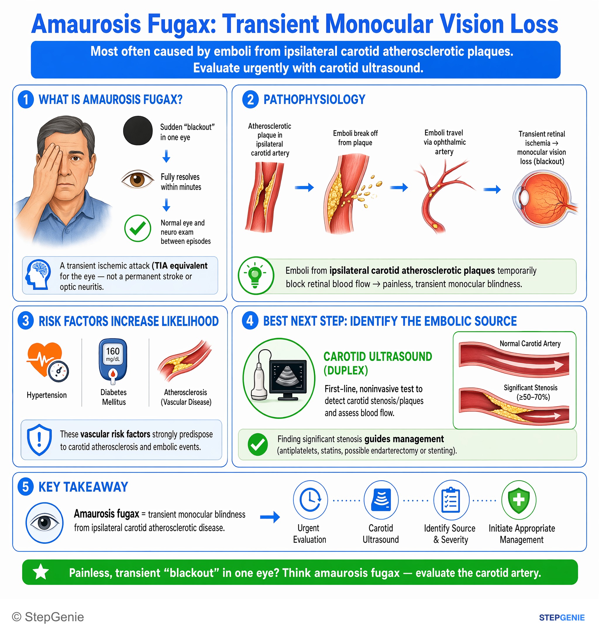
Amaurosis fugax from ipsilateral carotid atherosclerotic disease causes transient monocular blindness; evaluate urgently with carotid…
USMLE Step 2Frequently Asked Questions
How do you tell an anterior circulation stroke from a posterior circulation stroke?
Anterior circulation strokes come from the internal carotid, ACA or MCA and damage a cerebral hemisphere, so they cause unilateral cortical signs such as aphasia, neglect, gaze deviation and contralateral face-arm or leg weakness. Posterior circulation strokes come from the vertebral, basilar, cerebellar or posterior cerebral arteries and cause vertigo, nystagmus, ataxia, diplopia, dysarthria, dysphagia, crossed face-body findings, bilateral signs, reduced consciousness or a homonymous hemianopia. CT often misses posterior fossa infarcts, so MRI is used to confirm them.
What is the difference between a PICA stroke and an AICA stroke?
Both damage the lateral brainstem and cause vertigo, nystagmus, ipsilateral ataxia, crossed pain and temperature loss and Horner syndrome. PICA, or the vertebral artery that gives rise to it, supplies the lateral medulla, so Wallenberg syndrome adds hoarseness and dysphagia from nucleus ambiguus involvement, often with hiccups. AICA supplies the lateral pons, so it adds ipsilateral facial paralysis and hearing loss. Hoarseness and dysphagia point to PICA; facial palsy with hearing loss points to AICA.
What is the time window for tPA in ischemic stroke?
The 2026 AHA/ASA guideline recommends IV thrombolysis within 4.5 hours of symptom onset or last known well for eligible patients with disabling deficits, using either tenecteplase 0.25 mg/kg as a single bolus (maximum 25 mg) or alteplase 0.9 mg/kg (maximum 90 mg). The FDA labels for both drugs say within 3 hours. Selected patients who wake up with symptoms or are 4.5 to 9 hours out may be treated if advanced imaging shows salvageable tissue.
When is mechanical thrombectomy indicated for ischemic stroke?
Thrombectomy treats large vessel occlusions. Within 6 hours it is recommended for ICA or M1 occlusion with an NIHSS score of 6 or more, good prestroke function and ASPECTS 3 to 10. From 6 to 24 hours the 2026 AHA/ASA guideline recommends it for the same occlusions with ASPECTS 6 or more, and for selected patients younger than 80 with larger cores (ASPECTS 3 to 5) and no significant mass effect. For basilar artery occlusion it strongly recommends thrombectomy within 24 hours when the NIHSS score is 10 or more and brainstem damage on imaging is limited.
What blood pressure is needed before giving tPA?
Blood pressure must be below 185/110 mm Hg before IV thrombolysis starts and kept below 180/105 mm Hg for the first 24 hours afterward, because higher pressures raise the risk of intracranial hemorrhage. Without reperfusion therapy, pressures below 220/120 mm Hg are usually not treated in the first 48 to 72 hours unless another condition requires it; at 220/120 or more, an initial reduction of about 15% is advised. After successful thrombectomy, an intensive systolic target below 140 mm Hg is harmful according to the 2026 AHA/ASA guideline.
Why is the head CT often normal in an acute ischemic stroke?
Ischemic changes take hours to appear on noncontrast CT, and small infarcts in the brainstem and cerebellum are hard to see in the bony posterior fossa. CT is still done first because its job is to exclude hemorrhage before thrombolysis, and CT angiography looks for a large vessel occlusion that thrombectomy could treat. Diffusion-weighted MRI is the most sensitive test for acute ischemia and is the way to confirm a suspected posterior circulation stroke.