Lacunar infarcts are small (classically under 15 mm) ischemic strokes in deep structures (internal capsule, basal ganglia, thalamus, pons) caused by occlusion of a single small penetrating artery, usually from lipohyalinosis (hyaline arteriolosclerosis) driven by hypertension and diabetes. They cause one of five classic lacunar syndromes, pure motor hemiparesis, pure sensory stroke, sensorimotor stroke, ataxic hemiparesis and dysarthria-clumsy hand, with no cortical signs: no aphasia, neglect, visual field cut or gaze deviation. They make up about 20-30% of ischemic strokes (StatPearls). Early CT is often normal; MRI with diffusion-weighted imaging shows the lesion. Treatment is the same as for any ischemic stroke, with long-term blood pressure control at the center of prevention.
The q-bank question
A 62-year-old man with long-standing hypertension and type 2 diabetes has sudden slurred speech and clumsiness of his right hand. On examination he has dysarthria, mild right lower facial weakness, and a clumsy right hand on finger-to-nose testing and writing, with near-normal strength. Sensation is intact. He is alert, with no aphasia, neglect, gaze deviation or visual field defect. A noncontrast head CT is normal.
Which of the following is the most likely underlying pathology?
A. Rupture of Charcot-Bouchard microaneurysms B. Hyaline arteriolosclerosis (lipohyalinosis) of a small penetrating artery C. Cardioembolic occlusion of the superior division of the left middle cerebral artery D. Atherosclerotic plaque in the left internal carotid artery with artery-to-artery embolism E. Amyloid deposition in the walls of cortical vessels
Answer: B, lipohyalinosis of a penetrating artery
This is dysarthria-clumsy hand syndrome, one of the five classic lacunar syndromes, usually from a lacune in the pons or internal capsule. Three clues point to a lacune:
- Risk factors: long-standing hypertension and diabetes, the two most important risk factors for lacunar stroke (StatPearls).
- No cortical signs: speech is slurred (a motor problem, dysarthria), but language is intact (no aphasia); there is no neglect, field cut or gaze deviation.
- Normal early CT: CT often misses small infarcts early; MRI-DWI finds them (Merck).
The pathology. Lacunes come from nonatherothrombotic obstruction of small perforating arteries, usually lipohyalinosis: degeneration of the arterial media, which is replaced by lipids and collagen (Merck). StatPearls adds a second mechanism, microatheroma at the origin of a penetrating branch. Hyaline arteriolosclerosis is the pathology term you'll see on exams for the same hypertensive and diabetic small-vessel change. The name comes from the Latin for a pond or pit: the healed infarct leaves a small subcortical space filled with cerebrospinal fluid (StatPearls).
Why every other answer is wrong
A. Charcot-Bouchard microaneurysms. Same risk factor, opposite event. Chronic hypertension produces microaneurysms in small perforating arteries that rupture and bleed into the putamen (basal ganglia), thalamus, pons or cerebellum (Merck). That is a hemorrhage: sudden headache, often during activity, frequent loss of consciousness, vomiting and seizures, with blood visible on CT at once. A normal CT rules it out. See the hemorrhagic stroke guide.
C. Cardioembolic superior-division MCA occlusion. An embolus (think atrial fibrillation) to the left MCA superior division damages cortex: Broca (expressive) aphasia plus right face and arm weakness. Merck's MCA picture is contralateral weakness worse in the arm and face than the leg, hemianesthesia, homonymous hemianopia, and aphasia (dominant side) or neglect (nondominant side). This patient's language is intact. Compare the middle cerebral artery syndrome case.
D. Carotid atherosclerosis with artery-to-artery embolism. Atheromas form where flow is turbulent, particularly at the carotid bifurcation (Merck), and plaque there can throw emboli into the anterior circulation, including the MCA territory. So again you'd expect cortical signs, often preceded by TIAs or transient monocular blindness (amaurosis fugax) and sometimes a carotid bruit. The workup is carotid imaging, and symptomatic severe stenosis is treated with carotid endarterectomy or stenting.
E. Cerebral amyloid angiopathy. Amyloid in the walls of cortical vessels, usually in people older than 55 (MedlinePlus), causes lobar hemorrhages near the cortex, outside the basal ganglia, which may be multiple and recurrent (Merck). Again a bleed, and in the wrong place.
Sort the three small-vessel answers in one line: hypertension makes deep bleeds (Charcot-Bouchard) and deep infarcts (lipohyalinosis, lacunes); amyloid makes lobar bleeds.
The five classic lacunar syndromes
| Syndrome | Share of lacunar strokes (StatPearls) | Typical lesion location | Key features |
|---|---|---|---|
| Pure motor hemiparesis | About 45% (most common) | Posterior limb of internal capsule, corona radiata or ventral pons | Contralateral face, arm and leg weakness; mild dysarthria possible; no sensory loss |
| Sensorimotor stroke | About 20% | Thalamus and adjacent internal capsule (thalamocapsular), or putamen-capsule-caudate | Contralateral weakness plus sensory loss |
| Ataxic hemiparesis | About 10-18% | Internal capsule, pons or corona radiata | Contralateral limb ataxia is the prominent feature, with milder weakness of the same side |
| Pure sensory stroke | About 7% | Thalamus (VPL/VPM) | Contralateral numbness of face, arm and leg; poststroke thalamic pain (Dejerine-Roussy) possible |
| Dysarthria-clumsy hand | Not stated | Pons (basis pontis) or internal capsule | Dysarthria from weakness of tongue, larynx and facial muscles; a clumsy contralateral hand with preserved strength |
Cortical signs are absent in all five. Neglect, visual disturbances, aphasia and behavioral changes are typically absent in lacunar stroke (StatPearls). Merck's diagnostic rule is the same: no cortical deficits plus one of the lacunar syndromes.
Which arteries. Lacunes sit in the territory of small, deep penetrating branches from the circle of Willis (StatPearls): the lenticulostriate branches of the MCA (basal ganglia, internal capsule), thalamic perforators from the PCA, and paramedian pontine branches of the basilar artery. One blocked branch makes one small, deep infarct. Review the vessels with the Circle of Willis memory palace.
Why the internal capsule gives face, arm and leg together: the descending motor fibers for all three converge in the internal capsule (genu and posterior limb), so a tiny lesion hits all three. In the cortex those fibers fan out along the motor strip, and the leg area lies medially in ACA territory, so an MCA cortical stroke weakens the face and arm more than the leg.
Workup and treatment
- Noncontrast CT first, to exclude hemorrhage. A normal CT does not exclude a lacune; MRI-DWI separates acute from old infarcts, and MRI picks up small lesions that CT misses.
- Acute treatment is the same as for any ischemic stroke. For a disabling deficit, IV thrombolysis with tenecteplase or alteplase within 4.5 hours of onset, if eligible (2026 AHA/ASA guideline; StatPearls now calls tenecteplase the preferred agent). The US labels for both drugs still give a 3-hour window; the 4.5-hour window is the guideline's. For a non-disabling deficit, such as an isolated sensory syndrome, trials have not shown a benefit from thrombolysis, and the guideline prefers dual antiplatelet therapy. Mechanical thrombectomy targets large-vessel occlusions, which a lacune by definition is not. Run through the steps in the stroke management case.
- Minor stroke: aspirin plus clopidogrel started within 24 hours and continued for 21 days, then a single antiplatelet drug (Merck; StatPearls). Clopidogrel's US label carries a boxed warning: patients with two loss-of-function CYP2C19 alleles (poor metabolizers) get a diminished antiplatelet effect.
- Blood pressure: a systolic target under 130 mm Hg is beneficial after lacunar stroke (StatPearls).
- Statin, diabetes control and smoking cessation complete the risk-factor work.
- Prognosis: short term, survival is high, recurrence low and functional recovery relatively good; beyond the first year, the risk of death (mainly cardiovascular) rises, recurrence is as likely as after other stroke types, and dementia develops in 9-12% of patients by 9 years (StatPearls).
How USMLE tests it
- "Pure" anything (pure motor, pure sensory) with no cortical signs = lacune.
- Aphasia, neglect, field cut or gaze deviation = cortex, so think embolus or large-artery disease (carotid, MCA), not a lacune.
- Hypertension + deep hemorrhage on CT = Charcot-Bouchard; elderly + lobar hemorrhage = amyloid angiopathy.
- Pure sensory stroke, later pain on the numb side = thalamic (Dejerine-Roussy) pain syndrome.
- Pathology answer: hyaline arteriolosclerosis or lipohyalinosis (or microatheroma) of a penetrating artery, not "embolus" or "thrombus of the MCA trunk."
- Next best test after a normal CT in a suspected lacune: MRI with DWI.
Map any deficit to its artery with the stroke localization guide.
Key takeaways
- Lacune = small (under about 15 mm), deep ischemic infarct from occlusion of one penetrating artery.
- Cause: lipohyalinosis (hyaline arteriolosclerosis) or microatheroma; hypertension and diabetes are the main risk factors.
- Five syndromes: pure motor (posterior limb of internal capsule; most common), pure sensory (thalamus), sensorimotor (thalamocapsular), ataxic hemiparesis (pons or internal capsule), dysarthria-clumsy hand (pons or internal capsule).
- No cortical signs: no aphasia, neglect, field cut or gaze deviation.
- CT is often normal early; MRI-DWI shows the lesion.
- Distractors: Charcot-Bouchard = deep hypertensive hemorrhage; amyloid angiopathy = lobar hemorrhage; embolus = cortical signs.
- Treat as any ischemic stroke (thrombolysis within 4.5 hours for an eligible patient with a disabling deficit; dual antiplatelet therapy for a non-disabling one); prevent with antiplatelets, systolic BP under 130, a statin and diabetes control.
Sources
- Lacunar Stroke — StatPearls, NCBI Bookshelf (2026)
- Ischemic Stroke — Merck Manual Professional Edition (2025)
- Intracerebral Hemorrhage — Merck Manual Professional Edition (2025)
- Overview of Stroke — Merck Manual Professional Edition (2025)
- Cerebral amyloid angiopathy — MedlinePlus Medical Encyclopedia (2026)
- 2026 Guideline for the Early Management of Patients With Acute Ischemic Stroke: Top Things to Know — American Heart Association Professional Heart Daily (2026)
- New guideline expands stroke treatment for adults, offers first pediatric stroke guidance — American Heart Association Newsroom (2026)
- TNKase (tenecteplase) prescribing information — DailyMed, US National Library of Medicine (2025)
- Activase (alteplase) prescribing information — DailyMed, US National Library of Medicine (2026)
- Plavix (clopidogrel) prescribing information — DailyMed, US National Library of Medicine (2025)
Visual explanations from the StepGenie QBank
Infographics StepGenie drew to explain questions in its own QBank, matched to this article's topic. Each caption is the takeaway of the question it explains.
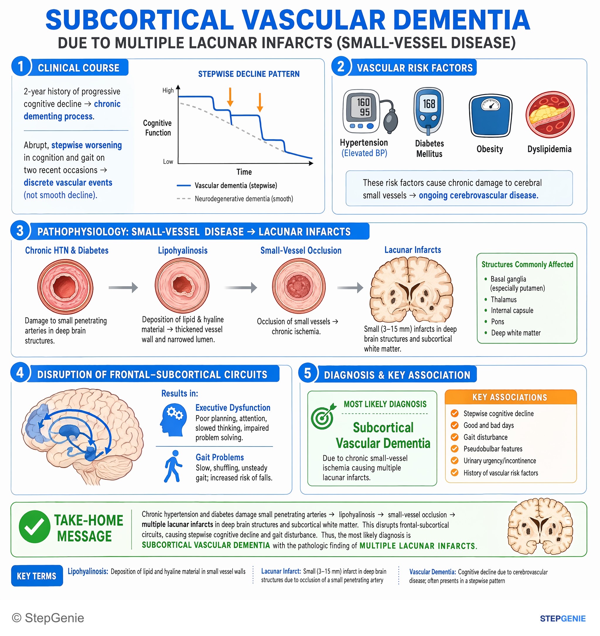
Vascular dementia presents with stepwise cognitive decline and gait disturbance, associated with multiple lacunar infarcts from chronic…
USMLE Step 2Frequently Asked Questions
What are the 5 lacunar syndromes?
The five classic lacunar syndromes are pure motor hemiparesis, the most common at about 45%, usually from the posterior limb of the internal capsule; sensorimotor stroke, from the thalamus and adjacent internal capsule; ataxic hemiparesis, from the internal capsule, pons or corona radiata; pure sensory stroke, from the thalamus; and dysarthria-clumsy hand syndrome, from the pons or internal capsule. None of them causes cortical signs such as aphasia or neglect.
What causes lacunar infarcts?
Lacunar infarcts are caused by occlusion of a single small penetrating artery, such as a lenticulostriate branch of the middle cerebral artery, a thalamic perforator or a paramedian pontine branch of the basilar artery. The usual mechanism is lipohyalinosis, also called hyaline arteriolosclerosis, in which the arterial wall degenerates and is replaced by lipid and collagen; microatheroma at the origin of the branch is another. Hypertension and diabetes are the main risk factors.
How do you tell a lacunar stroke from a cortical stroke?
Look for cortical signs. A lacunar stroke produces a pure motor, pure sensory, sensorimotor or ataxic deficit, or dysarthria with a clumsy hand, without aphasia, neglect, visual field loss, gaze deviation or behavioral change. A cortical stroke from an embolus or large-artery disease usually brings at least one of those. Weakness of face, arm and leg together also suggests the internal capsule, whereas a cortical MCA stroke weakens the face and arm more than the leg.
Why is the CT often normal in a lacunar stroke?
Lacunes are small, deep ischemic infarcts, classically under 15 mm, and CT often misses small infarcts early, so the first scan is frequently normal. The CT is still done first because it rules out hemorrhage before treatment. MRI with diffusion-weighted imaging is far more sensitive for small infarcts and separates a new lesion from old ones, so it is the next test when a lacune is suspected.
What is the difference between a lacunar infarct and a Charcot-Bouchard hemorrhage?
Both come from chronic hypertension damaging small penetrating arteries, but the events are opposite. In a lacunar infarct, lipohyalinosis narrows and blocks the vessel, causing a small ischemic stroke with a normal early CT. Charcot-Bouchard microaneurysms rupture and bleed into the putamen, thalamus, pons or cerebellum, causing sudden headache, vomiting and often loss of consciousness, with blood visible on CT. Cerebral amyloid angiopathy, by contrast, causes lobar hemorrhages.
How are lacunar strokes treated?
Acutely, a lacunar stroke is treated like any ischemic stroke. Under the 2026 AHA/ASA guideline, an eligible patient with a disabling deficit gets IV tenecteplase or alteplase within 4.5 hours of onset, while a non-disabling deficit, such as isolated numbness, gets dual antiplatelet therapy instead. Thrombectomy is for large-vessel occlusions, which lacunes are not. For a minor stroke, aspirin plus clopidogrel is started within 24 hours and given for 21 days. Long term, keep systolic pressure under 130 mm Hg, use a statin and control diabetes.