Posterior cord syndrome is damage to the dorsal (posterior) columns, the dorsal column–medial lemniscus pathway: vibration, joint position sense and fine touch are lost below the lesion, causing sensory ataxia and a positive Romberg sign. Strength, pain and temperature are spared, and the classic causes are B12 deficiency, tabes dorsalis, copper deficiency and compression.
It is the clinical picture of a posterior column lesion, sometimes called dorsal column syndrome, and the least common incomplete spinal cord injury pattern. It arises more often from metabolic, toxic or infectious disease than from trauma, and it is easy to miss because a standard strength and pinprick exam can be normal.
When the lateral corticospinal tracts are damaged as well, the picture becomes the posterolateral, or combined, pattern of subacute combined degeneration: dorsal column loss plus spastic weakness and Babinski signs.
What is posterior cord syndrome? The dorsal columns and the POST memory aid
The dorsal column–medial lemniscus (DCML) pathway carries fine touch, two-point discrimination, conscious proprioception and vibration from the whole body except the head. Remember what a posterior cord lesion takes away with POST:
- Position sense (proprioception) lost
- Oscillation, meaning vibration sense, lost
- Sensory ataxia with a positive Romberg sign
- Fine Touch and two-point discrimination lost
What it spares is just as testable: pain and temperature (spinothalamic tract) and voluntary strength (corticospinal tract).
The anatomy that drives localization:
- Two fasciculi. The fasciculus gracilis (medial) carries the legs and runs the whole length of the cord; the fasciculus cuneatus (lateral) carries the arms and exists only at T6 and above. A lesion below T6 spares the arms; a cervical lesion can affect arms and legs.
- Same side until the medulla. First-order fibers ascend on the same side to the gracile and cuneate nuclei in the caudal medulla; second-order fibers then cross as internal arcuate fibers and run in the medial lemniscus to the VPL nucleus of the thalamus. A cord lesion therefore causes ipsilateral loss below its level, bilateral when both columns are hit.
- Pain and temperature cross early. Second-order spinothalamic fibers cross in the anterior white commissure within 1–2 segments of entry and ascend in the anterolateral cord, outside the damaged zone.
- Blood supply. The posterior spinal arteries supply the posterior third of the cord; the anterior spinal artery supplies the anterior two-thirds.
Every tract is drawn side by side in the spinal cord tracts visual hack.
Posterior cord syndrome symptoms and signs
Patients report tingling or numbness, clumsy hands and unsteadiness that is worse in the dark or with the eyes closed, for example when washing the face. The posterior column signs to look for:
| Sign | What you see | How to test |
|---|---|---|
| Vibration loss | Cannot feel the tuning fork | 128-Hz tuning fork on a bony prominence, distal joints first |
| Joint position loss | Cannot say which way a toe or finger was moved | Small passive movements of the toe or finger joints, eyes closed |
| Sensory ataxia | Unaware of limb position; exaggerated steps with forceful ("stomping") ground contact | Gait exam |
| Positive Romberg | Steady with eyes open, sways or falls with eyes closed | Feet together, eyes open, then closed |
| Pseudoathetosis | Slow writhing of the fingers of outstretched hands when the eyes close | Arms out, eyes closed |
| Fine touch loss | Poor two-point discrimination; clumsy hands with cervical lesions | Two-point discrimination |
| Lhermitte sign | Electric shock down the spine and limbs on neck flexion | History; neck flexion |
Kept in a pure lesion: strength, pain and temperature. Reflexes show what else is involved: spasticity or Babinski signs add the lateral columns; areflexia usually adds the roots or nerves.
Lhermitte sign localizes to the cervical cord, not to one disease: multiple sclerosis (reported in 9–41% of patients), cervical spondylotic myelopathy, B12 deficiency, nitrous oxide misuse and radiation myelopathy all cause it.
Why it is missed. The ISNCSCI spinal cord injury exam tests light touch and pinprick; joint position sense is optional and vibration is not included, so these patients can look normal on paper.
Causes of posterior cord syndrome
Posterior column disease and degeneration come from a short list:
| Cause | What is damaged | Clues | Confirm |
|---|---|---|---|
| Vitamin B12 deficiency (subacute combined degeneration) | Dorsal columns plus lateral corticospinal and spinocerebellar tracts; neuropathy often coexists | Pernicious anemia, gastrectomy or bariatric surgery, ileal disease, metformin, PPIs, strict vegan diet; anemia can be absent | Low or low-normal B12 with raised MMA and homocysteine; T2 "inverted V" on MRI |
| Nitrous oxide | Same tracts, through inactivated B12 | Young adult inhaling "whippets" or balloons; hemoglobin and MCV usually normal | Raised MMA and homocysteine, even with a normal B12 |
| Copper deficiency | Same tracts as B12 deficiency | Upper GI or bariatric surgery, excess zinc (supplements, denture cream), malabsorption; anemia or neutropenia | Low serum copper and ceruloplasmin; normal B12 and MMA |
| Tabes dorsalis (tertiary syphilis) | Dorsal roots and dorsal columns | Lightning pains, areflexia, Argyll Robertson pupils, Charcot joints, urinary retention; decades after infection | Treponemal serology; CSF pleocytosis, raised protein, CSF-VDRL |
| Multiple sclerosis | Short, often dorsolateral, asymmetric plaques | Lhermitte sign, "useless hand," relapses, optic neuritis | MRI of brain and spine; CSF oligoclonal bands |
| HIV vacuolar myelopathy | Posterior and lateral columns, mainly thoracic | Advanced HIV with a low CD4 count; progressive spastic legs, sensory ataxia, autonomic symptoms | HIV test; a diagnosis of exclusion |
| Friedreich ataxia | Dorsal root ganglia, posterior columns, corticospinal and spinocerebellar tracts, cerebellum | Mean onset at 10–15 years; absent reflexes with upgoing toes, scoliosis, hypertrophic cardiomyopathy, diabetes | Biallelic GAA repeat expansion in FXN |
| Posterior spinal artery infarct | Posterior cord, often one side | Sudden onset, often with pain; mostly cervical; may lose reflexes at its level; can mimic a stroke | MRI |
| Compression or trauma | Back of the cord | Tumors, disc or spondylotic compression, a postoperative seroma; trauma such as neck hyperextension is uncommon | MRI |
Exam contrasts:
- Tabes dorsalis also damages the dorsal roots, so reflexes vanish and lightning pains appear, typically 10–30 years after infection; see the tabes dorsalis Q-bank breakdown.
- Copper deficiency is the B12 mimic with normal B12 and MMA (copper deficiency mnemonic).
- Friedreich ataxia is an autosomal recessive mixed ataxia, covered with the trinucleotide repeat disorders.
Posterolateral cord syndrome: when the lateral columns join in
A posterolateral (combined) cord syndrome adds lateral column damage to dorsal column loss: spastic weakness, hyperreflexia and extensor plantar responses. A coexisting neuropathy makes the reflexes mixed (brisk knees, absent ankle jerks, Babinski signs), and early B12 deficiency can even be hyporeflexic.
| Feature | B12 deficiency | Nitrous oxide | Copper deficiency | HIV vacuolar myelopathy | Friedreich ataxia |
|---|---|---|---|---|---|
| Typical patient | Older adult; pernicious anemia, GI surgery, vegan diet | Young recreational user | Bariatric surgery or zinc excess | Advanced HIV | Child or teenager |
| Blood clue | Macrocytic anemia, but can be absent | Hemoglobin and MCV usually normal | Anemia, neutropenia; can mimic myelodysplasia | Low CD4 count | Diabetes in up to 30% |
| Key test | High MMA and homocysteine | High MMA and homocysteine; B12 often normal | Low copper and ceruloplasmin | Exclusion of other causes | FXN GAA repeats |
| Spine MRI | Symmetric dorsal column T2 signal (inverted V) | Same, most often C3–C5 | Posterior T2 signal in about half | Cord atrophy, dorsal and lateral T2 signal | Cervical cord atrophy when advanced |
| Treatment | Parenteral B12 | Stop nitrous oxide; IM B12 | Copper; stop the zinc | Antiretroviral therapy, supportive care | Omaveloxolone, supportive care |
The full B12 pathway is laid out in the subacute combined degeneration flowchart.
The Romberg sign: sensory vs cerebellar ataxia
The patient stands with feet together, eyes open, then closed. Stance relies on vision, vestibular input and proprioception, so closing the eyes unmasks lost proprioception: a positive Romberg, the signature of sensory ataxia. Cerebellar patients are often unsteady with the eyes open too, so the Romberg is not a cerebellar test.
| Feature | Sensory (dorsal column) ataxia | Cerebellar ataxia |
|---|---|---|
| Romberg | Positive: balance falls apart only when the eyes close | Not the localizing sign; often unsteady with eyes open |
| Gait | Exaggerated steps, forceful ground contact | Wide base, arms out for balance, zigzag path |
| Other signs | Lost vibration and position sense, pseudoathetosis | Dysmetria, intention tremor, dysdiadochokinesia, nystagmus, dysarthria |
Caveats: uncompensated vestibular loss and anterior vermis lesions can also make the Romberg positive, and Friedreich ataxia mixes both kinds. The cerebellar side is in cerebellum function and lesions.
Posterior cord vs anterior cord, central cord and Brown-Séquard syndromes
ISNCSCI, the ASIA and ISCoS standard for spinal cord injury, describes five clinical syndromes in its current (2019) revision: central cord, Brown-Séquard, anterior cord, conus medullaris and cauda equina. They sit outside the exam and the AIS grade. Posterior cord syndrome is not among them (it has been missing since at least the 1996 standards, reportedly because it is rare), but it remains a standard teaching pattern.
| Syndrome | Damaged | Lost | Spared | Classic cause | Key fact |
|---|---|---|---|---|---|
| Posterior cord | Dorsal columns | Vibration, position sense, fine touch; positive Romberg | Strength, pain, temperature | B12 deficiency or nitrous oxide, copper deficiency, tabes dorsalis, compression | Least common incomplete pattern |
| Anterior cord | Anterior two-thirds (anterior spinal artery) | Strength, pain and temperature on both sides | Vibration and position sense | Anterior spinal artery infarction: aortic surgery, dissection | Poor prognosis: 10–20% chance of muscle recovery; 6.5% of traumatic SCI |
| Central cord | Central cervical cord | Strength, arms more than legs, hands worst; variable, often cape-like sensory loss; urinary retention | Sacral sensation, usually | Hyperextension injury in an older adult with cervical stenosis | Most common incomplete SCI; 14% of traumatic SCI |
| Brown-Séquard | One half of the cord | Same-side strength, vibration and position; opposite-side pain and temperature | The other modalities on each side | Penetrating trauma | 2% of traumatic SCI; best walking outcome, up to 90% walk unaided |
Anterior cord syndrome is the mirror image, taking exactly what posterior cord syndrome spares (anterior cord syndrome). Only a hemisection splits motor and pain loss across the two sides (Brown-Séquard syndrome overview), and bilateral, cape-like pain and temperature loss with intact dorsal columns points to a syrinx (syringomyelia).
Workup: MRI, the inverted V sign and targeted labs
- Examine what screening skips: vibration, position sense and the Romberg, then reflexes and plantar responses, the pupils (Argyll Robertson) and cerebellar signs.
- Image the cervical and thoracic cord. MRI excludes compression; in B12 and nitrous oxide myelopathy it shows symmetric T2 hyperintensity of the dorsal columns, the "inverted V" or "rabbit ears" sign on axial images. MS plaques are short (under 2 vertebral segments) and often dorsolateral. A normal MRI does not exclude a metabolic cause: only 47% of reported copper deficiency myelopathies had an abnormal scan.
- Send targeted blood tests: CBC and smear; B12 with MMA and homocysteine (MMA rises in B12 deficiency but not folate deficiency, and also in kidney failure); folate; copper, ceruloplasmin and zinc, especially when B12 and MMA are normal; syphilis serology including a treponemal test; HIV; vitamin E if all else is normal.
- Tap when indicated. With neurologic signs and positive syphilis serology, examine the CSF before treatment. Lymphocytic pleocytosis and raised protein support neurosyphilis; a reactive CSF-VDRL confirms it but has a sensitivity of only 67–72%, and a nonreactive CSF FTA-ABS makes it highly unlikely. Oligoclonal bands support MS.
- Test the gene (FXN GAA repeat) when ataxia starts young with areflexia and Babinski signs.
Treatment and prognosis by cause
- B12 deficiency: parenteral B12 without delay; StatPearls suggests cyanocobalamin 1,000 mcg IM every other day for 2 weeks, then monthly, for life if the cause persists. Never give folate alone: it may correct the anemia while neurologic damage progresses. Of treated patients, 86% improve but only 14% recover completely; deficits present for months or years become irreversible.
- Nitrous oxide: stop it and start IM B12; a UK practice review uses hydroxocobalamin 1 mg on alternate days for at least 2 weeks, continued until improvement plateaus.
- Copper deficiency: copper replacement, usually oral, and removal of the zinc source. Blood counts normalize; neurologic deficits improve or stabilize, but a review of 55 cases found no full neurologic recovery.
- Tabes dorsalis: aqueous crystalline penicillin G 18–24 million units a day IV, as 3–4 million units every 4 hours or a continuous infusion, for 10–14 days (CDC), plus HIV testing. Tabes tends to stay intractable even after treatment.
- Multiple sclerosis: a short corticosteroid course for relapses, plasma exchange if the response is poor, and disease-modifying therapy.
- HIV vacuolar myelopathy: antiretroviral therapy and supportive care; improvement is reported only in some cases.
- Friedreich ataxia: omaveloxolone, approved in the US and Europe from age 16, slows progression; screen the heart and glucose every year.
- Compression: decompress. Posterior spinal artery infarct: most reported patients were treated, usually with antiplatelet drugs; 82% walked, though nearly a third of them needed assistance.
In one rehabilitation series of 9 patients, two-thirds walked 150 feet with a rolling walker by discharge and 78% went home.
USMLE traps: classic teaching vs current evidence
| Trap | What's actually true |
|---|---|
| Posterior cord syndrome means a posterior spinal artery infarct | These infarcts are rare; most posterior cord syndromes are metabolic, toxic, infectious or compressive. Of 40 reported infarcts, 73% were cervical, 46% painful and 41% had hemiparesis or one-sided sensory loss |
| Posterior cord syndrome never causes weakness | The pure pattern is the exam ideal; in one rehabilitation series 7 of 9 patients had paraparesis, mostly from tumor compression |
| A positive Romberg always means dorsal column disease | Uncompensated vestibular loss and anterior vermis lesions can do it too |
| Normal hemoglobin or B12 rules out B12 myelopathy | Neurologic disease can occur without anemia; in nitrous oxide myelopathy, reviews find serum B12 low in only 20–70% of cases but MMA and homocysteine each raised in 80–95% |
| Folate is a reasonable first treatment | Folate alone can let the neurologic damage progress |
| The inverted V sign means B12 deficiency | Nitrous oxide gives the same picture, and copper deficiency and HIV vacuolar myelopathy also cause dorsal column T2 signal |
| A nonreactive RPR excludes tabes dorsalis | Nontreponemal tests can be negative in late or previously treated syphilis; treponemal tests usually stay positive |
| Absent ankle jerks exclude subacute combined degeneration | Absent ankle jerks with brisk knees and Babinski signs is the classic combined picture |
Worked USMLE-style vignettes
Vignette 1: localize it. A 45-year-old woman has 3 months of unsteadiness that is worse in the dark. She cannot feel a tuning fork at her toes or ankles or tell which way her big toes are moved; vibration and position sense in her fingers are normal. Strength, reflexes, pinprick and temperature are normal, and she sways when she closes her eyes with her feet together. Which structure is damaged?
Answer: The fasciculus gracilis on both sides.
Why not the others: The fasciculus cuneatus carries the arms, which are normal. A lateral spinothalamic lesion would remove pain and temperature. The dorsal spinocerebellar tract carries unconscious proprioception to the cerebellum, so its loss, like a vermis lesion, would leave bedside vibration and position sense intact.
Vignette 2: whippets. A 22-year-old man has 6 weeks of numb feet and hands and an unsteady gait at night. He inhales nitrous oxide most weekends. Vibration and position sense are lost in the feet, the Romberg sign is positive, knee reflexes are brisk, ankle jerks are absent and plantar responses are extensor; pinprick is normal. Hemoglobin and MCV are normal, and serum B12 is low-normal. Which result best supports the diagnosis?
Answer: A raised methylmalonic acid (with raised homocysteine): nitrous oxide–induced subacute combined degeneration. Treat with abstinence and IM B12.
Why not the others: Folate deficiency does not raise MMA and rarely causes neurologic disease. A low-normal B12 does not exclude the diagnosis. A positive treponemal test would not explain extensor plantars. Oligoclonal bands fit MS, which is relapsing and asymmetric. FXN testing fits childhood-onset ataxia with cardiomyopathy.
Vignette 3: the pupils. A 61-year-old man has 2 years of brief stabbing leg pains, a stamping gait that is worse in the dark and urinary retention. His pupils are small and irregular and constrict to near focus but not to light. Vibration and position sense are absent in the legs, knee and ankle reflexes are absent, strength is normal and the Romberg sign is positive. RPR is nonreactive; a treponemal test is reactive. What is the next step?
Answer: Lumbar puncture for CSF cell count, protein and VDRL, then IV aqueous penicillin G for 10–14 days, with HIV testing.
Why not the others: A nonreactive RPR does not exclude late syphilis. One IM dose of benzathine penicillin treats early syphilis, not neurosyphilis. B12 injections treat subacute combined degeneration, which brings spasticity and Babinski signs, not lightning pains and Argyll Robertson pupils. MRI for MS ignores the pupils and areflexia.
Vignette 4: normal B12, low neutrophils. A 52-year-old woman who had a Roux-en-Y gastric bypass 9 years ago uses large amounts of denture adhesive. Over 4 months she has developed a stiff, unsteady gait. Vibration and position sense are reduced in the legs, the Romberg sign is positive, and she has leg spasticity with extensor plantars. She has anemia and neutropenia; B12, MMA and homocysteine are normal, and MRI shows T2 signal in the posterior cervical cord. What is the most likely diagnosis?
Answer: Copper deficiency myelopathy from zinc excess and malabsorption. Confirm a low serum copper and ceruloplasmin (zinc may be high), stop the zinc and replace copper.
Why not the others: B12 deficiency is unlikely with a normal MMA. Calling the cytopenias myelodysplasia is a recognized misdiagnosis. HIV vacuolar myelopathy needs advanced HIV and remains a diagnosis of exclusion. MS is relapsing and asymmetric.
Key takeaways
- Posterior cord syndrome = dorsal columns (POST): vibration, position sense and fine touch lost below the lesion; pain, temperature and strength kept.
- Signs: positive Romberg, stomping sensory-ataxic gait, pseudoathetosis, and Lhermitte sign with cervical lesions; test vibration and position, because ISNCSCI does not.
- Causes: B12 deficiency or nitrous oxide, copper deficiency, tabes dorsalis, MS, HIV vacuolar myelopathy, Friedreich ataxia, compression and, rarely, posterior spinal artery infarction.
- Posterolateral pattern: spasticity and Babinski signs on top; MMA is high in B12 deficiency, normal in copper deficiency.
- Workup and treatment: cord MRI (inverted V), B12 with MMA and homocysteine, copper, syphilis serology, HIV; parenteral B12 fast, never folate alone; IV penicillin G for tabes.
More neuroanatomy reviews are on the neurology topic hub.
Sources
- Neuroanatomy, Posterior Column (Dorsal Column) — StatPearls, NCBI Bookshelf (2023)
- Spinal Cord Injuries — StatPearls, NCBI Bookshelf (2025)
- Spinal Cord Subacute Combined Degeneration — StatPearls, NCBI Bookshelf (2024)
- Vitamin B12 Deficiency — StatPearls, NCBI Bookshelf (2024)
- Romberg Test — StatPearls, NCBI Bookshelf (2023)
- Neurologic Exam — StatPearls, NCBI Bookshelf (2025)
- Cerebellar Dysfunction — StatPearls, NCBI Bookshelf (2024)
- Lhermitte Sign — StatPearls, NCBI Bookshelf (2023)
- Tabes Dorsalis — StatPearls, NCBI Bookshelf (2024)
- Multiple Sclerosis — StatPearls, NCBI Bookshelf (2024)
- Cervical Myelopathy — StatPearls, NCBI Bookshelf (2025)
- Central Cord Syndrome — StatPearls, NCBI Bookshelf (2026)
- Brown-Séquard Syndrome — StatPearls, NCBI Bookshelf (2024)
- Neuroanatomy, Spinothalamic Tract — StatPearls, NCBI Bookshelf (2023)
- Neuroanatomy, Spinocerebellar Dorsal Tract — StatPearls, NCBI Bookshelf (2023)
- Friedreich Ataxia — GeneReviews, NCBI Bookshelf (2025)
- Vitamin B12 Deficiency — Merck Manual Professional Edition (2026)
- Subacute Combined Degeneration — Merck Manual Professional Edition (2025)
- Syphilis — Merck Manual Professional Edition (2025)
- Spinal Cord Infarction — Merck Manual Professional Edition (2025)
- Neurosyphilis, Ocular Syphilis, and Otosyphilis — CDC STI Treatment Guidelines (2021)
- Rupp R, et al. International Standards for Neurological Classification of Spinal Cord Injury: Revised 2019 — Top Spinal Cord Inj Rehabil (2021)
- McKinley W, et al. Incidence and Outcomes of Spinal Cord Injury Clinical Syndromes — J Spinal Cord Med (2007)
- Cochrane M, et al. Posterior Cord Syndrome Associated With Postoperative Seroma — J Spinal Cord Med (2020)
- McKinley W, et al. Posterior Cord Syndrome: Demographics and Rehabilitation Outcomes — J Spinal Cord Med (2021)
- Engel-Haber E, et al. Incomplete Spinal Cord Syndromes: Current Incidence and Quantifiable Criteria for Classification — J Neurotrauma (2022)
- Paris A, et al. Nitrous Oxide-Induced Subacute Combined Degeneration of the Cord: Diagnosis and Treatment — Pract Neurol (2023)
- Ménétrier T, Denimal D. Vitamin B12 Status in Recreational Users of Nitrous Oxide — Antioxidants (2023)
- Jaiser SR, Winston GP. Copper Deficiency Myelopathy — J Neurol (2010)
- Garg RK, et al. Approach to a Case of Myeloneuropathy — Ann Indian Acad Neurol (2016)
- Beh SC, et al. Transverse Myelitis — Neurol Clin (2013)
- Wuliji N, et al. HIV-Associated Vacuolar Myelopathy and HIV-Associated Dementia as the Initial Manifestation of HIV/AIDS — Case Rep Infect Dis (2019)
- Tan YJ, et al. Clinical Features and Outcomes of Posterior Spinal Artery Infarctions: A Review — Neurol India (2026)
Visual explanations from the StepGenie QBank
Infographics StepGenie drew to explain questions in its own QBank, matched to this article's topic. Each caption is the takeaway of the question it explains.
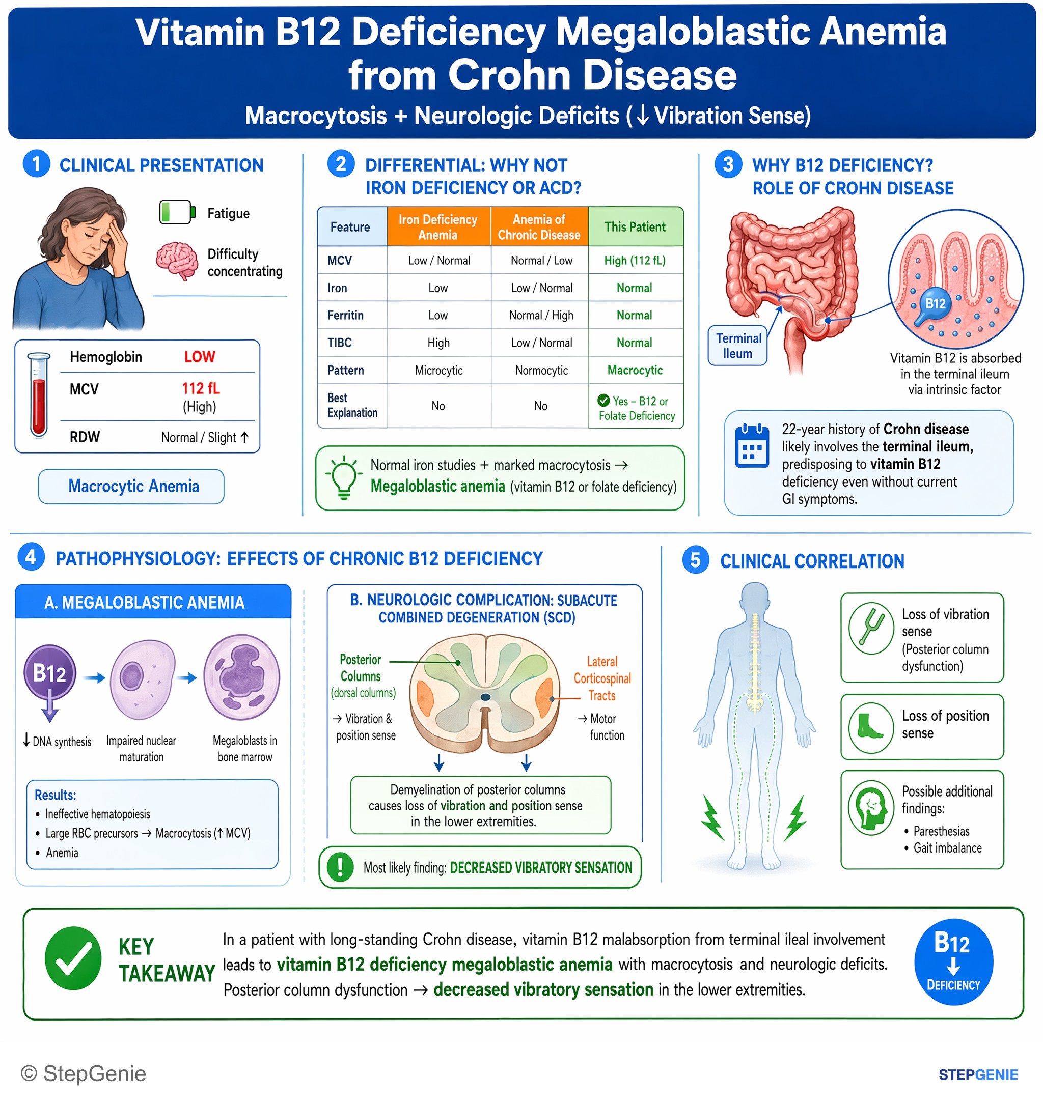
Vitamin B12 deficiency megaloblastic anemia from long-standing Crohn disease causes macrocytosis and neurologic deficits such as decreased…
USMLE Step 1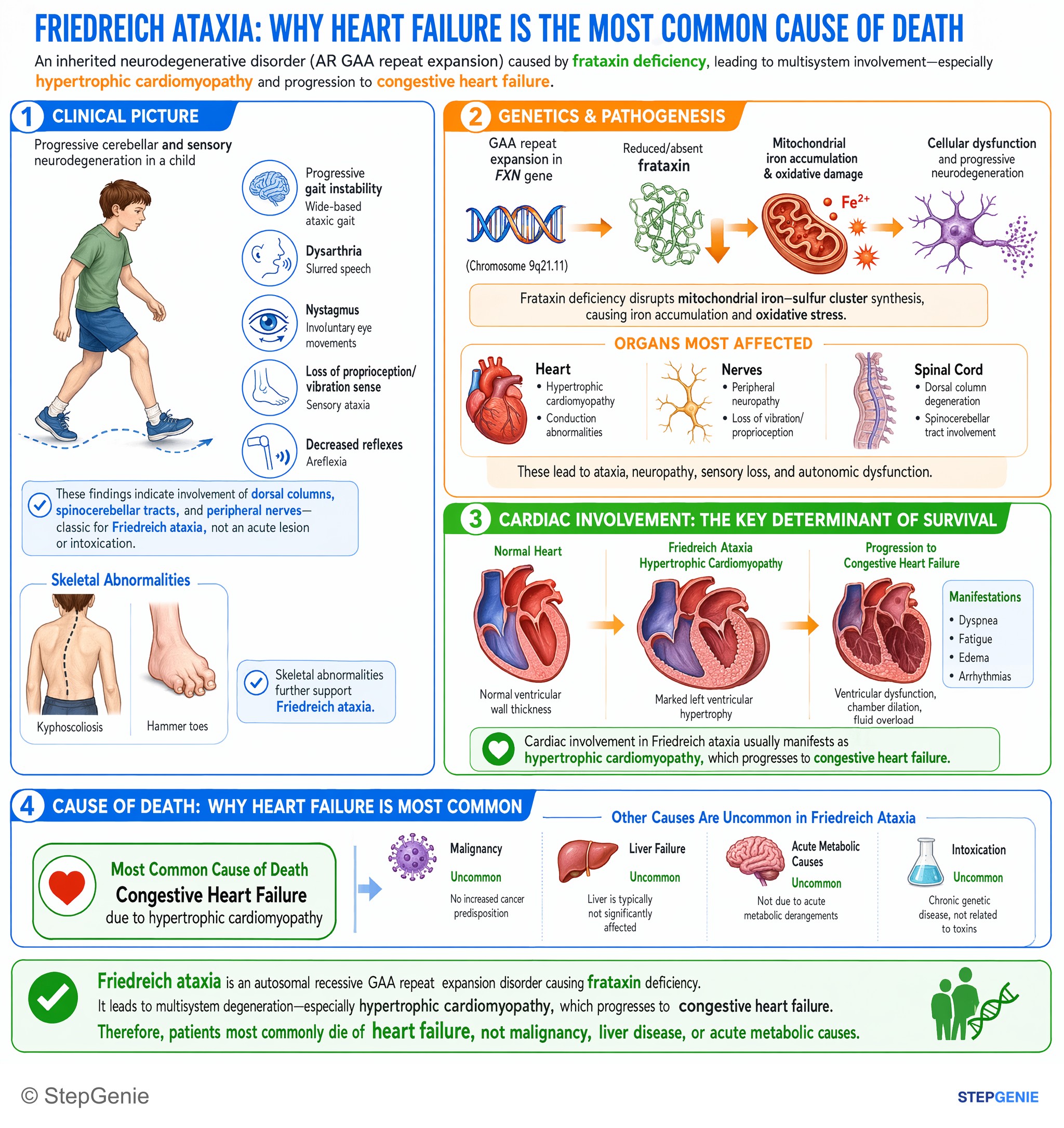
Friedreich ataxia (AR GAA repeat, frataxin deficiency) causes hypertrophic cardiomyopathy; patients most commonly die of congestive heart…
USMLE Step 1Frequently Asked Questions
What is posterior cord syndrome?
Posterior cord syndrome is damage to the dorsal (posterior) columns of the spinal cord, which carry the dorsal column-medial lemniscus pathway. Below the lesion the patient loses vibration sense, joint position sense and fine touch and develops sensory ataxia with a positive Romberg sign, while strength, pain and temperature sensation are preserved in a pure lesion. It is the least common incomplete spinal cord injury pattern and is more often metabolic, toxic or infectious than traumatic.
What are the signs and symptoms of posterior cord syndrome?
Patients describe tingling or numbness, clumsy hands and unsteadiness that is worse in the dark or with the eyes closed. On examination, vibration and joint position sense are lost, the Romberg sign is positive, and the gait is sensory ataxic, with exaggerated steps and forceful foot strikes. Outstretched fingers may writhe when the eyes close (pseudoathetosis), and cervical lesions can cause a Lhermitte sign. Strength, pain and temperature are spared in a pure lesion.
What causes posterior cord syndrome?
The classic causes are vitamin B12 deficiency (subacute combined degeneration), including functional deficiency from nitrous oxide misuse, copper deficiency and tabes dorsalis from tertiary syphilis. Others include multiple sclerosis, HIV vacuolar myelopathy, Friedreich ataxia, compression by tumors, cervical spondylosis or a postoperative seroma, and, rarely, posterior spinal artery infarction. The workup is spine MRI plus B12 with methylmalonic acid and homocysteine, copper and ceruloplasmin, syphilis serology and an HIV test.
What is posterolateral cord syndrome?
Posterolateral, or combined, cord syndrome adds damage to the lateral columns, which carry the corticospinal and spinocerebellar tracts, to dorsal column loss. Patients lose vibration and position sense and also develop spastic weakness, hyperreflexia and Babinski signs, often with absent ankle jerks from a coexisting neuropathy. The classic cause is subacute combined degeneration from vitamin B12 deficiency; nitrous oxide, copper deficiency, HIV vacuolar myelopathy and Friedreich ataxia produce similar patterns.
Why is the Romberg test positive in posterior cord syndrome?
Standing balance relies on vision, vestibular input and proprioception. When the dorsal columns are damaged, proprioception is lost and vision compensates, so the patient can stand with feet together and eyes open but sways or falls when the eyes close, which is a positive Romberg sign. Patients with cerebellar ataxia are often unsteady even with the eyes open, so the Romberg is a test of proprioception rather than of the cerebellum.
How is posterior cord syndrome different from anterior cord, central cord and Brown-Séquard syndromes?
Posterior cord syndrome removes vibration and position sense but spares strength, pain and temperature. Anterior cord syndrome, usually from anterior spinal artery infarction, is the mirror image: weakness and loss of pain and temperature on both sides with vibration and position sense preserved. Central cord syndrome weakens the arms more than the legs after a neck hyperextension injury. Brown-Séquard syndrome causes same-side weakness and dorsal column loss with opposite-side pain and temperature loss.