Craniopharyngioma is a benign (CNS WHO grade 1) epithelial tumor of the sellar region that arises from remnants of Rathke pouch, the embryonic precursor of the anterior pituitary. The classic USMLE picture is a child with headaches, bitemporal hemianopia, growth failure and sometimes central diabetes insipidus, plus a cystic, calcified suprasellar mass filled with yellow-brown, cholesterol-rich fluid (the classic "machine oil"). Since the 2021 WHO classification (CNS5, still the current edition) its two forms are separate tumor types: adamantinomatous (mostly children, calcified and cystic, CTNNB1/β-catenin) and papillary (mostly adults, solid, BRAF V600E). Treatment is surgery with or without radiation, and BRAF/MEK inhibitors shrink most papillary tumors.
Craniopharyngioma at a glance
| Feature | What to know |
|---|---|
| Origin | Epithelial remnants of Rathke pouch (oral ectoderm that forms the anterior pituitary) |
| Location | Suprasellar (sometimes with a sellar component), beside the optic chiasm, pituitary stalk, hypothalamus and third ventricle |
| Age | Bimodal: 5-14 years and 50-74 years; about half of cases are in adults |
| How common | 0.5-2 cases per million people per year; about 3% of intracranial tumors in children |
| Imaging | Mixed cystic and solid mass; calcification in most childhood tumors, best seen on CT; MRI maps the relationship to the chiasm and hypothalamus |
| Gross | Cysts with yellow-brown, cholesterol-rich fluid, classically described as "machine oil" (adamantinomatous type) |
| Microscopy | Adamantinomatous: palisading epithelium, stellate reticulum, wet keratin, calcification; papillary: well-differentiated squamous epithelium |
| Symptoms | Headache, bitemporal hemianopia, hypopituitarism (GH, TSH, ACTH, gonadotropins), central DI, hydrocephalus |
| Treatment | Surgery ± radiation; intracystic therapy for cysts; BRAF/MEK inhibitors for papillary tumors |
Why it causes those symptoms
A suprasellar mass sits on top of four structures, and each gives a cluster of findings:
- Optic chiasm → bitemporal hemianopia. The crossing nasal retinal fibers carry the temporal visual fields, so compression of the chiasm takes out both temporal fields; visual symptoms are reported in about 62-84% of patients.
- Pituitary and stalk → hypopituitarism. Growth hormone, thyroid and cortisol deficiencies are the most frequent in children; gonadotropin loss delays puberty or stops periods. Hormone deficits are found at diagnosis in 40-87%.
- Hypothalamus and posterior pituitary → central diabetes insipidus (about 20% at presentation), and after treatment hypothalamic obesity and behavior change.
- Third ventricle → obstructive hydrocephalus: morning headache, nausea and vomiting; headache occurs in about half of patients.
| Axis | Deficit | Clue in the stem |
|---|---|---|
| GH | Low | Falling off the growth curve, short stature |
| TSH | Low free T4 without the expected rise in TSH (central hypothyroidism) | Fatigue, cold intolerance, weight gain |
| ACTH | Low (secondary adrenal insufficiency) | Weakness, hypotension, hypoglycemia; no hyperpigmentation |
| LH and FSH | Low | Delayed puberty, amenorrhea, low libido |
| ADH | Low (central DI) | Polyuria, polydipsia, bed-wetting, dilute urine, high-normal or high sodium |
Review the axes in anterior pituitary hormones and the water-deprivation logic in central vs nephrogenic DI.
Adamantinomatous vs papillary (WHO CNS5)
The 2021 WHO classification lists adamantinomatous craniopharyngioma and papillary craniopharyngioma as two distinct tumor types under "tumors of the sellar region," each defined by its own molecular driver, rather than as variants of one tumor.
| Adamantinomatous (ACP) | Papillary (PCP) | |
|---|---|---|
| Typical patient | Children (also adults) | Adults |
| Driver | CTNNB1 mutation → β-catenin accumulates, WNT signaling on | BRAF V600E (about 95%) → MAPK pathway on |
| Look | Cystic and solid; calcified in about 90%; machine-oil fluid | Solid, well circumscribed; calcification rare |
| Histology | Squamous nests bordered by palisading columnar cells, stellate reticulum, wet keratin (pale eosinophilic keratin masses that may calcify) | Well-differentiated squamous epithelium; no wet keratin |
| Behavior | More often invades surrounding brain (hypothalamus), so complete removal is harder | Well circumscribed, invades brain much less often; responds to BRAF/MEK inhibitors |
Exam translation: "child + calcified cyst + wet keratin" = adamantinomatous; "adult + solid suprasellar mass + BRAF V600E" = papillary. BRAF V600E is the same mutation tested in melanoma and Langerhans cell histiocytosis, a good link to oncogenes vs tumor suppressors.
Diagnosis
- Imaging: MRI with contrast is the standard study; CT shows the calcifications better (a favorite exam detail). Most tumors show calcification plus a solid and cystic component.
- Visual fields and acuity at baseline.
- Full pituitary panel before surgery: morning cortisol (with ACTH testing as needed), TSH with free T4, IGF-1, LH, FSH and sex steroids, prolactin, and serum sodium, serum osmolality and urine osmolality if DI is suspected.
- Replace cortisol before thyroid hormone. Starting levothyroxine in someone with untreated adrenal insufficiency can precipitate an adrenal crisis.
- Tissue confirms the type; BRAF V600E testing identifies papillary tumors that may respond to targeted therapy.
Treatment and outcomes
- Surgery (endoscopic endonasal transsphenoidal or craniotomy) is the cornerstone. Gross-total resection is often technically hard because the tumor is surrounded by the chiasm, stalk and hypothalamus, so a limited (subtotal) resection followed by radiation is a standard alternative when complete removal would risk those structures.
- Radiation: fractionated external-beam treatment of about 50-54 Gy; proton therapy and intensity-modulated techniques reduce dose to the surrounding brain.
- Cystic tumors: cyst drainage or intracystic therapy (for example interferon, radioactive phosphorus or bleomycin).
- BRAF V600E papillary tumors: in the phase 2 Alliance A071601 trial (NEJM 2023), vemurafenib plus cobimetinib gave a partial or better response in 15 of 16 patients with newly diagnosed papillary craniopharyngioma, with a median tumor-volume reduction of 91%; all 5 patients in a small recurrent cohort also responded (2025). Some tumors regrow after the drugs are stopped, and a 2026 review reports that regrown tumors have responded again when treatment was restarted.
- US drug status (Oct 2026): no BRAF or MEK inhibitor has a craniopharyngioma-specific FDA indication. Vemurafenib is labeled for melanoma and Erdheim-Chester disease, and cobimetinib for melanoma and histiocytic neoplasms, so their use here is off-label. Dabrafenib plus trametinib has a tumor-agnostic accelerated approval for patients aged 1 year and older with unresectable or metastatic BRAF V600E solid tumors that have progressed after prior treatment and have no satisfactory alternatives.
- Adamantinomatous tumors have no proven targeted therapy; trials are testing MEK inhibition, IL-6 blockade (tocilizumab) and PD-1 inhibition.
- Outcomes: 5- and 10-year overall survival in children is about 80-90%, but morbidity is high: hypothalamic obesity, panhypopituitarism needing lifelong replacement (including desmopressin for DI), visual loss, behavioral and cognitive problems, seizures and vasculopathy with delayed strokes.
Differential: other sellar and suprasellar lesions
| Lesion | Usual patient | Key discriminator |
|---|---|---|
| Craniopharyngioma | Children (ACP) and adults (PCP) | Suprasellar, cystic and calcified (ACP), machine-oil fluid, bitemporal hemianopia plus hypopituitarism |
| Pituitary adenoma | Adults | The most common pituitary-region tumor; arises within the sella; often secretes hormones (prolactin most often), so think hormone excess |
| Rathke cleft cyst | Any age | Same Rathke pouch origin, but a benign, non-neoplastic epithelium-lined cyst with no solid tumor component; calcification is uncommon |
| Germinoma (suprasellar germ cell tumor) | Peak 10-19 years | DI is the sentinel symptom in 70-90%; check serum and CSF AFP and β-hCG (pure germinomas: negative or low β-hCG); very radiosensitive, over 90% 5-year survival |
| Langerhans cell histiocytosis | Children | DI with a thickened, nodular pituitary stalk and loss of the posterior pituitary bright spot; lytic bone lesions, rash; CD1a, langerin (CD207), S100; BRAF V600E in half or more |
| Empty sella | Adults | CSF fills the sella; no mass |
CRANIO: a memory hook
- Calcified, cystic suprasellar mass (calcification best on CT)
- Rathke pouch remnant (oral ectoderm, anterior pituitary precursor)
- Adamantinomatous in children (CTNNB1) vs papillary in adults (BRAF V600E)
- Near the chiasm: bitemporal hemianopia
- Insufficiency of pituitary hormones: GH, TSH, ACTH, LH/FSH, and ADH (central DI)
- Oily, machine-oil cyst fluid full of cholesterol
Practice question
A 9-year-old boy has 6 months of morning headaches, slipping school performance and bumping into door frames. His parents report increased thirst, frequent urination and new bed-wetting. His height has fallen from the 60th to the 15th percentile over the past year. Visual field testing shows bitemporal hemianopia. Serum sodium is 146 mEq/L with dilute urine. CT shows a suprasellar mass with cystic components and calcifications, and MRI shows a heterogeneous cystic and solid suprasellar mass compressing the optic chiasm. Which is the most likely diagnosis?
A. Adamantinomatous craniopharyngioma B. Germinoma C. Langerhans cell histiocytosis D. Papillary craniopharyngioma E. Pituitary adenoma F. Rathke cleft cyst
Answer: A. Child + suprasellar mass that is cystic and calcified + bitemporal hemianopia + growth failure + central DI = adamantinomatous craniopharyngioma, the type that dominates in children and calcifies in about 90%.
- B: suprasellar germinoma also causes DI (its sentinel symptom in 70-90%), visual loss and hypopituitarism in children and teenagers, but the cystic, calcified mass points to craniopharyngioma. Germinomas are worked up with serum and CSF AFP and β-hCG.
- C: Langerhans cell histiocytosis causes central DI by infiltrating the stalk, which looks thickened on MRI, usually with bone or skin lesions; it does not form a calcified cystic suprasellar mass.
- D: papillary craniopharyngioma is the right family but the wrong patient: an adult tumor that is usually solid, rarely calcified and driven by BRAF V600E.
- E: pituitary adenomas are mostly adult tumors that arise within the sella and often secrete hormones; a calcified, cystic suprasellar mass in a child is not the pattern.
- F: a Rathke cleft cyst shares the Rathke pouch origin but is a non-neoplastic cyst without a solid tumor component, and calcification is uncommon.
Key takeaways
- Craniopharyngioma = benign (grade 1) suprasellar tumor from Rathke pouch remnants; bimodal ages 5-14 and 50-74.
- Cystic + calcified (CT) + machine-oil fluid + wet keratin = adamantinomatous, the childhood type (CTNNB1).
- Papillary type: adults, solid, BRAF V600E; WHO CNS5 (2021) treats the two as separate tumor types.
- Mass effect: bitemporal hemianopia (chiasm), hypopituitarism (GH, TSH, ACTH, LH/FSH), central DI, hydrocephalus.
- Check cortisol before replacing thyroid hormone, and test all pituitary axes before surgery.
- Treatment: surgery ± radiation; BRAF/MEK inhibitors shrink most papillary tumors (off-label for this use in the US); hypothalamic obesity and panhypopituitarism are the big long-term problems.
Sources
- The 2021 WHO Classification of Tumors of the Central Nervous System: a summary — Neuro-Oncology (2021)
- WHO Classification of Tumours series: Central Nervous System Tumours, 5th edition — IARC Publications
- Childhood Craniopharyngioma Treatment (PDQ), Health Professional Version — National Cancer Institute (2024)
- Craniopharyngioma — StatPearls, NCBI Bookshelf (2023, archived)
- Systemic Molecularly Targeted Therapies for Neoadjuvant and Salvage Craniopharyngioma: A Contemporary Narrative Review — Biomedicines (2026)
- Alliance A071601: Phase II Trial of BRAF/MEK Inhibition in Recurrent Papillary Craniopharyngiomas — Neuro-Oncology abstract (2025)
- Tafinlar (dabrafenib) prescribing information — DailyMed (2026)
- Childhood Central Nervous System Germ Cell Tumors Treatment (PDQ), Health Professional Version — National Cancer Institute (2024)
- Langerhans Cell Histiocytosis Treatment (PDQ), Health Professional Version — National Cancer Institute (2025)
- Pituitary Tumors — Merck Manual Professional Edition (2026)
- Atypical Rathke's Cleft Cyst Associated with Ossification — American Journal of Neuroradiology (1999)
- Polyglandular Deficiency Syndromes — Merck Manual Professional Edition (2025)
Visual explanations from the StepGenie QBank
Infographics StepGenie drew to explain questions in its own QBank, matched to this article's topic. Each caption is the takeaway of the question it explains.
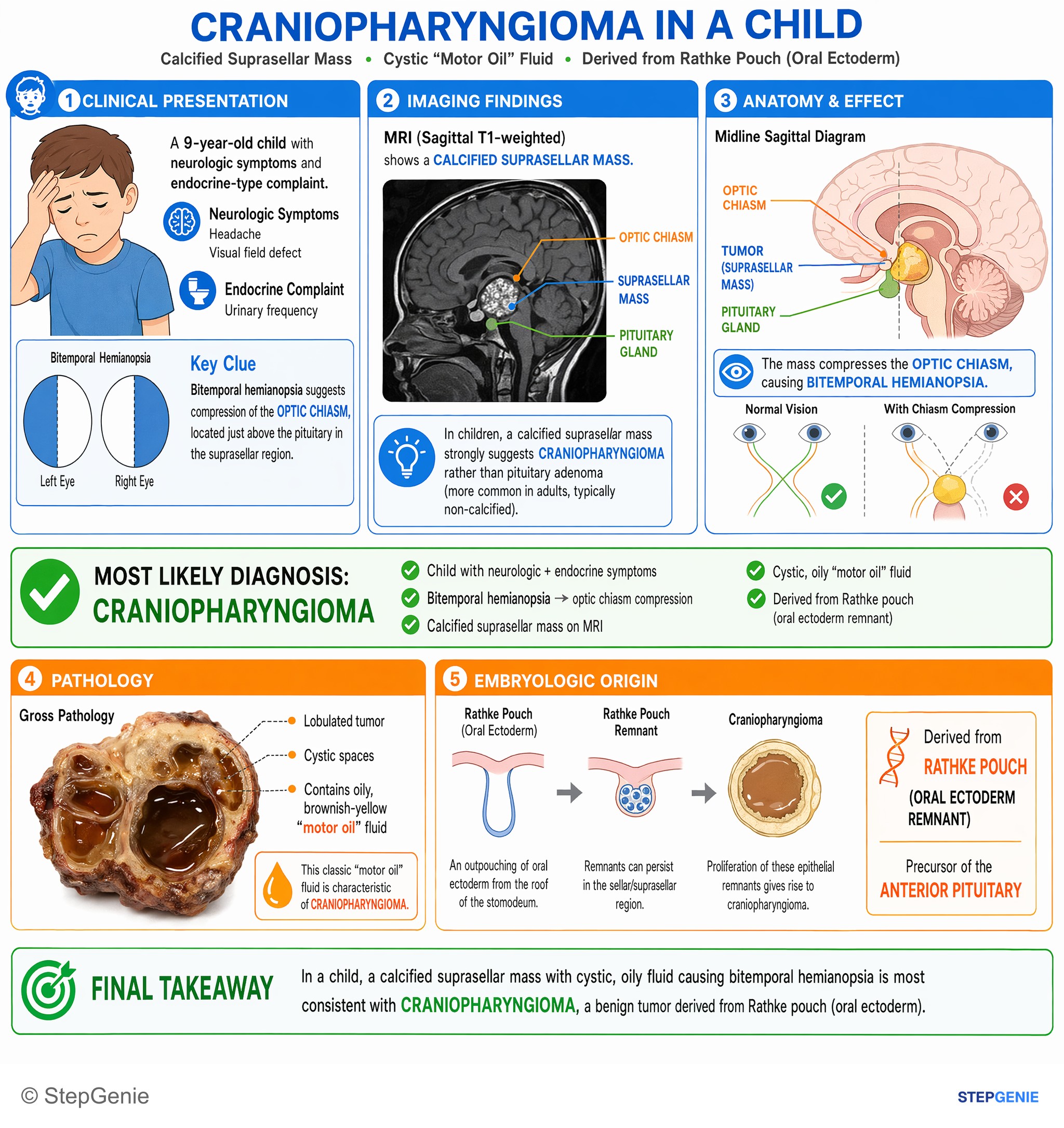
Craniopharyngioma in a child: calcified suprasellar mass with cystic, oily fluid; benign tumor derived from Rathke pouch (oral ectoderm…
USMLE Step 1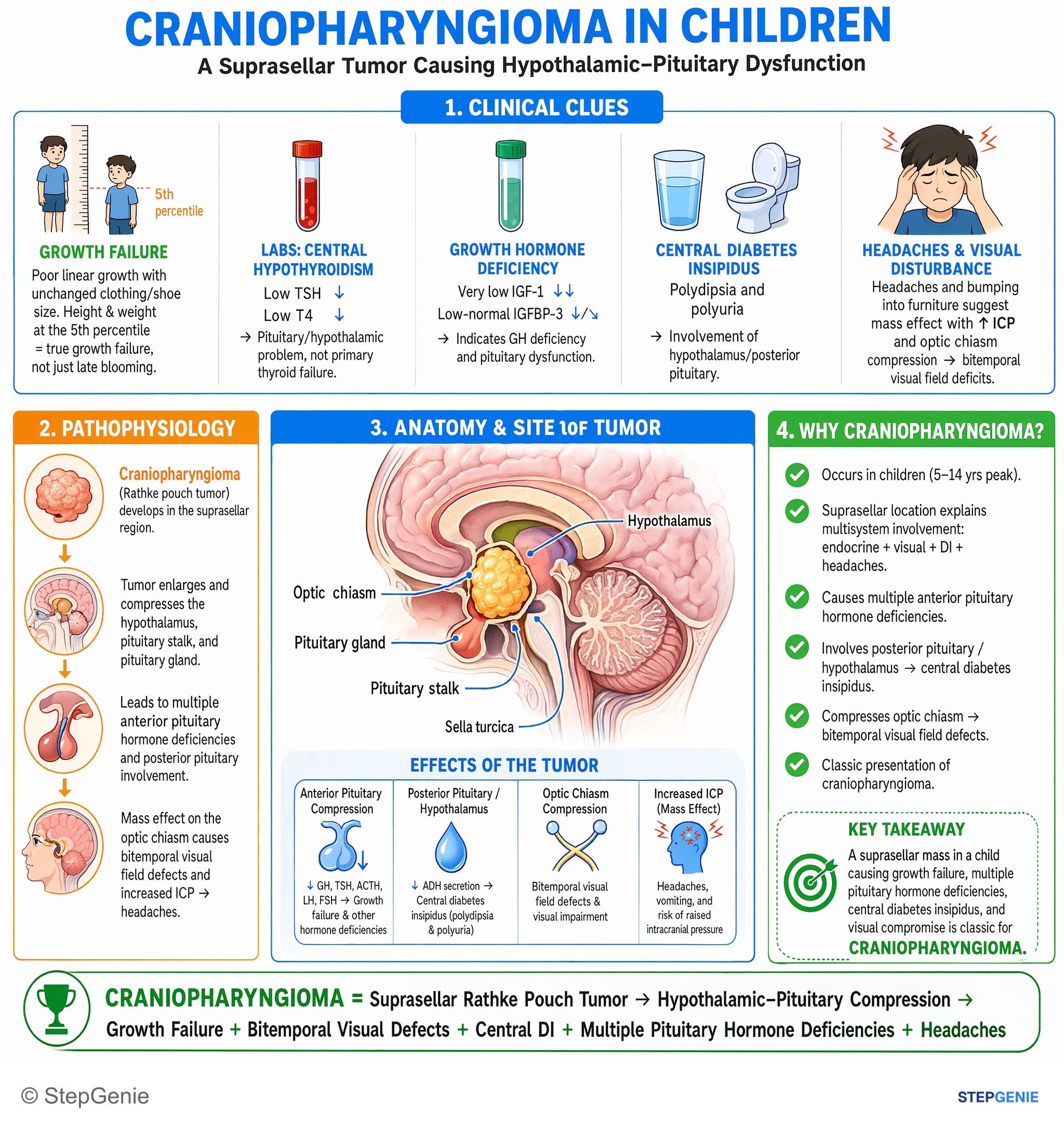
Craniopharyngioma in a child causes hypothalamic–pituitary compression → growth failure, bitemporal visual defects, headaches, central DI…
USMLE Step 2Frequently Asked Questions
What is a craniopharyngioma?
A craniopharyngioma is a benign (CNS WHO grade 1) epithelial tumor of the sellar region that arises from remnants of Rathke pouch, the embryonic tissue that forms the anterior pituitary. It usually sits above the sella, next to the optic chiasm, pituitary stalk and hypothalamus, so even though it does not spread it causes headache, visual loss and hormone deficiencies. It has a bimodal age distribution, peaking at 5 to 14 years and 50 to 74 years.
What is the difference between adamantinomatous and papillary craniopharyngioma?
Since the 2021 WHO classification they are separate tumor types. Adamantinomatous craniopharyngioma occurs mostly in children, is cystic and calcified in about 90% of cases, contains machine-oil fluid and wet keratin, and is driven by CTNNB1 mutations that activate beta-catenin. Papillary craniopharyngioma occurs mostly in adults, is solid and rarely calcified, and carries a BRAF V600E mutation in about 95% of cases, which makes it responsive to BRAF and MEK inhibitors.
Why does craniopharyngioma cause bitemporal hemianopia and hormone problems?
The tumor grows in the suprasellar space, directly above the optic chiasm and around the pituitary stalk and hypothalamus. Pressing on the chiasm damages the crossing nasal retinal fibers, which carry the temporal visual fields, causing bitemporal hemianopia. Pressure on the pituitary, stalk and hypothalamus causes deficiencies of growth hormone, TSH, ACTH and gonadotropins, and sometimes central diabetes insipidus. Blocking the third ventricle can also cause hydrocephalus with morning headache and vomiting.
What are the classic imaging findings of craniopharyngioma?
The classic finding is a suprasellar mass with both cystic and solid parts and calcifications, especially in children with the adamantinomatous type. CT is the best test for showing the calcifications, while MRI with contrast is the standard study for mapping the tumor's relationship to the optic chiasm, pituitary stalk, hypothalamus and third ventricle. The cysts contain yellow-brown, cholesterol-rich fluid, classically described as machine oil.
How is craniopharyngioma treated?
Surgery is the cornerstone, either removing the whole tumor or, when that would endanger the hypothalamus or optic pathways, a limited resection followed by radiation of about 50 to 54 Gy. Cystic tumors can be drained or treated with intracystic therapy. Papillary tumors with BRAF V600E respond to BRAF plus MEK inhibitors; in one trial 15 of 16 newly diagnosed patients responded to vemurafenib plus cobimetinib, although this use is off-label in the US. Lifelong hormone replacement is often needed.
How do you tell craniopharyngioma from a pituitary adenoma?
Pituitary adenomas are the most common pituitary-region tumors, occur mainly in adults, arise within the sella and often secrete hormones, especially prolactin, so the stem usually shows hormone excess. Craniopharyngioma classically appears in a child as a suprasellar mass that is cystic and calcified, with hormone deficiencies, growth failure, bitemporal hemianopia and sometimes central diabetes insipidus. Calcification on CT and machine-oil cyst fluid point strongly toward craniopharyngioma.