Glioblastoma is the kind of diagnosis that shows up in vignettes as “rapidly worsening neuro deficits + headache + seizures,” and on imaging as a gnarly, irregular mass that crosses brain structures like it owns the place. For Step 1, you don’t need to be a neuro-onc fellow—you need to recognize the pattern, know the classic histology and genetics, and understand why it behaves so aggressively.
Quick Definition (What It Is)
Glioblastoma (GBM) is a high-grade malignant astrocytoma and the most common primary malignant brain tumor in adults. It is:
- WHO grade 4 (the highest grade)
- Highly infiltrative (microscopic spread beyond what you can resect)
- Characterized by necrosis and microvascular proliferation
- Often located in the cerebral hemispheres (especially frontal/temporal)
Step 1 translation: aggressive adult brain tumor with pseudopalisading necrosis, ring enhancement, and butterfly midline crossing.
Epidemiology & High-Yield Associations
Who gets it?
- Typically older adults (peak ~55–75)
- Slight male predominance (board-relevant but not essential)
Classic board associations
- “Butterfly glioma”: tumor crosses the corpus callosum → bilateral hemispheric involvement
- Ring-enhancing lesion with central necrosis on MRI
- Pseudopalisading necrosis on histology
Genetics (the HY split: primary vs secondary GBM)
A very common Step-style question is to distinguish primary (de novo) vs secondary (progressed) glioblastoma.
| Feature | Primary GBM (most common) | Secondary GBM |
|---|---|---|
| Typical patient | Older | Younger |
| Arises from | De novo | Often from lower-grade astrocytoma |
| Key mutations | EGFR amplification, PTEN loss, TERT promoter | IDH1/IDH2 mutation, TP53 |
| Prognosis | Poor | Poor (often slightly better than primary) |
High-yield pearl: IDH mutation is a favorable prognostic marker in diffuse gliomas (including GBM when present), and it’s classically associated with secondary GBM.
Pathophysiology (Why It’s So Aggressive)
1) Infiltrative growth = not “one clean mass”
GBM cells diffusely invade surrounding brain tissue. Even if imaging shows one dominant lesion, tumor cells extend beyond the visible margins.
Clinical consequence: surgical resection is never truly curative.
2) Necrosis + angiogenesis
- Tumor outgrows its blood supply → central necrosis
- Hypoxia drives angiogenic signaling (e.g., VEGF) → microvascular proliferation
Histology consequence: classic pseudopalisading necrosis + glomeruloid (tufted) microvascular proliferation.
3) Mass effect and increased ICP
Expanding lesion + edema → increased intracranial pressure (ICP).
Step 1 consequence: headaches worse in the morning, vomiting, papilledema, focal deficits.
Clinical Presentation (How It Shows Up)
Symptoms depend on location, but these patterns are high-yield:
Common presenting features
- Headache (often worse in the morning)
- Seizures (new-onset adult seizure = brain tumor until proven otherwise)
- Focal neurologic deficits
- Weakness, aphasia, visual field deficits, personality changes (frontal lobe)
- Signs of increased ICP
- Nausea/vomiting, papilledema, altered mental status
“Butterfly” clue symptoms
If it crosses the corpus callosum, you may see rapid cognitive/behavioral changes plus bilateral signs.
Diagnosis (Boards Love the Imaging + Histology Combo)
Imaging
MRI with contrast is the go-to.
Classic imaging description:
- Irregular ring-enhancing mass
- Central necrosis
- Surrounding vasogenic edema
- May cross midline through corpus callosum (“butterfly glioma”)
Differential for ring-enhancing brain lesions (Step classic)
Use context:
- GBM: single irregular ring-enhancing lesion, necrosis, older adult
- Brain abscess: fever, leukocytosis, risk factors; diffusion restriction on MRI
- Toxoplasmosis: AIDS/CD4 < 100; multiple ring-enhancing lesions (basal ganglia)
- Metastases: multiple lesions at gray-white junction
- Tumefactive demyelination: incomplete ring enhancement, MS history
Histology (must-recognize features)
- Pseudopalisading necrosis: tumor cells lined up around necrotic center
- Microvascular proliferation: “glomeruloid” vessels
- Tumor cells often GFAP-positive (astrocytic lineage)
Board-style stem: “biopsy shows pseudopalisading necrosis and endothelial proliferation.”
Treatment (What You’re Expected to Know)
Even though prognosis is poor, Step questions often test the standard multimodal approach.
Core management
- Maximal safe surgical resection
- Radiation therapy
- Temozolomide (alkylating agent; oral)
Supportive/adjunct
- Dexamethasone for vasogenic edema (symptom relief)
- Antiseizure meds if seizures (not always prophylactic unless indicated clinically)
Prognosis
- Unfortunately poor; median survival often ~12–15 months with standard therapy (varies by molecular markers and treatment access)
HY prognostic marker: MGMT promoter methylation
- MGMT methylation → less DNA repair → better response to temozolomide (therefore better prognosis)
First Aid Cross-References (What to Flip To)
These are the sections typically aligned with GBM in First Aid for USMLE Step 1 (edition layouts vary slightly):
- Neuropathology: CNS tumors
- Astrocytoma/glioblastoma: pseudopalisading necrosis, ring enhancement, butterfly crossing corpus callosum
- Neuroimaging concepts
- Ring-enhancing lesions differential (often integrated with infections like toxoplasmosis/abscess)
- Oncology pharmacology (if integrated)
- Temozolomide (alkylating agent used for gliomas)
How to use FA efficiently: annotate the glioma table with:
- Primary vs secondary genetic associations (EGFR vs IDH)
- MGMT methylation → temozolomide response
- “Butterfly via corpus callosum” as a visual memory hook
High-Yield “You Will Be Asked This” Facts
Rapid-fire must-knows
- Most common primary malignant brain tumor in adults
- WHO grade 4 astrocytoma
- MRI: irregular ring enhancement + central necrosis
- Crosses corpus callosum → “butterfly glioma”
- Histology: pseudopalisading necrosis + microvascular proliferation
- Primary GBM: EGFR amplification (older adults)
- Secondary GBM: IDH mutation (younger, from lower-grade tumor)
- Treatment: resection + radiation + temozolomide
- MGMT promoter methylation predicts better temozolomide response
Mini-vignette pattern recognition
62-year-old with progressive headaches, new seizures, and focal weakness. MRI shows irregular ring-enhancing lesion with central necrosis crossing midline.
Answer: Glioblastoma.
Common Pitfalls (How NBME Tries to Trick You)
- Mistaking GBM for metastasis: metastases are often multiple and at the gray-white junction; GBM is often single, infiltrative, and can cross the corpus callosum.
- Mistaking GBM for toxoplasmosis: toxo usually in AIDS with multiple ring-enhancing lesions (classically basal ganglia).
- Forgetting primary vs secondary genetics: if they mention younger patient with a history of a lower-grade glioma → think IDH-mutant secondary GBM.
One-Table Summary (Perfect for Last-Minute Review)
| Category | Glioblastoma (GBM) |
|---|---|
| Tumor type | Malignant astrocytoma |
| WHO grade | IV |
| Age group | Usually older adults |
| Imaging | Ring-enhancing, central necrosis, edema; may cross corpus callosum (“butterfly”) |
| Histology | Pseudopalisading necrosis, microvascular proliferation |
| Key genetics | Primary: EGFR; Secondary: IDH, TP53 |
| Treatment | Resection + radiation + temozolomide (+ dexamethasone for edema) |
| Prognosis | Poor; MGMT methylation improves response to temozolomide |
Visual explanations from the StepGenie QBank
Infographics StepGenie drew to explain questions in its own QBank, matched to this article's topic. Each caption is the takeaway of the question it explains.
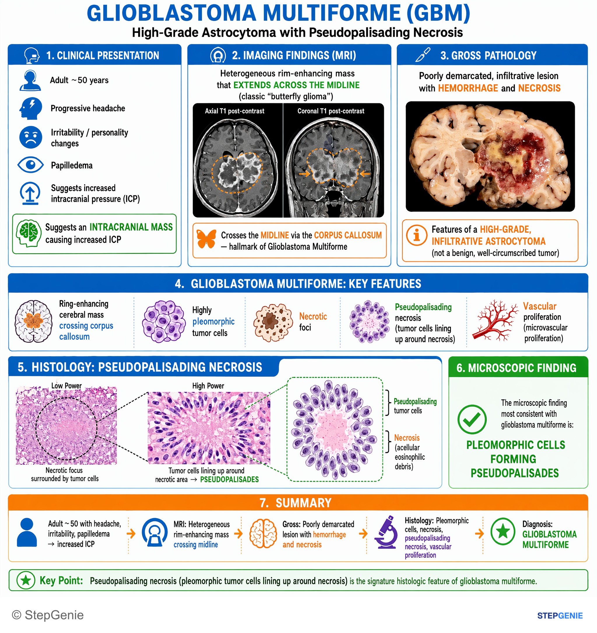
Glioblastoma multiforme presents with ring-enhancing cerebral mass crossing corpus callosum; histology shows pleomorphic tumor cells with…
USMLE Step 1Frequently Asked Questions
Why is glioblastoma so hard to cure with surgery alone?
Glioblastoma is hard to cure surgically because it is highly infiltrative. Even when MRI shows one dominant mass, tumor cells diffusely invade surrounding brain tissue beyond the visible margins, so resection cannot remove every malignant cell. That infiltrative behavior is a key reason surgery is never truly curative and why treatment typically combines maximal safe resection with radiation and temozolomide.
How do you recognize glioblastoma on imaging and histology?
Glioblastoma is classically recognized by an irregular ring-enhancing brain mass with central necrosis and surrounding vasogenic edema on contrast MRI. It may cross the midline through the corpus callosum, creating the classic “butterfly glioma” pattern. On histology, the must-know findings are pseudopalisading necrosis and microvascular or glomeruloid endothelial proliferation, reflecting its aggressive biology.
How do you tell glioblastoma from other ring-enhancing brain lesions on Step 1?
You distinguish glioblastoma by combining the lesion pattern with the clinical context. Glioblastoma usually presents in an older adult as a single irregular ring-enhancing lesion with necrosis. Brain abscess is suggested by fever, leukocytosis, risk factors, and diffusion restriction on MRI. Toxoplasmosis usually causes multiple basal ganglia lesions in AIDS with very low CD4 count, while metastases are often multiple at the gray-white junction.
What symptoms should make you think of glioblastoma in a vignette?
Glioblastoma should come to mind with rapidly worsening neurologic deficits, headache, seizures, and signs of increased intracranial pressure. High-yield clues include morning-predominant headache, nausea or vomiting, papilledema, altered mental status, and new-onset adult seizure. Depending on tumor location, patients may also have weakness, aphasia, visual field deficits, or personality changes, especially with frontal lobe involvement.
What are the high-yield genetics and prognostic markers for glioblastoma?
The key split is primary versus secondary glioblastoma. Primary glioblastoma, which is more common and usually occurs in older patients, is associated with EGFR amplification, PTEN loss, and TERT promoter changes. Secondary glioblastoma tends to occur in younger patients, often arises from a lower-grade astrocytoma, and is associated with IDH1 or IDH2 mutation and TP53. IDH mutation is favorable, and MGMT promoter methylation predicts better response to temozolomide.