A schwannoma is a benign, encapsulated tumor of Schwann cells that pushes its nerve aside instead of infiltrating it. The Step 1 version is the vestibular schwannoma (acoustic neuroma): slowly progressive unilateral sensorineural hearing loss with tinnitus and imbalance, plus an enhancing mass at the cerebellopontine angle (CPA) that extends into the internal auditory canal on gadolinium MRI. Histology shows cellular Antoni A areas with Verocay bodies alternating with loose Antoni B areas, and the tumor cells stain diffusely for S-100 and SOX10. Bilateral vestibular schwannomas mean NF2-related schwannomatosis (the 2022 name for neurofibromatosis type 2), caused by loss of the NF2 tumor suppressor on chromosome 22, which encodes merlin.
What a schwannoma is
- Cell of origin: Schwann cells, which wrap and insulate peripheral nerve fibers. About 95% of vestibular schwannomas are unilateral and occur without an inherited predisposition, yet sporadic and NF2-related tumors alike are driven by loss of function of the NF2 tumor suppressor.
- Growth: slow, well-circumscribed and eccentric, displacing the nerve rather than infiltrating it.
- Behavior: malignant transformation is very rare.
- "Acoustic neuroma" is a double misnomer: it almost always arises from the vestibular division of CN VIII, and it is a Schwann cell tumor, not a neuroma.
- How common: vestibular schwannomas are about 8% of intracranial tumors and the most common CPA tumor, ahead of meningioma and epidermoid cyst. Sporadic tumors usually cause symptoms between ages 30 and 60.
Histology: Antoni A, Antoni B and Verocay bodies
| Feature | What it looks like | Exam point |
|---|---|---|
| Antoni A | Dense, hypercellular spindle cells | Where nuclear palisading and Verocay bodies appear |
| Verocay bodies | Palisading nuclei arranged around pink (eosinophilic) zones | The schwannoma buzzword |
| Antoni B | Loose, hypocellular, myxoid tissue | Alternates with Antoni A |
| S-100 and SOX10 | Diffuse, strong staining of the tumor cells | Schwann cell markers; a neurofibroma mixes Schwann cells with fibroblasts and other cells |
How a vestibular schwannoma presents
Early, while the tumor is small:
- Unilateral or asymmetric sensorineural hearing loss, usually slowly progressive. It can also be abrupt or fluctuate, so sudden hearing loss does not rule a schwannoma out.
- Speech discrimination worse than the pure-tone loss suggests.
- Tinnitus in the same ear, and dizziness or dysequilibrium.
Later, as it grows into the CPA:
- CN V: facial numbness or tingling and an impaired corneal reflex; some patients have pain resembling trigeminal neuralgia.
- CN VII: facial weakness is uncommon and late; significant facial palsy is rare even with large tumors.
- Cerebellum and brainstem: unsteady gait and ataxia; large tumors can cause hydrocephalus and become life-threatening.
Diagnosis
- Audiogram first: asymmetric sensorineural hearing loss with poor speech discrimination.
- MRI with gadolinium is the preferred test and confirms the diagnosis: an enhancing mass that fills and widens the internal auditory canal and, as it grows, projects into the CPA.
- Auditory brainstem response may show absent waveforms or longer latencies.
- Genetic testing for NF2 for anyone younger than 30 with a unilateral vestibular schwannoma and for everyone with bilateral tumors.
NF2-related schwannomatosis (formerly neurofibromatosis type 2)
In 2022 an international consensus (Plotkin et al.) renamed neurofibromatosis type 2 NF2-related schwannomatosis, a name that better reflects updated diagnostic criteria combining clinical features with genetic testing. Many references still say NF2; the disease is the same.
- Genetics: autosomal dominant. NF2 at 22q12.2 encodes merlin (schwannomin), a tumor suppressor (see the oncogenes vs tumor suppressors cheat sheet). About half of cases are inherited and half arise from a new variant, and many people with a de novo variant are mosaic, carrying it in only some of their cells. Tumors form after a second, somatic hit in the remaining copy.
- Tumors: bilateral vestibular schwannomas (average onset 18–24 years; most affected people have both by age 30), schwannomas of other cranial, spinal and peripheral nerves, meningiomas and ependymomas (see the ependymoma Q-bank breakdown).
- Other clues: juvenile cataract (posterior subcapsular lens opacity), retinal hamartomas, skin tumors and childhood mononeuropathy such as facial palsy.
- Diagnosis: bilateral vestibular schwannomas alone, the same NF2 variant in two anatomically distinct tumors, or set combinations of major and minor criteria (for example, a unilateral vestibular schwannoma plus two or more meningiomas).
| NF1 | NF2-related schwannomatosis | Schwannomatosis | |
|---|---|---|---|
| Gene | NF1 (neurofibromin) | NF2 on 22q12.2 (merlin) | SMARCB1 or LZTR1 |
| Inheritance | Autosomal dominant; about half new variants | Autosomal dominant; about half de novo | Familial in 15–25%, then autosomal dominant |
| Signature nerve tumor | Neurofibromas, including plexiform | Bilateral vestibular schwannomas | Multiple non-vestibular schwannomas |
| Other clues | Café-au-lait spots, armpit and groin freckling, Lisch nodules, optic gliomas; risk of MPNST | Meningiomas, ependymomas, juvenile cataract | Chronic pain; vestibular schwannomas almost never |
Schwannoma vs neurofibroma
| Schwannoma | Neurofibroma | |
|---|---|---|
| Capsule | Encapsulated | Unencapsulated |
| Relation to nerve | Eccentric; displaces the nerve | Axons run through the tumor |
| Cells | Almost exclusively neoplastic Schwann cells | A mix of Schwann cells, fibroblasts, neural cells and mast cells |
| Syndrome | NF2-related schwannomatosis; schwannomatosis | NF1 (multiple or plexiform neurofibromas) |
| Malignant change | Very rare | Malignant peripheral nerve sheath tumor (MPNST), mainly from plexiform neurofibromas in NF1 |
Management at Step 1 and Step 2 CK depth
Whether to operate or irradiate depends on residual hearing, tumor size, age, general health and patient preference.
| Option | Typically used for | Key points |
|---|---|---|
| Observation with serial MRI | Small, asymptomatic tumors that are not growing | "Wait and scan": treat if the tumor grows or causes symptoms |
| Microsurgery | Growing or symptomatic tumors, and large tumors with significant brainstem compression that radiosurgery doesn't suit | Middle fossa or retrosigmoid approach to preserve hearing, translabyrinthine if no useful hearing remains; risks include facial palsy and hearing loss |
| Stereotactic radiosurgery (Gamma Knife or LINAC) | Small or medium tumors without significant brainstem compression | Sometimes preferred for older patients, poor surgical candidates, bilateral tumors or a tumor in the only hearing ear |
| Bevacizumab (anti-VEGF antibody) | Rapidly growing vestibular schwannomas in NF2-related schwannomatosis | Can shrink tumors, slow hearing loss and delay surgery |
In NF2-related schwannomatosis, radiation is used cautiously because it may induce, accelerate or transform tumors, and it is avoided in children; hearing is supported with hearing aids or cochlear or auditory brainstem implants.
Look-alikes at the cerebellopontine angle
| Answer choice | What its stem shows | Why it is not a vestibular schwannoma |
|---|---|---|
| Meningioma | Second most common CPA tumor: dura-based, with a broad dural attachment and a dural tail; may be calcified | Canal widening favors schwannoma, though meningiomas sometimes extend into the canal; both enhance and both occur in NF2, so rely on the imaging |
| Epidermoid cyst | Third most common CPA lesion: a keratin-filled cyst lined by squamous epithelium | Little or no enhancement, and bright (restricted) on diffusion-weighted imaging |
| Vestibular neuritis | A single attack of severe vertigo lasting days, presumed viral | No hearing loss or tinnitus, and no mass |
| Labyrinthitis | Acute vertigo with hearing loss, tinnitus or both | An acute illness rather than months of progression; no mass on MRI |
| Ménière disease | Vertigo attacks lasting 20 minutes to 12 hours, fluctuating low-frequency sensorineural hearing loss, tinnitus, aural fullness | Episodic; MRI of the internal auditory canals is done to exclude a schwannoma |
The rest of the meningioma picture is in the meningioma guide.
Exam traps
- Bilateral vestibular schwannomas = NF2-related schwannomatosis, not NF1; a unilateral tumor before age 30 also calls for NF2 testing.
- Sudden hearing loss can still be a schwannoma; asymmetric sensorineural loss is worked up with an audiogram and gadolinium MRI.
- Facial weakness is late; as the tumor enlarges, look for trigeminal signs such as facial numbness or an impaired corneal reflex.
- Antoni A + Antoni B + Verocay bodies + S-100/SOX10 = schwannoma; an unencapsulated tumor with axons running through it is a neurofibroma.
Worked example
Vignette. A 52-year-old woman has had progressive right-sided hearing loss and tinnitus for 8 months, with intermittent imbalance and trouble understanding speech in noisy rooms. She hears finger rub poorly on the right and has a mildly unsteady gait; she has no fever or headache. An audiogram shows right-sided sensorineural hearing loss with poor word recognition. MRI with gadolinium shows a well-circumscribed, enhancing mass at the right cerebellopontine angle extending into the internal auditory canal. What is the most likely diagnosis?
A. Meningioma
B. Epidermoid cyst
C. Vestibular neuritis
D. Vestibular schwannoma
E. Ménière disease
Answer: D. Months of one-sided sensorineural loss with tinnitus, imbalance and poor word recognition, plus an enhancing CPA mass entering the internal auditory canal, is a vestibular schwannoma.
Why the others are wrong:
- A: a meningioma can sit at the CPA and enhance, but it is dura-based with a dural tail and less often enters the internal auditory canal.
- B: an epidermoid cyst would show little or no enhancement and would be bright on diffusion-weighted imaging.
- C: vestibular neuritis is a single acute attack of vertigo without hearing loss or a mass.
- E: Ménière disease causes episodic vertigo with fluctuating low-frequency hearing loss and aural fullness, and MRI shows no mass.
Likely follow-up questions: the finding that best supports the diagnosis is S-100 positivity, Verocay bodies or Antoni A and B areas; bilateral tumors would point to NF2-related schwannomatosis (NF2, chromosome 22, merlin); and the imaging feature that would favor the top distractor, meningioma, is a dural tail.
Key takeaways
- Schwannoma: benign, encapsulated Schwann cell tumor; Antoni A (with Verocay bodies) and Antoni B; S-100 and SOX10 positive; malignant change is very rare.
- Vestibular schwannoma: progressive unilateral sensorineural hearing loss, tinnitus, imbalance; enhancing CPA mass entering the internal auditory canal.
- Workup: audiogram, then gadolinium MRI; NF2 testing if bilateral or younger than 30.
- NF2-related schwannomatosis (formerly NF2): autosomal dominant, NF2 on 22q12.2, merlin; bilateral vestibular schwannomas, meningiomas, ependymomas, juvenile cataract.
- Management: serial MRI, microsurgery or radiosurgery; bevacizumab for growing NF2-related tumors.
- Look-alikes: dural tail = meningioma; DWI-bright with little or no enhancement = epidermoid.
Sources
- Vestibular Schwannoma — Merck Manual Professional Edition
- Vestibular Schwannoma (Acoustic Neuroma) and Neurofibromatosis — NIDCD
- Neurofibromatosis — Merck Manual Professional Edition
- Evans DGR. Neurofibromatosis 2 — Genetics in Medicine, 2009
- Neurofibromatosis type 2 — MedlinePlus Genetics
- Schwannomatosis — MedlinePlus Genetics
- Helbing DL, Schulz A, Morrison H. Pathomechanisms in schwannoma development and progression — Oncogene, 2020
- Pekmezci M, et al. Morphologic and immunohistochemical features of malignant peripheral nerve sheath tumors and cellular schwannomas — Modern Pathology, 2015
- Gao K, et al. Meningiomas of the cerebellopontine angle: radiological differences in tumors with internal auditory canal involvement — PLOS One, 2015
- Pop MM, et al. Intracranial epidermoid cysts: benign entities with malignant behavior — Scientific Reports, 2023
- Vestibular Neuronitis — Merck Manual Professional Edition
- Meniere Disease — Merck Manual Professional Edition
Visual explanations from the StepGenie QBank
Infographics StepGenie drew to explain questions in its own QBank, matched to this article's topic. Each caption is the takeaway of the question it explains.
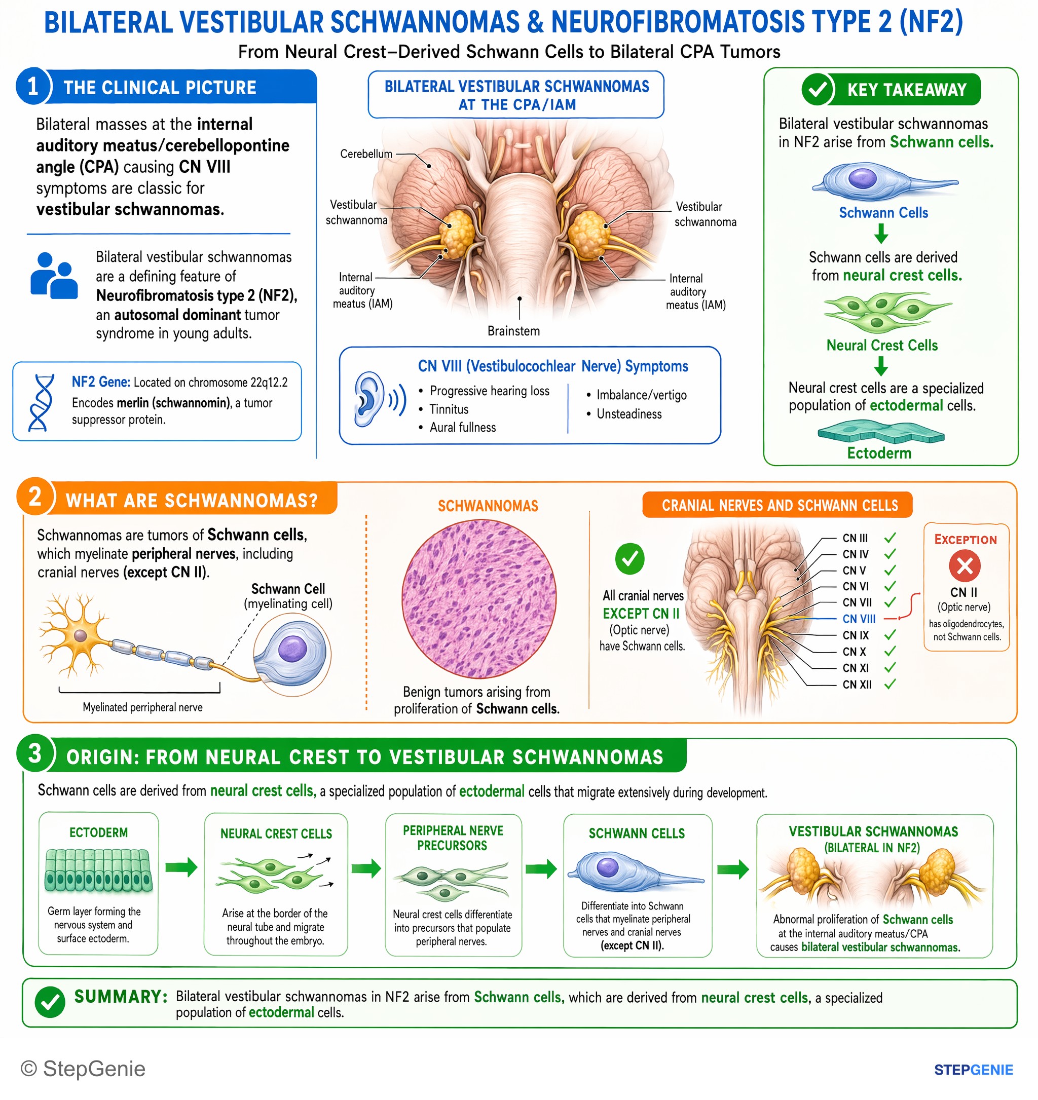
Bilateral vestibular schwannomas in NF2 arise from Schwann cells, which are derived from neural crest ectoderm.
USMLE Step 1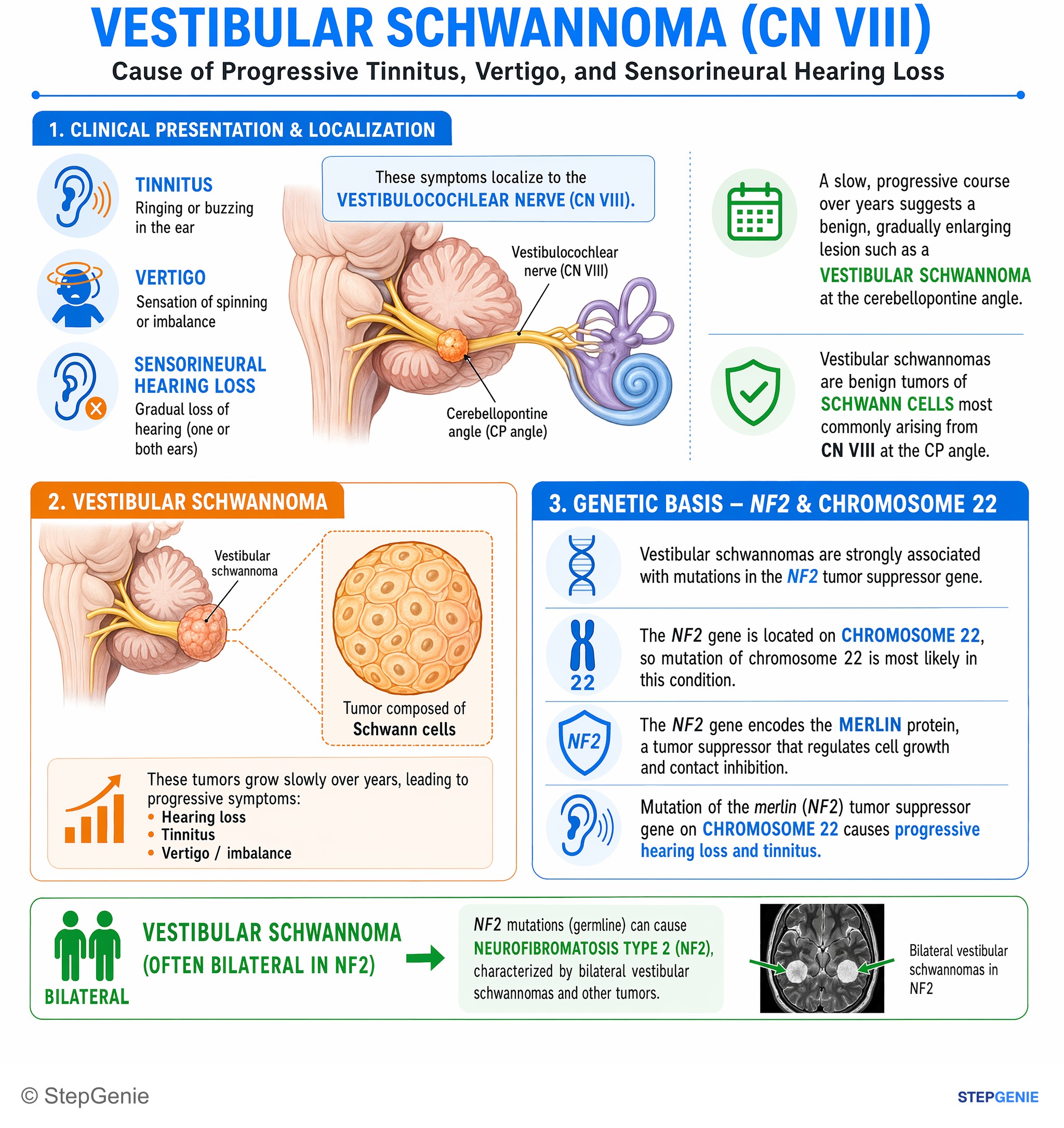
Vestibular schwannoma (often bilateral in NF2) involves mutation of the merlin tumor suppressor gene on chromosome 22 causing progressive…
USMLE Step 1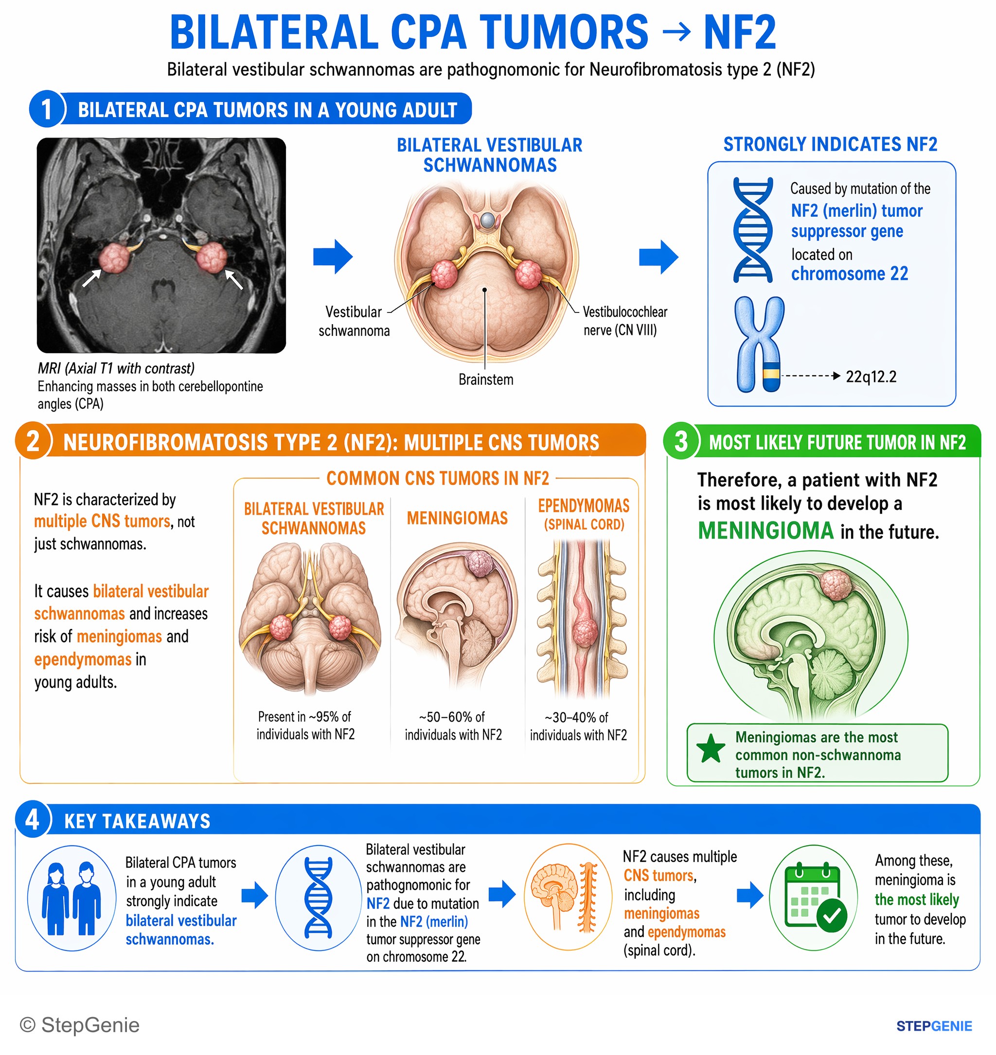
Neurofibromatosis type 2 causes bilateral vestibular schwannomas and increases risk of meningiomas and ependymomas in young adults.
USMLE Step 1Frequently Asked Questions
How do you recognize a vestibular schwannoma in a USMLE question?
Look for months of one-sided sensorineural hearing loss with tinnitus and imbalance in an adult, often with speech understanding worse than the hearing thresholds suggest. The imaging clue is an enhancing mass at the cerebellopontine angle that enters and widens the internal auditory canal. Larger tumors add facial numbness, an impaired corneal reflex, gait unsteadiness or hydrocephalus.
What is the difference between a schwannoma and a neurofibroma?
A schwannoma is encapsulated, sits eccentrically on its nerve and is made almost entirely of Schwann cells that stain diffusely for S-100 and SOX10. A neurofibroma is unencapsulated and mixes Schwann cells with fibroblasts, neural cells and mast cells. Schwannomas go with NF2-related schwannomatosis; multiple or plexiform neurofibromas suggest NF1 and carry a risk of malignant peripheral nerve sheath tumor.
What are Antoni A, Antoni B and Verocay bodies?
They are the classic histologic patterns of schwannoma. Antoni A areas are dense and cellular, made of spindle cells whose nuclei can line up in palisades; Verocay bodies are palisading nuclei arranged around pink, eosinophilic zones within that tissue. Antoni B areas are loose, hypocellular and myxoid. The two patterns alternate, and together with S-100 and SOX10 staining they point to schwannoma.
Why was neurofibromatosis type 2 renamed NF2-related schwannomatosis?
A 2022 international consensus (Plotkin et al.) recommended the new name because it better reflects the updated diagnostic criteria, which combine clinical features with genetic testing. The disease itself is unchanged: autosomal dominant loss of the NF2 tumor suppressor on chromosome 22, which encodes merlin, causing bilateral vestibular schwannomas, meningiomas and ependymomas. Many references and exam questions still use the term NF2.
How do you tell a vestibular schwannoma from a meningioma at the cerebellopontine angle?
Both can enhance and both occur in NF2-related schwannomatosis, so use the imaging. A vestibular schwannoma typically fills and widens the internal auditory canal as it grows into the angle. A meningioma is dura-based with a broad attachment and a dural tail, may be calcified, and less often extends into the canal. Vestibular schwannoma is also the more common CPA tumor.