You’re cruising through a neuro q-bank, feeling good… and then a pediatric brain tumor question hits you with four answer choices that all sound vaguely familiar. This is exactly where Step-style test writers live: the “right” diagnosis is only half the battle—the real points come from knowing why the other options are wrong. Let’s break down a classic ependymoma vignette and turn every distractor into a memory anchor.
Tag: Neurology > CNS Tumors & Infections
The Clinical Vignette (Q-Bank Style)
A 4-year-old child is brought to the ED for progressive morning headaches and vomiting for several weeks. Parents report the child has become clumsier and occasionally “tilts” while walking. Exam shows truncal ataxia. MRI reveals a well-circumscribed mass in the fourth ventricle causing obstructive hydrocephalus. Biopsy shows perivascular pseudorosettes.
Question: What is the most likely diagnosis?
Correct Answer: Ependymoma
Why it’s ependymoma
This vignette stacks multiple high-yield clues:
- Child + posterior fossa symptoms (ataxia)
- Fourth ventricle mass → obstructive hydrocephalus
- Histology: perivascular pseudorosettes (tumor cells arranged around blood vessels with an anuclear zone)
High-yield facts you should know cold
- Cell of origin: ependymal cells lining ventricles/central canal
- Classic location:
- Kids: 4th ventricle (posterior fossa)
- Adults: more often spinal cord
- Key complication: noncommunicating (obstructive) hydrocephalus
- Histology buzzwords:
- Perivascular pseudorosettes
- True ependymal rosettes can occur (less commonly tested)
- Gross clue (often in explanations): can extend through the foramina of Luschka and Magendie
- Genetics associations (Step-relevant):
- Can be associated with NF2 (especially spinal ependymomas)
The Mechanism: Why Hydrocephalus Happens Here
A tumor in the 4th ventricle blocks CSF flow out of the ventricular system, leading to increased intracranial pressure.
- CSF pathway: Lateral ventricles → 3rd ventricle → cerebral aqueduct → 4th ventricle → foramina of Luschka/Magendie → subarachnoid space
- Ependymoma in 4th ventricle = “plug at the drain” → noncommunicating hydrocephalus
The Distractors (And Why They’re Tempting)
Below is how these answer choices typically show up—and how to kill them quickly on test day.
Quick Comparison Table
| Tumor | Typical Age | Common Location | Key Clues | Histology |
|---|---|---|---|---|
| Ependymoma | Children | 4th ventricle | Hydrocephalus, truncal ataxia | Perivascular pseudorosettes |
| Medulloblastoma | Children | Cerebellar vermis | Ataxia, can drop metastases | Small blue cells, Homer Wright rosettes |
| Pilocytic astrocytoma | Children/teens | Cerebellum | Cystic mass w/ mural nodule | Rosenthal fibers |
| Subependymal giant cell astrocytoma (SEGA) | Children/teens | Foramen of Monro | Hydrocephalus + TSC signs | Giant cells; TSC association |
Distractor #1: Medulloblastoma
Why they want you to pick it: It’s also a pediatric posterior fossa tumor with ataxia.
How to rule it out:
- Location matters: medulloblastoma classically arises in the cerebellar vermis, not the 4th ventricle itself (though it can compress it).
- Spread: notorious for CSF “drop metastases” → spinal seeding.
- Histology: Homer Wright rosettes (neuroblastic tumor), not perivascular pseudorosettes.
- Extra high-yield association: c-MYC amplification (classically linked in many resources) and is a primitive neuroectodermal tumor (PNET)-type malignancy.
Exam-day takeaway:
If the stem gives you 4th ventricle + perivascular pseudorosettes, don’t let “child + ataxia” pull you into medulloblastoma.
Distractor #2: Pilocytic Astrocytoma
Why they want you to pick it: It’s the most common benign pediatric brain tumor, often in the cerebellum.
How to rule it out:
- Imaging clue: classic is a cystic lesion with a mural nodule in the cerebellar hemisphere.
- Histology: Rosenthal fibers (thick, corkscrew eosinophilic fibers) and “piloid” astrocytes.
- Hydrocephalus: can happen, but it’s not the signature mechanism like a 4th ventricle ependymoma.
Exam-day takeaway:
If you see cyst + mural nodule + Rosenthal fibers, think pilocytic astrocytoma. If you see 4th ventricle obstruction + pseudorosettes, think ependymoma.
Distractor #3: Subependymal Giant Cell Astrocytoma (SEGA)
Why they want you to pick it: It’s near ventricles and causes hydrocephalus—sounds close.
How to rule it out:
- Location: SEGA arises near the foramen of Monro (lateral ventricle region), not the 4th ventricle.
- Syndrome clue: strongly associated with tuberous sclerosis complex (TSC).
- Look for: seizures, developmental delay, facial angiofibromas, ash leaf spots, shagreen patches.
- Stem vibe: SEGA questions usually include obvious TSC features or mention “foramen of Monro.”
Exam-day takeaway:
Hydrocephalus + ventricular tumor is not enough. Which ventricle (Monro vs 4th) is the discriminator.
Distractor #4 (Common in Real Q-Banks): Choroid Plexus Papilloma
Even if not listed in your specific question, this is a frequent hydrocephalus distractor worth having in your mental “ventricle tumor” set.
Why it’s tempting: Also involves ventricles and can cause hydrocephalus.
How to rule it out:
- Mechanism differs: causes hydrocephalus by increasing CSF production, not by obstruction.
- Locations:
- Children: often lateral ventricles
- Adults: more often 4th ventricle
- Histology: papillary fronds (epithelial) rather than rosettes.
Exam-day takeaway:
Hydrocephalus from overproduction → think choroid plexus papilloma. Hydrocephalus from blockage at 4th ventricle + pseudorosettes → ependymoma.
High-Yield “One-Liners” for Test Day
- Ependymoma: Kids, 4th ventricle, obstructive hydrocephalus, perivascular pseudorosettes.
- Medulloblastoma: Child, cerebellar vermis, highly malignant, CSF drop metastases, Homer Wright rosettes.
- Pilocytic astrocytoma: Benign cerebellar tumor, cyst + mural nodule, Rosenthal fibers.
- SEGA: TSC + foramen of Monro mass causing hydrocephalus.
How to Answer Faster Next Time (Pattern Recognition)
When you see a pediatric posterior fossa mass, force yourself to answer these two questions before picking:
-
Where exactly is the lesion?
- 4th ventricle → ependymoma
- Vermis → medulloblastoma
- Cerebellar hemisphere/cystic mural nodule → pilocytic astrocytoma
- Foramen of Monro → SEGA
-
What is the histologic “tell”?
- Perivascular pseudorosettes → ependymoma
- Homer Wright rosettes → medulloblastoma
- Rosenthal fibers → pilocytic astrocytoma
That’s how you turn a “gotcha” vignette into free points.
Visual explanations from the StepGenie QBank
Infographics StepGenie drew to explain questions in its own QBank, matched to this article's topic. Each caption is the takeaway of the question it explains.
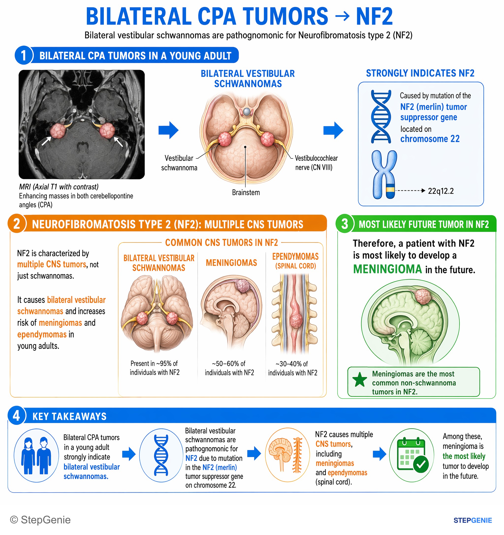
Neurofibromatosis type 2 causes bilateral vestibular schwannomas and increases risk of meningiomas and ependymomas in young adults.
USMLE Step 1Frequently Asked Questions
How do you recognize ependymoma in a pediatric brain tumor vignette?
Ependymoma is suggested by a child with posterior fossa symptoms, a fourth ventricle mass, obstructive hydrocephalus, and perivascular pseudorosettes on biopsy. The classic vignette includes morning headaches, vomiting, and truncal ataxia from a well-circumscribed fourth ventricle tumor. The histology clue that seals the diagnosis is tumor cells arranged around blood vessels with an anuclear zone, which are perivascular pseudorosettes.
Why does a fourth ventricle ependymoma cause noncommunicating hydrocephalus?
A fourth ventricle ependymoma causes noncommunicating hydrocephalus by blocking cerebrospinal fluid outflow. CSF normally travels from the lateral ventricles to the third ventricle, through the cerebral aqueduct, into the fourth ventricle, and then exits through the foramina of Luschka and Magendie. When a tumor sits in the fourth ventricle, it acts like a plug at the drain, increasing intracranial pressure and producing obstructive hydrocephalus.
How do you tell ependymoma from medulloblastoma on Step-style questions?
The best way to distinguish them is by location and histology. Ependymoma classically arises in the fourth ventricle and shows perivascular pseudorosettes, whereas medulloblastoma classically arises in the cerebellar vermis and shows small blue cells with Homer Wright rosettes. Both can occur in children and cause ataxia, so if a stem gives fourth ventricle obstruction plus pseudorosettes, ependymoma is the better answer.
How do you distinguish ependymoma from pilocytic astrocytoma?
Ependymoma is favored when the stem describes fourth ventricle obstruction with perivascular pseudorosettes. Pilocytic astrocytoma instead is classically a cerebellar cystic lesion with a mural nodule and histology showing Rosenthal fibers and piloid astrocytes. Although both can occur in children and involve the cerebellar region, the combination of a ventricular mass causing obstructive hydrocephalus points more strongly to ependymoma.
How do you tell ependymoma from SEGA or choroid plexus papilloma?
The discriminator is the exact ventricular location and the mechanism of hydrocephalus. Ependymoma in children is classically a fourth ventricle tumor causing obstructive hydrocephalus and showing perivascular pseudorosettes. Subependymal giant cell astrocytoma arises near the foramen of Monro and usually comes with tuberous sclerosis clues such as facial angiofibromas or ash leaf spots. Choroid plexus papilloma causes hydrocephalus by increasing CSF production and shows papillary fronds rather than rosettes.