Radiculopathy is compression or irritation of a spinal nerve root, most often by a herniated disc, causing pain that radiates along a dermatome plus deficits in that root's territory: dermatomal sensory loss and lower motor neuron signs (myotomal weakness, a reduced reflex). The anchors are C6 = thumb and biceps/brachioradialis reflex, C7 = middle finger and triceps, C8 = little finger and grip; L4 = medial ankle, knee extension and patellar reflex, L5 = dorsum of the foot, big-toe extension and foot drop with no routine reflex, S1 = lateral foot, plantarflexion and ankle jerk. A posterolateral disc usually hits the root named for the lower vertebra of the pair (C5-C6 → C6, L4-L5 → L5). C7 is the most commonly affected cervical root, and about 95% of lumbar herniations are at L4-L5 or L5-S1. Most cases settle with conservative care; red flags or a progressive deficit call for MRI.
The acronym: ROOTS
Run this checklist before you name a level.
| Letter | Stands for | What to remember |
|---|---|---|
| R | Radiating, dermatomal pain | Neck or back pain shooting into one limb along one dermatome, with paresthesias in the same strip |
| O | One level | Sensory loss, weakness and reflex change all fit a single root (tables below) |
| O | Only lower motor neuron signs | Weakness, atrophy, fasciculations, reduced reflex. Hyperreflexia or a Babinski sign means the cord (myelopathy), not a root |
| T | Tests that load the root | Spurling test for the neck; straight-leg raise and crossed straight-leg raise for the leg |
| S | Start conservative, scan for red flags | Most improve within weeks; MRI for red flags, a progressive deficit, or symptoms persisting beyond about 6 weeks when surgery or another intervention is being considered |
Cervical roots: count your fingers
Make a "six-shooter" with your thumb and index finger: C6 runs to the thumb. Point with the middle finger: C7. The little finger, last in line: C8.
| Root | Disc level | Pain and sensory loss | Weakness | Reflex |
|---|---|---|---|---|
| C5 | C4-C5 | Neck and shoulder | Deltoid, biceps, rotator cuff | Biceps |
| C6 | C5-C6 | Trapezius ridge and shoulder tip, radiating to the thumb | Biceps, wrist extensors | Biceps, brachioradialis |
| C7 | C6-C7 | Shoulder blade and axilla, radiating to the middle finger | Triceps | Triceps |
| C8 | C7-T1 | Little finger | Finger flexors (grip) | No routine reflex |
- Numbering rule: cervical roots exit above the vertebra of the same number, and C8 exits between C7 and T1. That is why a C5-C6 disc compresses C6 and a C6-C7 disc compresses C7.
- Most common: C7 accounts for more than half of cervical radiculopathies and C6 for roughly a quarter.
- Spurling test: axial compression with the neck extended and rotated toward the painful side reproduces the arm pain. It is highly specific but only moderately sensitive.
- Natural history: more than 85% of acute cervical radiculopathy resolves within 8-12 weeks without specific treatment.
- Myelopathy warning: hand clumsiness, gait instability, urinary urgency, heavy legs, hyperreflexia or a Babinski sign means cord compression, which is localized with the spinal cord tracts, not the root tables.
Lumbar roots: 4-5-1 = knee, toe, sole
L4 kneels (knee extension, patellar reflex, medial ankle). L5 lifts the big toe (dorsum of the foot, foot drop, no routine reflex). S1 stands on its sole (plantarflexion, lateral foot, ankle jerk).
| Root | Usual disc (posterolateral) | Pain and sensory loss | Weakness | Reflex |
|---|---|---|---|---|
| L3 | L2-L3 | Anterior and medial thigh, knee | Quadriceps | Patellar |
| L4 | L3-L4 | Medial lower leg, medial malleolus | Quadriceps (knee extension), ankle dorsiflexion | Patellar |
| L5 | L4-L5 | Buttock, posterolateral thigh and calf, dorsum of the foot and big toe | Tibialis anterior and extensor hallucis longus (foot drop), tibialis posterior (inversion), peroneals (eversion) | No routine reflex (medial hamstring at most) |
| S1 | L5-S1 | Posterior leg and buttock, lateral foot and lateral malleolus | Gastrocnemius (plantarflexion) | Achilles (ankle jerk) |
- Numbering rule: lumbar roots exit below the vertebra of the same number. A posterolateral (paracentral) disc catches the traversing root on its way to the foramen below, so L4-L5 → L5 and L5-S1 → S1.
- Far-lateral exception: an extreme lateral herniation compresses the exiting root instead, so a far-lateral L4-L5 disc causes an L4 radiculopathy.
- Where and who: about 95% of lumbar herniations occur at L4-L5 or L5-S1, so most lumbar radiculopathies are L5 or S1. Disc herniation peaks between ages 30 and 50 and is about twice as common in men.
- Straight-leg raise: lifting the straight leg of a supine patient sends pain down the posterior thigh to below the knee, pointing to the lower lumbar roots. The crossed straight-leg raise (lifting the good leg reproduces pain in the bad one) is less sensitive but about 90% specific for a herniated disc.
- Central herniation can cause bilateral leg pain and, if large, cauda equina compression.
The reflex rhyme
1, 2 buckle my shoe (S1: Achilles) · 3, 4 kick the door (L3-L4: patellar) · 5, 6 pick up sticks (C5-C6: biceps; C6 also brachioradialis) · 7, 8 lay them straight (triceps, mainly C7). L5 and C8 have no routine reflex of their own, so the standard reflex exam is usually normal with those roots (L5 can reduce the medial hamstring reflex).
Look-alikes the exam uses
| Look-alike | How it differs |
|---|---|
| Common peroneal (fibular) neuropathy vs L5 | Both cause foot drop. Peroneal palsy (compression at the fibular neck; leg crossing, weight loss) weakens dorsiflexion and eversion and numbs the anterolateral leg and dorsum of the foot. Weak inversion, radiating back or buttock pain, or a positive straight-leg raise points to L5 |
| Carpal tunnel syndrome vs C6 | Numbness of the palmar first 3 fingers, worse at night, with weak thumb abduction and opposition; no neck pain and no biceps or brachioradialis reflex change (carpal tunnel visual hack) |
| Ulnar neuropathy vs C8 | Numbness of the little finger and the medial half of the ring finger, weak interossei, thumb adductor and little-finger abductor, claw hand when severe; no neck pain. Electrodiagnostic testing separates nerve from root |
| Cauda equina syndrome | Urinary retention or incontinence, constipation or fecal incontinence, perianal (saddle) sensory loss, erectile dysfunction and loss of rectal tone and sphincter reflexes: urgent MRI and decompression (cauda equina flowchart) |
| Radiculopathy without a disc | Osteoarthritic or rheumatoid bone changes, tumors, meningiomas, abscesses, herpes zoster, Lyme disease, tuberculosis, and diabetes-related nerve root ischemia (diabetic nerve damage overall is in the diabetic complications question) |
Workup and treatment
- No imaging in the first 4-6 weeks if there are no red flags.
- Red flags: fever, unexplained weight loss, night sweats, cancer, IV drug use, immunosuppression, prolonged steroid use, thoracic pain, bowel or bladder dysfunction, and a progressive or severe neurologic deficit.
- MRI is the test of choice, usually without contrast: as soon as possible for a deficit suggesting root or cord compression, and for symptoms persisting after 6 weeks of optimal care in a patient who is a candidate for surgery or another intervention. For suspected cauda equina syndrome, cancer, infection or immunosuppression, ACR rates MRI with and without IV contrast as usually appropriate too (ACR 2021). Electrodiagnostic testing localizes the affected root but cannot identify the cause.
- Conservative care: acetaminophen or NSAIDs, light activity as tolerated and strengthening exercises; prolonged bed rest and traction are not indicated. Systemic or epidural corticosteroids give modest, temporary relief at best. Options for chronic radicular pain include gabapentinoids, tricyclic antidepressants, SNRIs and topical lidocaine or capsaicin.
- Surgery is for progressive weakness, sphincter dysfunction, pain that stays intractable despite medication, or cord or cauda equina compression.
- Course: about half of lumbosacral radiculopathies resolve within 1-2 weeks and 90% within 6-12 weeks.
Practice question
A 42-year-old man has had 3 weeks of low back pain that shoots down the back of his left thigh and the outside of his calf to the top of his foot; it began after he lifted a heavy box. He has no bladder or bowel symptoms. He has weak left big-toe extension, ankle dorsiflexion and ankle inversion, and decreased pinprick sensation over the dorsum of the left foot and big toe. Patellar and ankle reflexes are normal and symmetric. Raising his straight left leg reproduces the pain below the knee. Which is the most likely cause?
A. Compression of the common fibular (peroneal) nerve at the fibular neck B. L3-L4 posterolateral disc herniation C. L4-L5 posterolateral (paracentral) disc herniation D. L4-L5 far-lateral disc herniation E. L5-S1 posterolateral disc herniation
Answer: C. This is an L5 radiculopathy: dorsum-of-foot and big-toe numbness, weak extensor hallucis longus and ankle dorsiflexion, weak inversion (tibialis posterior), normal patellar and ankle reflexes (L5 has no routine reflex) and a positive straight-leg raise. A paracentral L4-L5 disc compresses the traversing L5 root.
- A: peroneal palsy weakens dorsiflexion and eversion, but it does not explain weak inversion, radiating back pain or a positive straight-leg raise.
- B: an L3-L4 disc compresses L4: medial leg and ankle numbness, quadriceps weakness and a reduced patellar reflex, which is normal here.
- D: a far-lateral L4-L5 disc compresses the exiting L4 root, giving the same L4 picture as B.
- E: an L5-S1 disc compresses S1: lateral foot numbness, plantarflexion weakness and a reduced ankle jerk, which is normal here.
Key takeaways
- ROOTS: radiating dermatomal pain, one level, only lower motor neuron signs, tests that load the root, start conservative and scan for red flags.
- Cervical: C6 thumb (biceps, brachioradialis), C7 middle finger (triceps), C8 little finger (grip, no routine reflex); C7 is the most common cervical root.
- Lumbar 4-5-1: L4 knee and patellar reflex; L5 big toe and foot drop, no routine reflex; S1 sole, plantarflexion and ankle jerk.
- Disc rules: cervical roots exit above their vertebra (C5-C6 → C6); a posterolateral lumbar disc hits the traversing root (L4-L5 → L5); a far-lateral disc hits the exiting root (L4-L5 → L4).
- Spurling is specific; the straight-leg raise is sensitive and the crossed straight-leg raise specific.
- Upper motor neuron signs mean myelopathy; saddle numbness with bladder or bowel dysfunction means cauda equina syndrome and urgent MRI.
Sources
- Nerve Root Disorders — Merck Manual Professional Edition (2026)
- Cervical Radiculopathy — StatPearls, NCBI Bookshelf (2025)
- Lumbosacral Radiculopathy — StatPearls, NCBI Bookshelf (2024)
- Lumbar Disc Herniation — StatPearls, NCBI Bookshelf (2023)
- Nucleus Pulposus Herniation — StatPearls, NCBI Bookshelf (2025)
- Anatomy, Skin, Dermatomes — StatPearls, NCBI Bookshelf (2023)
- Lumbar Disc Herniation — Merck Manual Professional Edition (2024)
- Cervical Disc Herniation — Merck Manual Professional Edition (2024)
- Overview of Neck and Back Pain — Merck Manual Professional Edition (2024)
- Mononeuropathies — Merck Manual Professional Edition (2026)
- ACR Appropriateness Criteria: Low Back Pain — American College of Radiology (2021)
Visual explanations from the StepGenie QBank
Infographics StepGenie drew to explain questions in its own QBank, matched to this article's topic. Each caption is the takeaway of the question it explains.
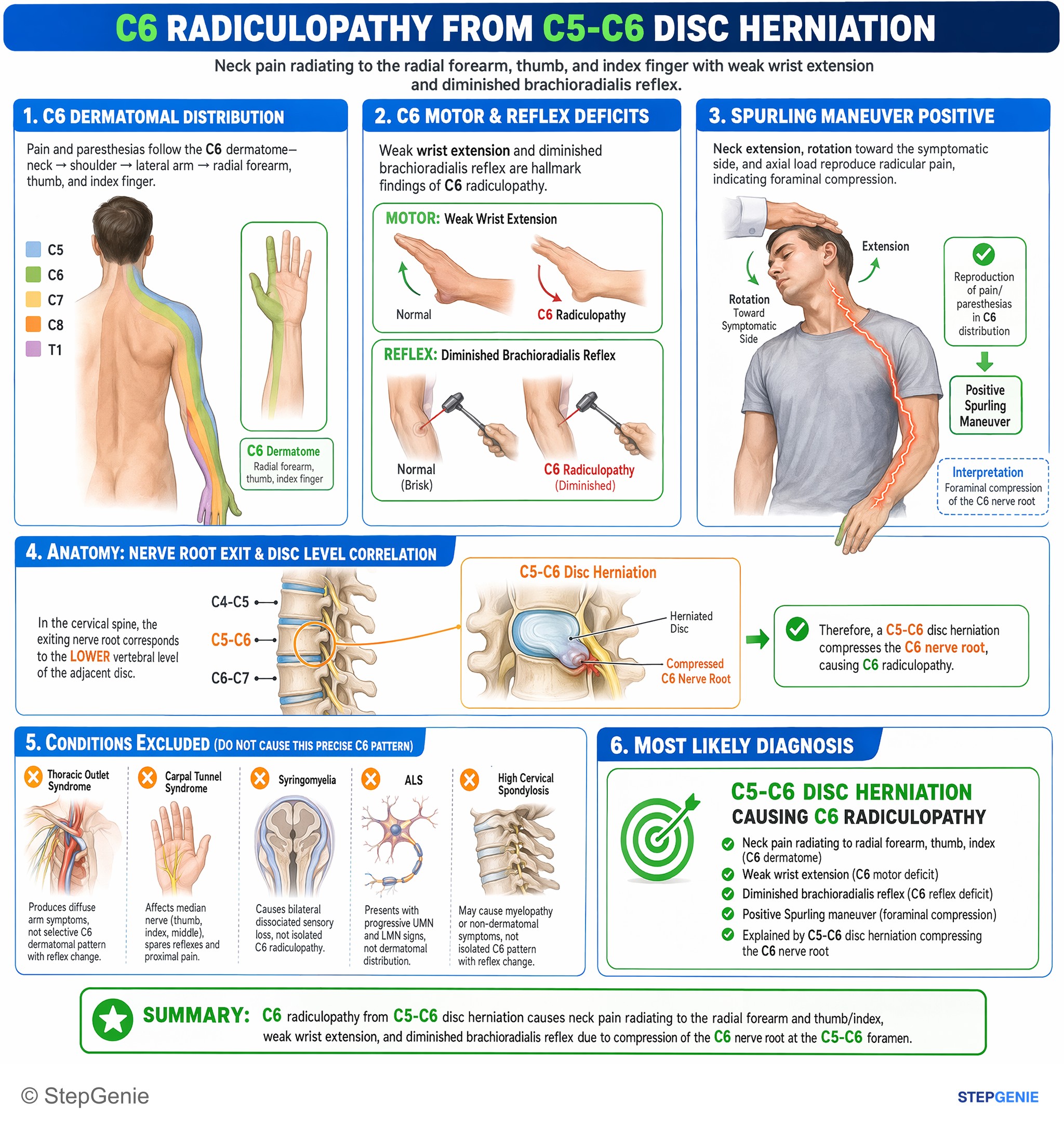
C6 radiculopathy from C5-C6 disc herniation causes neck pain radiating to radial forearm/thumb, weak wrist extension, and diminished…
USMLE Step 2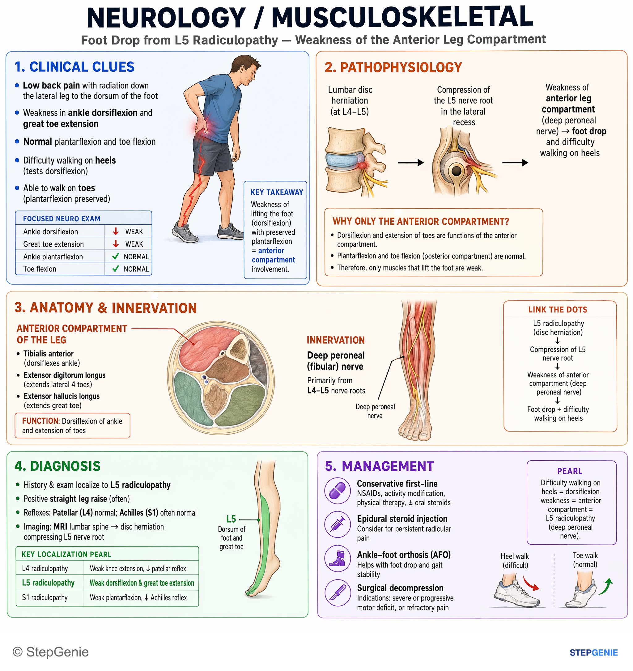
L5 radiculopathy from disc herniation → weakness of anterior leg compartment (deep peroneal nerve) → foot drop and difficulty walking on…
USMLE Step 1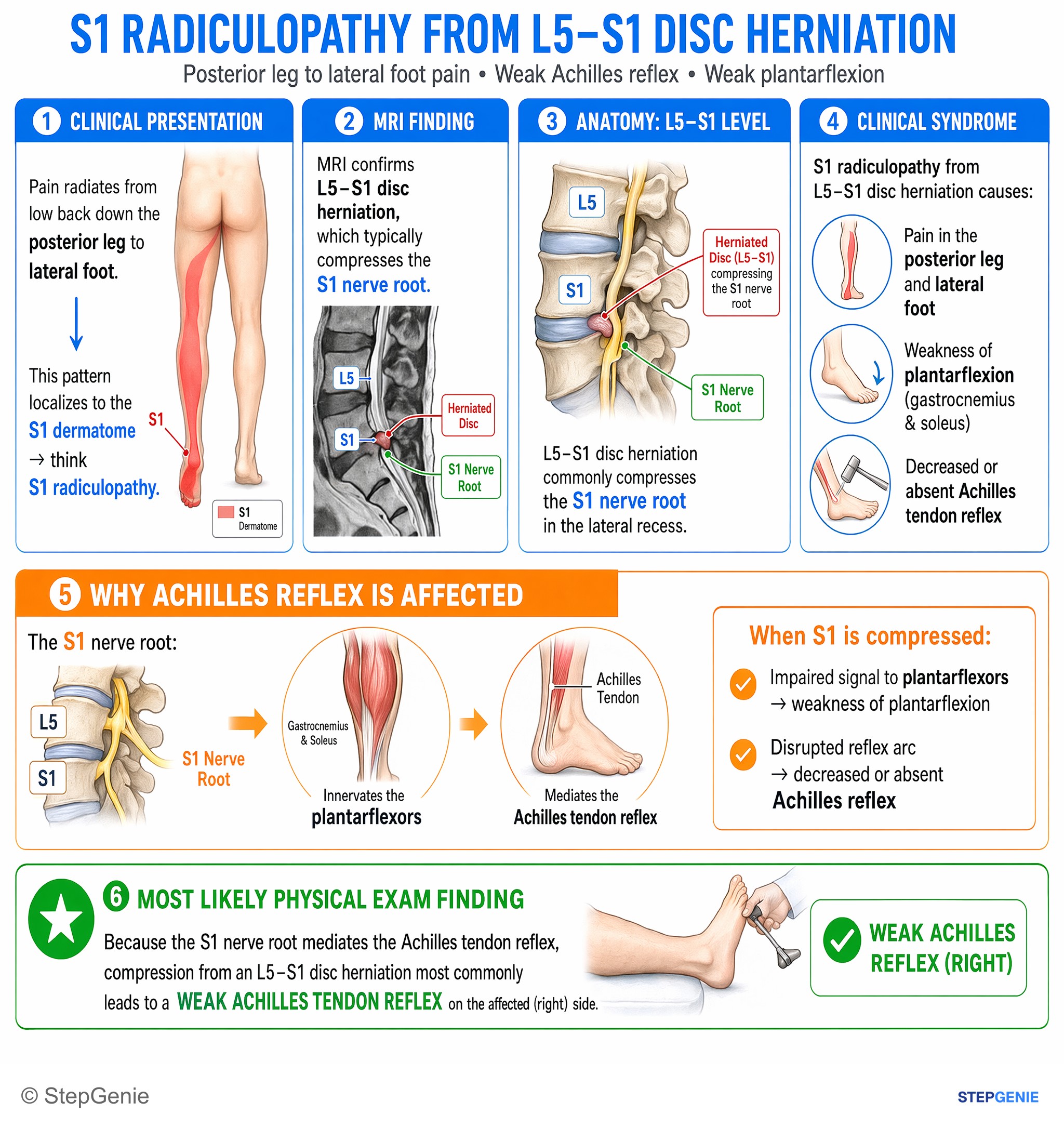
S1 radiculopathy from L5–S1 disc herniation causes posterior leg/lateral foot pain with weak Achilles reflex and impaired plantarflexion.
USMLE Step 1Frequently Asked Questions
How do you remember the cervical radiculopathy levels?
Count your fingers. Make a six-shooter with thumb and index finger: C6 runs to the thumb and affects the biceps and brachioradialis reflexes and wrist extension. Point with the middle finger for C7, which weakens the triceps and reduces the triceps reflex. The little finger is C8, which weakens grip and has no routine reflex. C5 affects the deltoid, biceps and rotator cuff, with the biceps reflex.
How do you tell L4, L5 and S1 radiculopathy apart?
Use 4-5-1 = knee, toe, sole. L4 numbs the medial lower leg and medial ankle, weakens knee extension and reduces the patellar reflex. L5 numbs the dorsum of the foot and big toe, weakens big-toe extension and ankle dorsiflexion (foot drop) and has no routine reflex. S1 numbs the lateral foot, weakens plantarflexion and reduces the ankle jerk.
Which nerve root does a herniated disc compress?
In the neck, roots exit above the vertebra of the same number and C8 exits between C7 and T1, so a C5-C6 disc compresses C6 and a C6-C7 disc compresses C7. In the lumbar spine, a posterolateral disc compresses the traversing root headed for the foramen below, so L4-L5 affects L5 and L5-S1 affects S1. A far-lateral herniation compresses the exiting root instead, so a far-lateral L4-L5 disc affects L4.
What is the most common cervical radiculopathy?
C7 is the most commonly affected cervical root, accounting for more than half of cases, and C6 accounts for roughly a quarter. C7 radiculopathy causes pain from the shoulder blade and axilla radiating to the middle finger, triceps weakness and a reduced triceps reflex. More than 85 percent of acute cervical radiculopathy resolves within 8 to 12 weeks without specific treatment.
How do you tell L5 radiculopathy from peroneal nerve palsy?
Both cause foot drop. Common peroneal palsy, usually from compression at the fibular neck in people who cross their legs or have lost weight, weakens ankle dorsiflexion and eversion and numbs the anterolateral leg and dorsum of the foot. L5 radiculopathy also weakens ankle inversion, usually comes with back or buttock pain radiating down the leg, and often gives a positive straight-leg raise. Electrodiagnostic testing can confirm the level.
When is MRI needed for radiculopathy?
Imaging is usually unnecessary in the first 4 to 6 weeks if there are no red flags, because most radiculopathy improves with conservative care. MRI, usually without contrast, is indicated promptly for a neurologic deficit suggesting root or cord compression or suspected cauda equina syndrome, and for symptoms persisting after about 6 weeks of optimal treatment in a patient being considered for surgery or another intervention. When cancer or infection is suspected, MRI with and without contrast is an equally appropriate choice.