Cauda equina syndrome (CES) is compression of two or more lumbar and sacral nerve roots below the conus medullaris, the tip of the spinal cord at about L1-L2; the most common cause is a large lumbar disc herniation. The picture is low back pain or sciatica (often bilateral) plus urinary retention, saddle anesthesia, bowel or sexual dysfunction and lower motor neuron leg weakness with reduced reflexes. It is a surgical emergency: emergency MRI of the lumbosacral spine (CT myelography if MRI is impossible), then urgent surgical decompression. Outcomes are best when surgery comes early, ideally before painless urinary retention sets in; AANS cites a significant advantage when patients are treated within 48 hours after onset.
The flowchart: from back pain to decompression
Step 1. Spot the red flags in the history. The UK national suspected CES pathway (NHS GIRFT, updated March 2026) asks for back or leg pain plus recent (14 days or less) onset or worsening of any of:
- Difficulty starting to urinate, or reduced sensation of urine flow
- Altered perianal, perineal or genital sensation (S2-S5 dermatomes)
- Severe or progressive weakness or numbness in both legs
- Loss of the feeling of rectal fullness
- Sexual dysfunction (new erectile or ejaculatory failure)
Also a warning sign: sciatica that starts in both legs at once, or spreads from one leg to both. Without any of the symptoms above, the UK pathway asks for urgent review within two weeks rather than an emergency referral.
Step 2. Examine the saddle, the legs and the bladder.
- Saddle sensation: pinprick and light touch around the anus, perineum and genitals
- Legs: weakness, hypotonia and reduced or absent ankle and knee reflexes are lower motor neuron signs
- Anal tone and bulbocavernosus reflex: both reduced or absent in CES (StatPearls). Merck and StatPearls still list rectal tone among the exam findings; the UK pathway now says a digital rectal exam is not necessary, but the patient's perianal sensation should be recorded.
- Bladder scan for post-void residual (PVR)
Step 3. Read the PVR. In the UK pathway, a PVR under 200 mL does not exclude CES; over 200 mL makes CES 20 times more likely; over 600 mL, catheterize and document whether bladder sensation is present.
Step 4. Get an emergency MRI. MRI of the lumbosacral spine is the test of choice. Merck: MRI immediately if available, CT myelography if it is not (StatPearls adds: or if MRI is contraindicated). StatPearls sets an ideal goal of one hour from presentation; the UK pathway requires MRI as soon as possible and within 4 hours of the request, around the clock. The UK pathway treats a sagittal T2 sequence as the single sequence needed to screen for compression; StatPearls describes sagittal and axial T1 and T2 images as the gold standard.
Step 5. Act in parallel; don't wait for the scan to call for help.
- Urgent spine surgery consult (neurosurgery or orthopedic spine)
- Drain the bladder if it is retaining
- Treat the cause while decompression is arranged: antibiotics for an abscess, reversal of anticoagulation for a hematoma, dexamethasone for a metastasis
Step 6. Decompress. Laminectomy with or without discectomy (StatPearls). StatPearls links surgery within 48 hours of the initial presentation with a better prognosis, and AANS reports a significant advantage when patients are treated within 48 hours after onset. The UK pathway operates on incomplete CES (CES-I) as quickly as possible and on CES with retention (CES-R) within 24 hours of the MRI.
The anatomy behind every sign
The spinal cord ends at the upper lumbar vertebrae, usually between L1 and L2, as the conus medullaris (Merck); below it, the lumbar and sacral roots run down the spinal canal as the cauda equina ("horse's tail"). Compressing roots is a lower motor neuron lesion, so expect flaccid weakness, hypotonia, atrophy and reduced or absent reflexes, not spasticity.
The sacral roots explain the rest:
- S2-S4 parasympathetic fibers drive detrusor contraction. Lose them and you get a flaccid bladder: large volume, low pressure, no contractions, so the patient retains urine and dribbles constantly (overflow incontinence) (Merck).
- S2-S4 somatic fibers (pudendal nerve) supply perineal sensation, the external sphincters and the bulbocavernosus reflex: saddle anesthesia, lax anal tone, fecal incontinence.
- S2-S4 also control erection, so new erectile dysfunction is a red flag.
How often each appears (StatPearls): back pain and sciatica in 97%, saddle anesthesia in up to 93%, bladder dysfunction in up to 92%, bowel dysfunction in up to 72%. Painless urinary retention often has the greatest predictive value, but it is a late sign that often means irreversible damage; perineal numbness with bladder dysfunction typically marks the start of CES, and that is when the clock starts. To pin down which root a unilateral sciatica involves, see the radiculopathy acronym trick.
Cauda equina vs conus medullaris
| Feature | Conus medullaris syndrome | Cauda equina syndrome |
|---|---|---|
| What is compressed | Terminal spinal cord (about T12-L2) | Lumbar and sacral nerve roots below the conus |
| Neuron type | Mixed UMN + LMN | Pure LMN |
| Onset | Sudden, bilateral | Gradual, unilateral |
| Pain | More low back pain; radicular pain less severe | Severe radicular pain (sciatica) |
| Sensory loss | Perianal, symmetric and bilateral | Saddle area; asymmetric, may be unilateral |
| Weakness | Symmetric, distal, less marked; may be hyperreflexic | Asymmetric, more marked, areflexic; atrophy |
| Reflexes | Knee jerks preserved, ankle jerks affected | Knee and ankle jerks both affected |
| Bladder and bowel | Early | Later in the course |
| Erectile dysfunction | Frequent | Less frequent |
This is the classic exam contrast (Medscape's comparison table; StatPearls). Real patients overlap: StatPearls notes that CES leg deficits are usually bilateral, though they can be unilateral and asymmetric, especially when the injury is incomplete, and conus lesions can show UMN signs such as spasticity and hyperreflexia. The workup and the urgency are the same for both: emergency MRI and decompression. The one-line rule: conus = early sphincter loss, symmetric, mixed UMN/LMN; cauda = painful, asymmetric, pure LMN. For other cord patterns, see the spinal cord tracts visual hack.
What compresses the cauda equina
| Cause | Board-style clue | Cause-specific step |
|---|---|---|
| Lumbar disc herniation (most common; about 45% of CES in StatPearls) | Lifting injury, acute back pain and sciatica, then bladder symptoms; most lumbar ruptures hit the L5 or S1 roots | Discectomy or laminectomy |
| Spinal epidural abscess | Back pain, fever and percussion tenderness, then deficits over hours to days; bacteremia, injection drug use, instrumentation; S. aureus in about 60% | MRI; blood cultures; antibiotics, plus surgical drainage if deficits; lumbar puncture is contraindicated |
| Spinal epidural hematoma | Anticoagulant or thrombolytic therapy, trauma, lumbar puncture with a bleeding diathesis; deficits over minutes to hours | Reverse the anticoagulant; immediate surgical drainage |
| Metastatic tumor | Known cancer, weight loss, night pain, percussion tenderness | Dexamethasone, then surgery or radiation |
| Spinal stenosis, trauma, diskitis | Chronic neurogenic claudication; fracture with bone retropulsion | Decompression; stabilize fractures |
The abscess and osteomyelitis overlap is covered in the osteomyelitis guide.
Why every hour counts
The longer the compression lasts, the worse the permanent damage (StatPearls). Incomplete injuries do better, and even after surgery many patients keep some bladder, bowel or sexual dysfunction or saddle numbness. The UK pathway splits patients by the bladder:
- CES-I (incomplete): urinary symptoms such as difficulty starting the stream or reduced urinary sensation, short of painless retention. This group has the better outlook, so it goes to surgery as an emergency.
- CES-R (retention): painless urinary retention with overflow incontinence. The prognosis is poorer, yet about 70% still benefit from decompression.
Untreated CES can leave permanent paralysis and incontinence (AANS).
How USMLE tests it
- "Next step" with red flags = emergency MRI of the lumbosacral spine, never physical therapy, NSAIDs and follow-up, or outpatient imaging.
- Overflow incontinence means retention. A high PVR in a patient with back pain is CES until proven otherwise; don't call it a UTI or BPH (compare the BPH and urinary retention case).
- Back pain without red flags gets conservative care first: analgesics and light activity, since a herniated disc tends to reabsorb over 6-9 months (Merck). Neurologic deficits flip that to urgent imaging.
- Fever + injection drug use + back pain = epidural abscess; anticoagulants, or a lumbar puncture in a patient with a bleeding disorder = hematoma; cancer history = metastasis.
- UMN signs below the waist point to the conus or higher in the cord (compare the Brown-Séquard syndrome guide). The differential for non-compressive cord disease includes transverse myelitis, spinal cord infarct, multiple sclerosis, syringomyelia and spinal AVM.
Practice question
A 42-year-old man has had severe low back pain since lifting a sofa 3 days ago. Since this morning the pain radiates down both legs, he has trouble starting his urine stream, and he says toilet paper "feels numb." He is afebrile. Pinprick sensation is reduced around the anus and perineum, ankle plantar flexion is weak bilaterally, and both ankle reflexes are absent. A bladder scan after voiding shows 650 mL. Which is the most appropriate next step?
A. NSAIDs, physical therapy and review in 2 weeks B. Plain radiographs of the lumbar spine C. Emergency MRI of the lumbosacral spine D. Nerve conduction studies and electromyography E. Lumbar puncture
Answer: C. Bilateral sciatica, saddle anesthesia, urinary retention (PVR over 600 mL) and LMN leg signs after a lifting injury add up to cauda equina syndrome from a disc herniation until proven otherwise. Get an emergency MRI, call spine surgery, and catheterize in parallel.
- A: conservative care is for sciatica without red flags; saddle numbness and retention make this an emergency.
- B: plain films cannot show a disc compressing nerve roots; they are only an add-on when a traumatic fracture is suspected.
- D: electrodiagnostic tests cannot show the compressing lesion and would only delay decompression.
- E: a lumbar puncture does not image the canal, and it is contraindicated when a spinal epidural abscess is possible.
Key takeaways
- CES = compression of the lumbosacral roots below the conus (L1-L2); a large lumbar disc herniation is the most common cause.
- Red flags: bilateral or progressing sciatica, saddle anesthesia, urinary retention or overflow, fecal incontinence, new sexual dysfunction.
- Painless urinary retention often has the greatest predictive value but is a late sign; check a PVR (in the UK pathway, over 200 mL makes CES 20 times more likely).
- Pure LMN signs: flaccid weakness, reduced reflexes, lax anal tone, absent bulbocavernosus reflex.
- Emergency MRI (CT myelography if MRI is impossible), then urgent decompression: outcomes are better within 48 hours, and the longer the compression lasts, the worse the permanent damage.
- Conus medullaris: early, symmetric sphincter loss, mixed UMN/LMN signs, less radicular pain.
- Fever = abscess (no LP); anticoagulants = hematoma; cancer = metastasis (dexamethasone).
Sources
- Cauda Equina Syndrome — Merck Manual Professional Edition (2025)
- Cauda Equina and Conus Medullaris Syndromes — StatPearls, NCBI Bookshelf (2023)
- National Suspected Cauda Equina Syndrome (CES) Pathway — NHS Getting It Right First Time (updated 2026)
- Cauda Equina Syndrome — American Association of Neurological Surgeons (2024)
- Cauda Equina and Conus Medullaris Syndromes — Medscape Reference (2016)
- Overview of Spinal Cord Disorders — Merck Manual Professional Edition (2025)
- Spinal Cord Compression — Merck Manual Professional Edition (2025)
- Spinal Epidural Abscess — Merck Manual Professional Edition (2025)
- Spinal Subdural or Epidural Hematoma — Merck Manual Professional Edition (2025)
- Lumbar Disc Herniation — Merck Manual Professional Edition (2024)
- Neurogenic Bladder — Merck Manual Professional Edition (2026)
Visual explanations from the StepGenie QBank
Infographics StepGenie drew to explain questions in its own QBank, matched to this article's topic. Each caption is the takeaway of the question it explains.
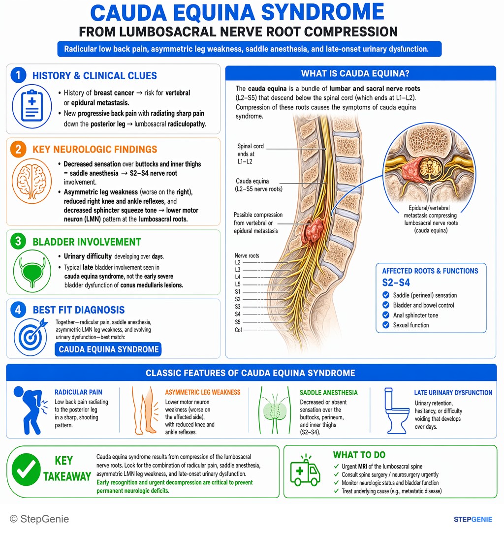
Cauda equina syndrome from lumbosacral nerve root compression: radicular low back pain, asymmetric leg weakness, saddle anesthesia, and…
USMLE Step 2Frequently Asked Questions
What are the red flags of cauda equina syndrome?
The red flags are back or leg pain with new difficulty starting to urinate or reduced sensation of urine flow, numbness around the anus, perineum or genitals (saddle anesthesia), loss of the feeling of rectal fullness or fecal incontinence, new erectile or ejaculatory failure, and severe or progressive weakness or numbness in both legs. Sciatica that starts in both legs at once, or spreads from one leg to both, is also a warning sign that needs urgent review even without the other symptoms.
What is the most predictive symptom of cauda equina syndrome?
Painless urinary retention often has the greatest predictive value, but it is a late sign that often means irreversible damage; perineal numbness together with bladder dysfunction typically marks the start of the syndrome. A bladder scan helps: in the UK national pathway, a post-void residual over 200 mL makes cauda equina syndrome about 20 times more likely, although a residual under 200 mL does not exclude it. Retention may show up as overflow incontinence.
What is the first test for suspected cauda equina syndrome?
Emergency MRI of the lumbosacral spine is the test of choice and should not wait for other tests. The UK national pathway asks for MRI within 4 hours of the request, and StatPearls sets an ideal goal of one hour from presentation. If MRI is unavailable or contraindicated, CT myelography is used. Plain radiographs cannot show nerve root compression and are added only when a fracture is suspected.
How quickly does cauda equina syndrome need surgery?
As soon as possible. Surgical decompression, usually laminectomy with or without discectomy, is linked to better results when done within 48 hours of onset or presentation (AANS, StatPearls). The UK national pathway operates on incomplete cauda equina syndrome as an emergency, as quickly as possible, and on patients with painless retention within 24 hours of the MRI. Longer compression means more permanent bladder, bowel and sexual dysfunction.
What is the difference between cauda equina syndrome and conus medullaris syndrome?
Conus medullaris syndrome damages the tip of the spinal cord at about T12 to L2, so it can mix upper and lower motor neuron signs; it classically starts suddenly and symmetrically, with early bladder and bowel loss, frequent erectile dysfunction and less radicular pain. Cauda equina syndrome compresses nerve roots, so it is purely lower motor neuron, with severe sciatica, asymmetric areflexic weakness and later sphincter loss. Both need emergency MRI.
What causes cauda equina syndrome?
A large lumbar disc herniation is the most common cause, accounting for about 45% of cases in StatPearls. Other causes include spinal epidural abscess (fever, injection drug use, bacteremia), spinal epidural hematoma (anticoagulants, trauma, or lumbar puncture with a bleeding disorder), metastatic tumors, severe spinal stenosis, trauma with fracture fragments pushed into the canal, and complications of spinal surgery.