Hereditary spherocytosis (HS) is an inherited defect in red cell membrane proteins (most often ankyrin; also spectrin, band 3 or protein 4.2) that makes red cells shed membrane, round up into spherocytes and get destroyed in the spleen. The Step 1 picture is anemia, jaundice, splenomegaly and pigment gallstones, often with a family history, plus spherocytes on the smear, a high MCHC, reticulocytosis and a negative direct antiglobulin (Coombs) test. The negative Coombs test is what separates HS from warm autoimmune hemolytic anemia, which also shows spherocytes. Confirm it with an eosin-5′-maleimide (EMA) binding test or an osmotic fragility test, give folic acid, and reserve splenectomy (after vaccination, ideally after age 6) for severe disease, deciding case by case in moderate disease.
Mechanism: why the red cell becomes a sphere
The red cell's lipid bilayer is anchored to a spectrin skeleton through ankyrin, band 3 and protein 4.2. When one of these links is defective, the membrane is unstable and bits of it are lost. The cell keeps its hemoglobin but loses surface area, so it can no longer stay a flexible biconcave disc:
Defective membrane protein → membrane loss → less surface area for the same contents → rigid spherocyte → trapped in the splenic cords → eaten by macrophages (extravascular hemolysis).
- Genes: ANK1 (ankyrin) accounts for about half of cases; the others are SPTB and SPTA1 (β- and α-spectrin), SLC4A1 (band 3) and EPB42 (protein 4.2).
- Inheritance: about 75% of cases are autosomal dominant. The rest are autosomal recessive or new (de novo) mutations, and variable penetrance can skip generations, so a negative family history does not exclude HS.
- Who gets it: about 1 in 2,000 people of Northern European ancestry; it is the most common inherited anemia in that population.
- Why the MCHC rises: the cell loses membrane but not hemoglobin, so hemoglobin is more concentrated and the MCHC is increased. The MCV is usually normal; the cell is narrower, not smaller in volume.
How it presents
Severity varies widely. MedlinePlus estimates that 20–30% of patients have mild disease, 60–70% moderate, about 10% moderate-to-severe and 3–5% severe.
- Mild: little or no anemia; HS may surface only through gallstones, family screening or an aplastic crisis.
- Moderate: anemia, jaundice and splenomegaly, usually appearing in childhood.
- Severe: transfusion-dependent anemia, sometimes with short stature, delayed sexual development and skeletal changes.
- Splenomegaly is almost invariable.
- Pigment gallstones are common and may be the presenting problem; about half of patients develop gallstones. Stone types are compared in the cholelithiasis mnemonic.
- Aplastic crisis: parvovirus B19 briefly shuts down red cell production. A patient who depends on a high reticulocyte count then has a sharp fall in hemoglobin with a low reticulocyte count. The virus is reviewed in the parvovirus B19 Q-bank breakdown.
Labs and smear
| Test | Result in HS | Why |
|---|---|---|
| Hemoglobin | Normal to low | Marrow compensation varies |
| Reticulocytes | High (low during an aplastic crisis) | Marrow response to hemolysis |
| MCV | Usually normal | The cell is narrower, not smaller in volume |
| MCHC | High | Same hemoglobin, less membrane |
| Indirect bilirubin and LDH | High | Red cell breakdown |
| Haptoglobin | Often low | Hemolysis |
| Smear | Spherocytes: small, round cells with no central pallor | Membrane loss |
| Direct antiglobulin test | Negative | No antibody on the cells |
| Osmotic fragility | Increased | Spherocytes lyse sooner in hypotonic saline |
| EMA binding (flow cytometry) | Reduced | Less fluorescent dye binds as membrane protein is lost |
| Genetic panel | Membrane-gene variant | Confirms the diagnosis; especially useful before splenectomy |
Hereditary spherocytosis vs look-alikes
| Hereditary spherocytosis | Warm autoimmune hemolytic anemia | G6PD deficiency | Hereditary elliptocytosis | |
|---|---|---|---|---|
| Cause | Membrane protein defect (ankyrin, spectrin, band 3, protein 4.2) | IgG autoantibodies; associated with SLE, CLL and methyldopa | X-linked enzyme deficiency; oxidant triggers | Defects in spectrin, protein 4.1 or glycophorin C |
| Smear | Spherocytes | Microspherocytes | Bite and blister cells; Heinz bodies on special stain | Oval or cigar-shaped elliptocytes |
| Direct antiglobulin test | Negative | Positive | Negative | Negative |
| Pattern | Chronic and lifelong; family history | Acquired, at any age | Episodes after drugs, infection or fava beans | Usually little or no hemolysis |
| Treatment | Folic acid; splenectomy if severe | Glucocorticoids first line; rituximab next | Avoid triggers; supportive care | Usually none |
Osmotic fragility is increased in both HS and warm autoimmune hemolysis, so it cannot separate them; the direct antiglobulin test does. The antibody side is covered in the autoimmune hemolytic anemia overview, and oxidant hemolysis in the G6PD deficiency guide.
Management
- Folic acid, because long-term hemolysis raises folate needs.
- Transfusion for severe anemia or an aplastic crisis; many aplastic crises resolve as the infection clears.
- Splenectomy is the only specific treatment. Merck lists symptomatic hemolysis or complications such as biliary colic or a persistent aplastic crisis as indications. The European Hematology Association red cell working group (Iolascon et al., 2017) matches the decision to severity:
- Severe HS (transfusion-dependent, or hemoglobin below 8 g/dL): splenectomy indicated, delayed if possible until after age 6.
- Moderate HS: individualized, based on spleen size and quality of life.
- Mild HS: not indicated.
- Why wait: overwhelming post-splenectomy infection is most likely in young children, so splenectomy is normally avoided before age 5. Partial splenectomy is used in some young children with severe disease, but the experts did not reach a consensus on it.
- Vaccinate first against Streptococcus pneumoniae, Neisseria meningitidis (MenACWY, plus MenB from age 10) and Haemophilus influenzae type b; CDC's schedules advise giving any needed Hib dose preferably at least 14 days before an elective splenectomy. The infection risk is highest in the first year but probably lifelong, and splenectomy also raises the long-term risk of thrombosis.
- Gallbladder: remove it during the same operation if symptomatic gallstones are present.
- After splenectomy: spherocytes remain and osmotic fragility stays high, but the cells survive longer, so anemia, jaundice and reticulocytosis improve. In one large series the median hemoglobin rose by about 3 g/dL.
Exam traps
- Spherocytes do not equal HS. Warm autoimmune hemolytic anemia also shows spherocytes and increased osmotic fragility; a positive direct antiglobulin test means immune hemolysis.
- No family history does not rule HS out: about a quarter of cases are de novo or recessive.
- A low reticulocyte count in a known HS patient means an aplastic crisis (think parvovirus B19), not recovery.
- A normal osmotic fragility test can occur in mild HS unless the blood is first incubated for 24 hours; EMA binding or genetic testing helps.
- Gallstones in a child or adolescent should make you think of chronic hemolysis.
- Splenectomy does not fix the membrane: spherocytes persist afterward.
Worked example
Vignette. A 7-year-old boy is brought in for fatigue and pallor a week after a mild febrile illness; his younger sister has just had a slapped-cheek rash. His father had his spleen removed as a teenager. He has scleral icterus and a palpable spleen. Hemoglobin is 5.9 g/dL (10.8 g/dL a year ago), the reticulocyte count is 0.3%, the MCHC is high and the direct antiglobulin test is negative. The smear shows many spherocytes. What explains the sudden drop in hemoglobin?
Answer: A parvovirus B19 aplastic crisis in hereditary spherocytosis. Transfuse as needed and give supportive care; the crisis usually settles as the infection clears. Afterward, confirm HS with EMA binding or genetic testing, start folic acid and discuss splenectomy, after vaccination, if his disease proves severe.
Why not the others?
- Warm autoimmune hemolytic anemia: the direct antiglobulin test would be positive, and the marrow would respond with a high reticulocyte count.
- G6PD deficiency: X-linked episodes after an oxidant, with bite and blister cells rather than spherocytes and a rising reticulocyte count.
- Iron deficiency anemia: microcytic red cells, no spherocytes and no signs of hemolysis such as jaundice or a high reticulocyte count.
Key takeaways
- HS = ankyrin, spectrin, band 3 or protein 4.2 defect → spherocytes → extravascular hemolysis in the spleen.
- Clues: anemia, jaundice, splenomegaly, pigment gallstones, family history, high MCHC and a negative Coombs test.
- Confirm with EMA binding or increased osmotic fragility; genetic testing helps before splenectomy.
- Parvovirus B19 causes an aplastic crisis with a low reticulocyte count.
- Treatment: folic acid, transfusion when needed, and splenectomy for severe disease (case by case in moderate disease), after vaccination and ideally after age 6; the spherocytes persist afterward.
Sources
- Hereditary Spherocytosis and Hereditary Elliptocytosis — Merck Manual Professional Edition
- Hereditary spherocytosis — MedlinePlus Genetics
- Iolascon A, et al. Recommendations regarding splenectomy in hereditary hemolytic anemias — Haematologica 2017
- Overview of Hemolytic Anemia — Merck Manual Professional Edition
- Autoimmune Hemolytic Anemia — Merck Manual Professional Edition
- Glucose-6-Phosphate Dehydrogenase (G6PD) Deficiency — Merck Manual Professional Edition
- Evaluation of Anemia — Merck Manual Professional Edition
- Adult Immunization Schedule Notes (asplenia and elective splenectomy) — CDC
- Child and Adolescent Immunization Schedule Notes (asplenia and elective splenectomy) — CDC
- Erythema Infectiosum (Parvovirus B19 Infection) — Merck Manual Professional Edition
Visual explanations from the StepGenie QBank
Infographics StepGenie drew to explain questions in its own QBank, matched to this article's topic. Each caption is the takeaway of the question it explains.
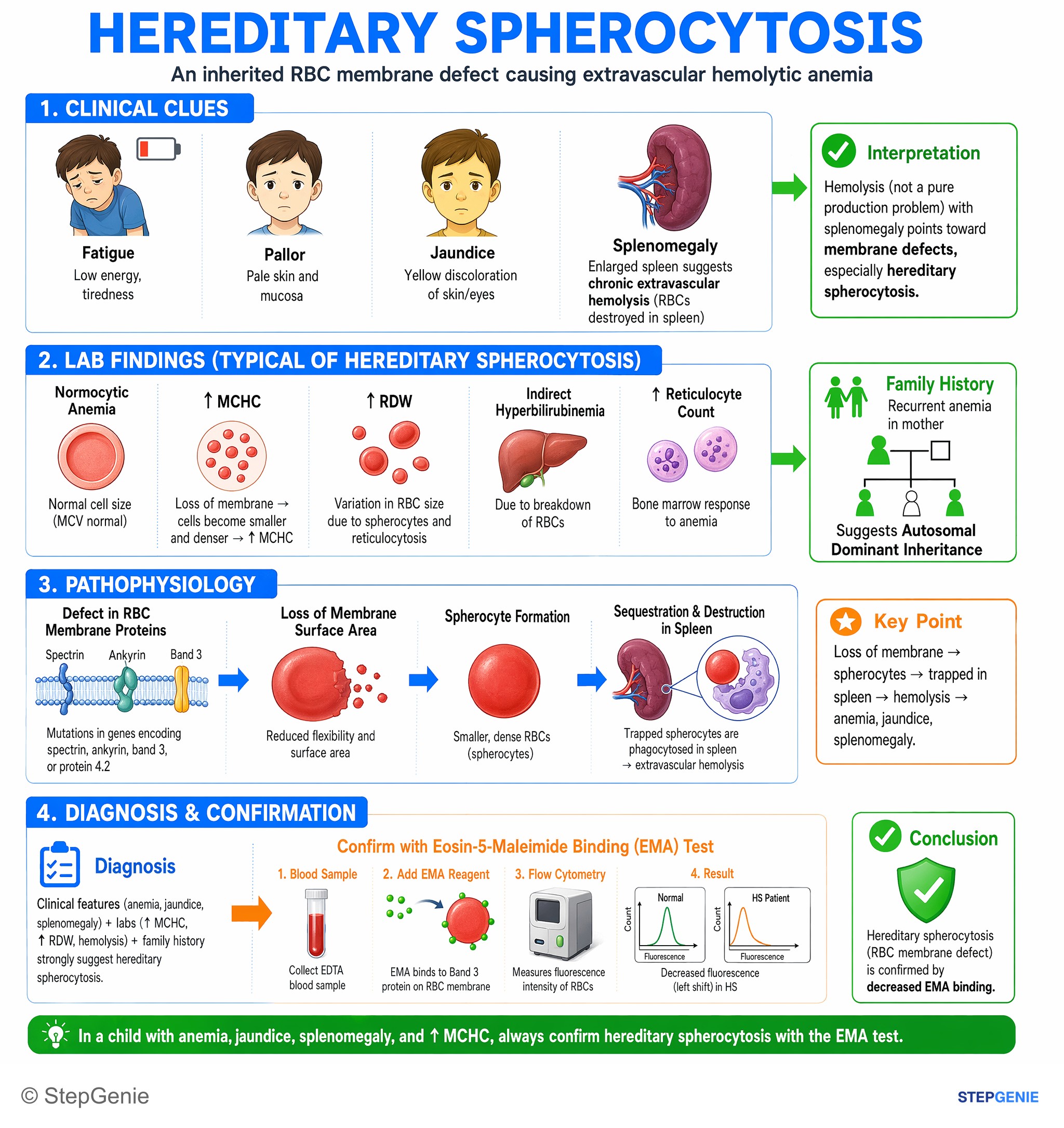
Hereditary spherocytosis in a child with anemia, jaundice, splenomegaly, ↑MCHC → confirm with eosin-5-maleimide binding (EMA) test for…
USMLE Step 1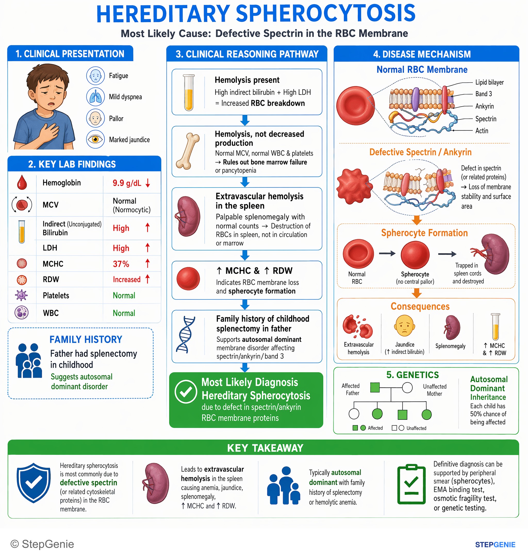
Hereditary spherocytosis due to spectrin/ankyrin defect → extravascular hemolysis, splenomegaly, ↑MCHC, jaundice; often autosomal dominant…
USMLE Step 1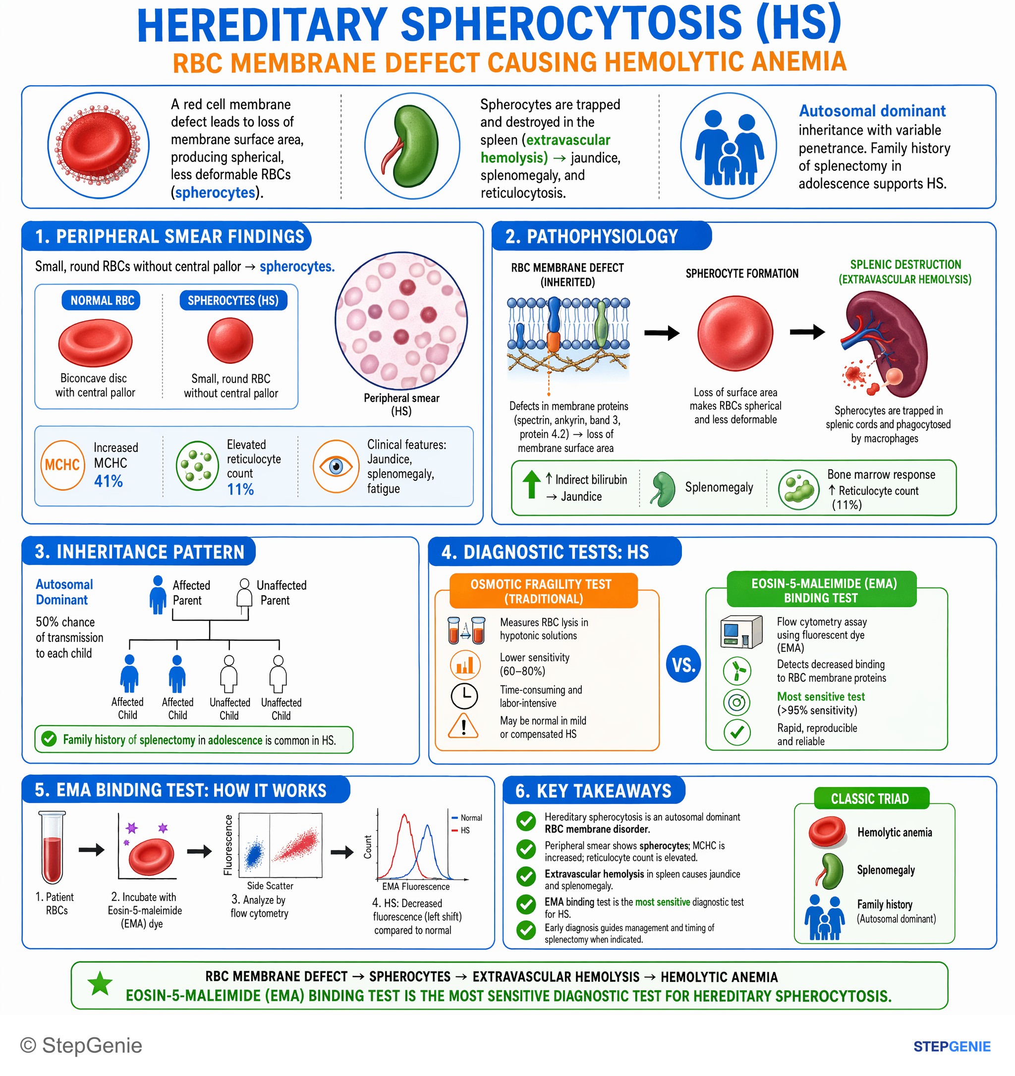
Hereditary spherocytosis from RBC membrane defect; causes hemolytic anemia with spherocytes. Eosin-5-maleimide binding test is most…
USMLE Step 1Frequently Asked Questions
Why is the MCHC high in hereditary spherocytosis?
The defective membrane skeleton sheds bits of membrane, so each red cell keeps its hemoglobin but loses surface area and rounds up into a small sphere. The hemoglobin is therefore more concentrated inside the cell, and the mean corpuscular hemoglobin concentration rises while the MCV usually stays normal. A high MCHC together with spherocytes and a negative direct antiglobulin test is a classic hereditary spherocytosis clue.
How do you tell hereditary spherocytosis from warm autoimmune hemolytic anemia?
Both show spherocytes, extravascular hemolysis, splenomegaly and increased osmotic fragility, so the smear alone cannot separate them. The direct antiglobulin (Coombs) test does: it is negative in hereditary spherocytosis and positive in warm autoimmune hemolytic anemia, where IgG autoantibodies coat the red cells. A lifelong course and a family history favor spherocytosis; lupus, CLL or methyldopa favor autoimmune hemolysis.
What test confirms hereditary spherocytosis?
Once hemolysis is established and the direct antiglobulin test is negative, a red cell membrane test confirms the diagnosis. Eosin-5′-maleimide (EMA) binding measured by flow cytometry is reduced, and the osmotic fragility test shows cells that lyse too easily in hypotonic saline, although it can be normal in mild cases. Genetic panels for membrane disorders are also used, especially when splenectomy is being considered.
When is splenectomy recommended for hereditary spherocytosis?
European expert recommendations advise splenectomy for severe, transfusion-dependent disease, a case-by-case decision in moderate disease, and no splenectomy in mild disease. When possible it is delayed until after age 6, because overwhelming infection is most likely in young children. Patients are vaccinated beforehand against pneumococcus, meningococcus and Hib, and symptomatic gallstones are removed during the same operation.
What does splenectomy change in hereditary spherocytosis?
Splenectomy removes the main site of red cell destruction, so the cells live longer, anemia and reticulocytosis improve and jaundice lessens. It does not repair the membrane defect: spherocytes remain on the smear and osmotic fragility stays high. The trade-offs are a lifelong risk of overwhelming infection with encapsulated bacteria and a higher long-term risk of thrombosis.