You’re doing a Q-bank block and you hit a classic: microcytic anemia. The stem screams iron deficiency anemia (IDA)… but the answer choices are designed to punish pattern recognition. The fastest way to get these questions right consistently is to understand why each distractor is wrong—and what clue would make it right.
Tag: Heme/Onc > RBC Disorders & Anemias
The Vignette (Typical Q-bank Style)
A 29-year-old woman presents with fatigue and decreased exercise tolerance for 3 months. She reports heavy menstrual periods and craving ice. Vitals are normal. Exam shows conjunctival pallor. Labs:
| Test | Value |
|---|---|
| Hemoglobin | 8.9 g/dL |
| MCV | 72 fL |
| RDW | Increased |
| Platelets | Increased |
| Ferritin | Low |
| Serum iron | Low |
| TIBC | High |
| Transferrin saturation | Low |
Peripheral smear: microcytosis and hypochromia.
Question: What is the most likely diagnosis / best next step?
The Correct Answer: Iron Deficiency Anemia (IDA)
Why this is IDA
This vignette has multiple “lock-in” clues:
- Microcytic anemia (low MCV)
- Increased RDW (iron availability fluctuates → RBC sizes vary)
- Pica (pagophagia = ice craving) is very classic
- Reactive thrombocytosis can occur in IDA
- Iron studies:
- Low ferritin (best single marker of low iron stores)
- Low serum iron
- High TIBC (liver makes more transferrin to scavenge iron)
- Low transferrin saturation
High-yield physiology
- Ferritin reflects iron stores. In pure IDA, ferritin is low.
- The liver responds to low iron by increasing transferrin production → TIBC goes up.
- RBCs become hypochromic because there’s less heme.
“Next best step” pearl (Step 2 flavor)
- In premenopausal women, heavy menses is a common cause, but you still want to think about:
- pregnancy, dietary deficiency, and occult GI blood loss depending on story.
- In men and postmenopausal women, IDA is colon cancer until proven otherwise → colonoscopy/EGD workup.
How to Systematically Destroy the Distractors
Below is the key: microcytic anemia has a limited differential. Most distractors will mimic some aspect of IDA but will break on iron studies, smear, or clinical context.
Quick Comparator Table (Microcytic Anemias)
| Condition | Ferritin | Serum Iron | TIBC | RDW | Smear/Clues |
|---|---|---|---|---|---|
| Iron deficiency anemia | ↓ | ↓ | ↑ | ↑ | Hypochromic microcytes; pica; thrombocytosis |
| Anemia of chronic disease (inflammation) | ↑ | ↓ | ↓ | Normal/↓ | Hepcidin-mediated iron sequestration |
| Thalassemia trait | Normal/↑ | Normal/↑ | Normal | Normal | Target cells; very low MCV with mild anemia |
| Sideroblastic anemia | ↑ | ↑ | Normal/↓ | ↑ | Ring sideroblasts; basophilic stippling |
| Lead poisoning | Normal/↑ | Variable | Variable | Variable | Basophilic stippling + neuro/GI symptoms |
Distractor 1: Anemia of Chronic Disease (ACD)
Why it’s tempting
ACD is also often microcytic (or normocytic early) and has low serum iron.
Why it’s wrong here
- ACD typically has:
- High ferritin (acute-phase reactant + iron trapped in macrophages)
- Low TIBC (liver downregulates transferrin during inflammation)
High-yield mechanism
Inflammation → IL-6 increases hepcidin (from the liver) → hepcidin blocks ferroportin → decreased iron absorption and decreased iron release from macrophages.
Board clue for ACD: chronic infection, autoimmune disease (RA), cancer, CKD; labs show low iron + low TIBC + high ferritin.
Distractor 2: Thalassemia Trait (Alpha or Beta)
Why it’s tempting
Thalassemia trait causes dramatic microcytosis, and Q-banks love to test “microcytosis = not always iron deficiency.”
Why it’s wrong here
- Thalassemia trait usually has:
- Normal RDW (uniformly small RBCs)
- Often normal or increased RBC count despite low MCV
- Iron studies are typically normal
- The vignette’s low ferritin and high TIBC are not thalassemia.
High-yield smear and testing
- Target cells suggest thalassemia.
- Hemoglobin electrophoresis:
- Beta-thal trait: ↑HbA2 (and sometimes ↑HbF)
- Alpha-thal trait: often normal electrophoresis (diagnosis is clinical/genetic)
Board clue for thal trait: very low MCV (sometimes <70) with only mild anemia; Mediterranean/African/SE Asian ancestry; family history.
Distractor 3: Sideroblastic Anemia (Including B6 Deficiency)
Why it’s tempting
It’s microcytic and involves heme synthesis—so it feels like “iron problem.”
Why it’s wrong here
Sideroblastic anemia is a problem of incorporating iron into heme, not acquiring iron.
- Iron studies tend to show:
- High serum iron
- High ferritin
- TIBC normal or low
High-yield associations
- Alcohol use disorder
- Vitamin B6 (pyridoxine) deficiency (e.g., isoniazid without supplementation)
- Lead poisoning (overlaps conceptually; see below)
- Myelodysplastic syndrome
Signature finding
- Bone marrow: ring sideroblasts (iron-laden mitochondria around nucleus)
- Smear: may show basophilic stippling (not specific, but common in lead/sideroblastic)
Distractor 4: Lead Poisoning
Why it’s tempting
Lead can cause microcytic anemia and basophilic stippling—very “Step 1-ish.”
Why it’s wrong here
The vignette is dominated by iron deficiency clues (pica + low ferritin + high TIBC). Lead poisoning usually has a different symptom cluster:
- Abdominal pain, constipation
- Peripheral neuropathy (wrist/foot drop)
- Cognitive issues (kids)
- Risk factors: old paint, industrial exposure, contaminated water
High-yield mechanism
Lead inhibits:
- ALA dehydratase
- Ferrochelatase → impaired heme synthesis → anemia.
Board clue for lead: basophilic stippling + neuro/GI symptoms + exposure history; may see elevated free erythrocyte protoporphyrin.
Distractor 5: Hereditary Spherocytosis / Hemolytic Anemia (Often Included to Trick You)
Why it’s tempting
Fatigue + anemia can make people choose hemolysis reflexively.
Why it’s wrong here
Hemolytic anemia is usually normocytic with reticulocytosis and hemolysis labs:
- ↑LDH, ↑indirect bilirubin, ↓haptoglobin
- Smear: spherocytes/schistocytes depending on cause
IDA is about production failure due to low iron, not destruction.
Board clue for hemolysis: jaundice, dark urine, splenomegaly, elevated retics.
The “Ferritin Trap” (High-Yield Test-Taking Pearl)
Ferritin is an acute-phase reactant. So:
- Low ferritin is highly specific for iron deficiency.
- Normal or high ferritin does not rule out IDA if there’s inflammation.
If the stem includes chronic inflammatory disease and “normal ferritin,” check:
- transferrin saturation
- CRP/ESR context
- TIBC pattern
High-Yield Takeaways You’ll Actually Use on Test Day
When you see microcytosis, run this mental algorithm
- Check RDW
- ↑RDW → think iron deficiency (or mixed anemia)
- Normal RDW → think thalassemia trait
- Check ferritin
- ↓ ferritin → IDA
- ↑ ferritin → ACD or sideroblastic/iron overload states
- Check TIBC
- ↑ TIBC → IDA
- ↓ TIBC → ACD
- Look for clinical anchors
- Pica, koilonychia, heavy menses → IDA
- Chronic inflammation/CKD → ACD
- Ethnicity + target cells + normal iron → thal
- Alcohol/INH/MDS + high iron → sideroblastic
- GI/neuro + exposure + stippling → lead
Don’t miss the “cause” question (Step 2 classic)
- Men/postmenopausal women with IDA: evaluate for occult GI bleeding/malignancy
- Premenopausal women: consider menstrual blood loss, but don’t ignore GI symptoms/red flags
Mini Rapid-Fire: Common Q-bank Follow-Ups
- Why platelets high in IDA? Reactive thrombocytosis can occur due to increased marrow activity and cytokine signaling.
- What happens to reticulocytes in IDA? Often low/normal until iron is replaced (production is limited).
- What does iron therapy do first? Reticulocyte count rises in ~7–10 days; hemoglobin improves over weeks; ferritin repletes last.
Visual explanations from the StepGenie QBank
Infographics StepGenie drew to explain questions in its own QBank, matched to this article's topic. Each caption is the takeaway of the question it explains.
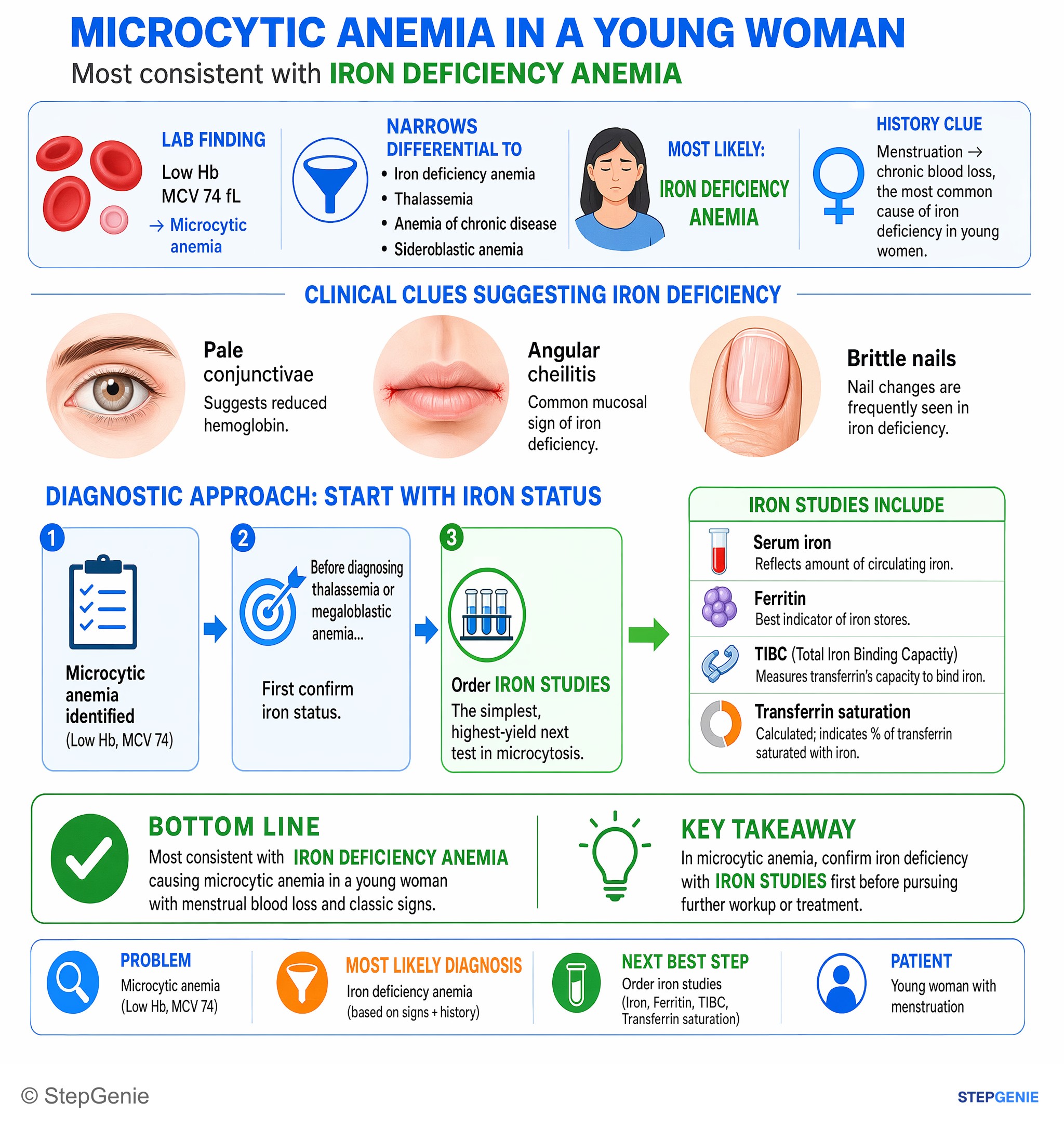
Iron deficiency anemia causing microcytic anemia in a young woman; first confirm with iron studies before further workup or treatment.
USMLE Step 2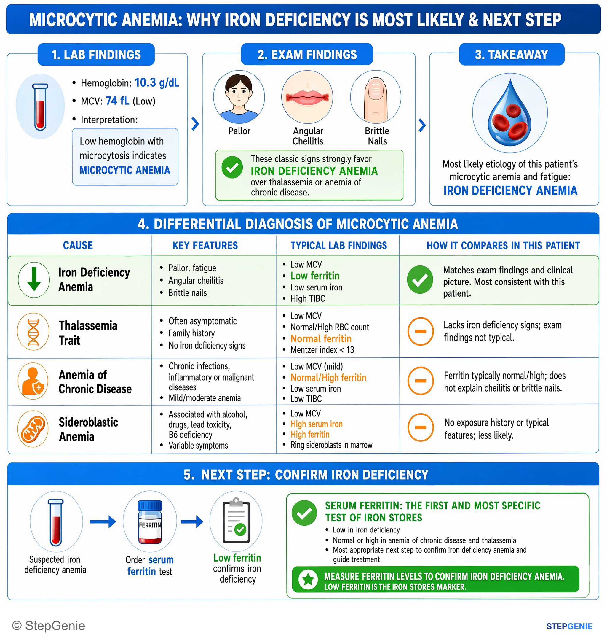
Iron deficiency anemia causing microcytic anemia and fatigue; first confirm with low ferritin as iron stores marker.
USMLE Step 2Frequently Asked Questions
How do you tell iron deficiency anemia from anemia of chronic disease on iron studies?
Iron deficiency anemia shows low ferritin, low serum iron, high TIBC, and low transferrin saturation, whereas anemia of chronic disease typically shows high ferritin, low serum iron, and low TIBC. The key difference is that ferritin is low in pure iron deficiency because iron stores are depleted, while chronic inflammation raises hepcidin and traps iron in macrophages, making ferritin high and transferrin production lower.
Why does iron deficiency anemia cause high TIBC?
Iron deficiency anemia causes high TIBC because the liver increases transferrin production when iron is low. This is a compensatory attempt to scavenge more circulating iron. In the same pattern, ferritin is low because iron stores are depleted, serum iron is low, and transferrin saturation is low, which together strongly support iron deficiency rather than another microcytic anemia.
What clues make microcytic anemia most likely to be iron deficiency anemia?
The strongest clues are low MCV, increased RDW, pica such as ice craving, reactive thrombocytosis, and iron studies showing low ferritin, low serum iron, high TIBC, and low transferrin saturation. A peripheral smear with microcytosis and hypochromia also fits. In this setting, low ferritin is especially useful because it is the best single marker of low iron stores in pure iron deficiency anemia.
How do you tell iron deficiency anemia from thalassemia trait in a question stem?
Iron deficiency anemia is suggested by increased RDW and abnormal iron studies, especially low ferritin and high TIBC, while thalassemia trait usually has normal RDW and typically normal iron studies. Thalassemia trait often shows target cells, a very low MCV with only mild anemia, and sometimes a normal or increased RBC count despite the microcytosis. Those findings help separate it from classic iron deficiency.
What is the next best step when a patient has iron deficiency anemia?
The next best step depends on the clinical context and who the patient is. In premenopausal women, heavy menstrual bleeding is a common cause, but pregnancy, dietary deficiency, and occult GI blood loss should still be considered depending on the story. In men and postmenopausal women, iron deficiency anemia should prompt evaluation for colon cancer, with colonoscopy and EGD workup.