Autosomal recessive (AR) inheritance means a person is affected only when both copies of an autosomal gene carry a pathogenic variant, one inherited from each parent; carriers with a single copy are usually healthy. When two carriers have children, every pregnancy has a 1/4 chance of an affected child, a 1/2 chance of a carrier and a 1/4 chance of a noncarrier. An unaffected sibling of an affected child is a carrier with probability 2/3: knowing the sibling is unaffected rules out the affected box, and two of the three remaining equally likely outcomes are carriers. On a pedigree, look for affected siblings of unaffected parents, males and females affected equally, and consanguinity.
Autosomal recessive inheritance at a glance
| Feature | What to expect |
|---|---|
| Affected genotype | Two pathogenic variants in the same gene: homozygous (aa) or compound heterozygous (two different variants) |
| Carriers (Aa) | Usually healthy, because one working copy typically makes enough protein |
| Unaffected parents of an affected child | Both are obligate carriers |
| Pedigree shape | Horizontal: affected siblings in one generation; the trait seems to skip generations |
| Sex ratio | Equal |
| Carrier × carrier | 1/4 affected, 1/2 carriers, 1/4 noncarriers, in every pregnancy |
| Unaffected sibling of an affected person | 2/3 chance of being a carrier |
| Affected parent | Noncarrier partner: all children carriers, none affected; carrier partner: 1/2 affected (pseudodominance) |
| What raises the risk | Consanguinity, founder populations, heterozygote advantage (sickle cell trait protects against malaria) |
The acronym: A RARE
Use A RARE to recognize an autosomal recessive pedigree and remember what the question will ask next.
| Letter | Clue | What it looks like in a question |
|---|---|---|
| A | Affected siblings, unaffected parents | A horizontal pedigree: several affected children in one generation, healthy parents, a trait that seems to skip generations |
| R | Related parents | Consanguinity raises the chance that both parents carry a variant inherited from a shared ancestor |
| A | All sexes equally | Sons and daughters affected in equal numbers, because the gene is on an autosome |
| R | Risk math | 1/4 affected and 1/2 carriers for two carrier parents; 2/3 carrier for an unaffected sibling |
| E | Enzyme deficiencies | Most inborn errors of metabolism are AR |
The first A is the recognition step: if two unaffected parents have an affected child, both are obligate carriers, and every probability below builds on that. The E has exceptions: ornithine transcarbamylase (OTC) deficiency, Hunter syndrome and Fabry disease are X-linked, and acute intermittent porphyria is autosomal dominant.
The 2/3 rule: why an unaffected sibling is a carrier
Draw the Punnett square for two carrier parents, where A is the normal allele and a the pathogenic one.
| Mother's A | Mother's a | |
|---|---|---|
| Father's A | AA: noncarrier | Aa: carrier |
| Father's a | Aa: carrier | aa: affected |
- Start with four equally likely outcomes: 1 AA, 2 Aa, 1 aa.
- Use what the stem tells you. The sibling is unaffected, so the aa box is impossible for this person. Cross it out.
- Count again. Three equally likely boxes remain (AA, Aa, Aa) and two are carriers: 2/3. The chance of being a noncarrier is 1/3.
- Check with conditional probability: P(carrier given unaffected) = P(carrier) ÷ P(unaffected) = (1/2) ÷ (3/4) = 2/3.
The tempting wrong answer is 1/2, the carrier risk of a child whose status is unknown. "Unaffected" shrinks the denominator from four boxes to three.
When 2/3 is the wrong number:
- Status unknown (a fetus, or an untested newborn): carrier 1/2, affected 1/4.
- Not a sibling. A parent's sibling (the affected child's aunt or uncle) has a 1/2 chance: the carrier parent got the allele from one grandparent, who passed it to each child with probability 1/2. A first cousin of the affected child has 1/4.
- New information. A carrier test result replaces 2/3; a sibling who has an affected child is an obligate carrier.
- Incomplete penetrance. "Unaffected" rules out aa only if aa would show. In HFE hemochromatosis many homozygotes never develop disease, so first-degree relatives are tested with ferritin and HFE genotyping instead.
Chaining probabilities in three steps
- Find each prospective parent's carrier probability: 1 for an obligate or test-proven carrier, 2/3 for an unaffected sibling of an affected person, 1/2 for an affected child's aunt or uncle, and the population carrier frequency for an unrelated partner. An affected parent always passes on a pathogenic allele.
- Multiply the two parents' probabilities, once each.
- Multiply by the transmission risk: 1/4 if both are carriers; 1/2 if one parent is affected and the other a carrier.
| Scenario | Risk per child | Arithmetic |
|---|---|---|
| Carrier × carrier | 1/4 affected | Aa × Aa |
| Carrier couple who already have an affected child: next child | Still 1/4 | Each pregnancy is independent |
| Unaffected sibling × known carrier | 1/6 | 2/3 × 1 × 1/4 |
| Two unaffected siblings of affected people, unrelated families | 1/9 | 2/3 × 2/3 × 1/4 |
| Unaffected sibling × partner with carrier frequency 1/25 | 1/150 | 2/3 × 1/25 × 1/4 |
| Unaffected sibling × affected partner | 1/3 | 2/3 × 1/2 |
| Unaffected sibling × noncarrier: child is a carrier | 1/3 (none affected) | 2/3 × 1/2 |
| Affected × noncarrier | None affected; all carriers | aa × AA |
| Affected × carrier | 1/2 affected | aa × Aa (pseudodominance) |
| Wilson disease: affected × unrelated partner (carrier frequency about 1/90) | 1/180 | 1/90 × 1/2 |
| Affected × affected, same gene | All affected | aa × aa |
Hardy-Weinberg: where the partner's carrier frequency comes from
In a large, randomly mating population without selection, mutation or migration, allele frequencies p (normal) and q (pathogenic), where p + q = 1, give stable genotype frequencies of p² (AA) + 2pq (Aa) + q² (aa) = 1. For an AR disease:
- q² = frequency of the disease, so q = √(disease frequency).
- Carrier frequency = 2pq ≈ 2q, because p is close to 1 when the allele is rare.
- Consanguinity violates random mating, so related couples need family-specific numbers (Problem 3).
Worked cystic fibrosis example. MedlinePlus Genetics puts CF at 1 in 2,500 to 3,500 White newborns in the United States. Using 1 in 2,500, q² = 1/2,500, so q = 1/50 and p = 49/50, and the carrier frequency is 2pq = 2 × (49/50) × (1/50) = 98/2,500, about 1 in 25 (the shortcut 2q = 2/50 agrees). Merck agrees: carriers are about 4% (1 in 27) of the White population, and its US incidence of 1 in 3,300 White births gives 2q ≈ 1 in 29.
| Published figure | Hardy-Weinberg step | Result |
|---|---|---|
| Tay-Sachs carriers about 1 in 30 in Ashkenazi Jewish people (GeneReviews) | 2q = 1/30, so q = 1/60 | Predicted incidence q² = 1/3,600 |
| HFE homozygotes about 1 in 200 in people of Northern European ancestry (Merck) | q = √(1/200) ≈ 0.071 | 2pq ≈ 0.13, about 1 in 8, matching Merck's heterozygote figure; 2q would give 1 in 7 |
| Carrier frequency 1 in 100 (ACOG's panel threshold) | q = 1/200 | Disease incidence 1/40,000, as ACOG states |
The HFE row is the warning: 2q is safe only for rare alleles; at q ≈ 0.07 it overestimates carriers by about 8%. More examples are in Hardy-Weinberg equilibrium for Step 1.
Reading the pedigree: AR vs AD, X-linked and mitochondrial
"Skips generations" is not unique to AR: X-linked recessive pedigrees skip too. The sex ratio, father-to-son transmission and the children of affected fathers settle most questions.
| Feature | Autosomal recessive | Autosomal dominant | X-linked recessive | X-linked dominant | Mitochondrial |
|---|---|---|---|---|---|
| Pedigree shape | Horizontal; skips generations | Vertical; usually every generation | Affected males linked through carrier females | Every generation; affected females outnumber males | Passed only through mothers; can appear in every generation |
| Sex ratio | Equal | Equal | Nearly all affected people are male | More females; males often more severe | Equal |
| Children of an affected father | None affected if the mother is a noncarrier (all carriers) | 1/2 affected | Sons unaffected; all daughters carriers | All daughters affected; no sons | None affected |
| Children of an affected or carrier mother | None affected if the father is a noncarrier | 1/2 affected | Carrier mother: 1/2 of sons affected, 1/2 of daughters carriers | Heterozygous mother: 1/2 of children of either sex | All at risk; severity varies with heteroplasmy |
| Father-to-son transmission | Only if the mother also carries the allele | Yes | Never | Never | Never |
| Examples | CF, sickle cell disease, PKU, Tay-Sachs | Huntington disease, Marfan syndrome, ADPKD | Hemophilia, red-green color blindness, Hunter syndrome, G6PD deficiency | X-linked hypophosphatemic rickets, Rett syndrome | LHON, MELAS, MERRF |
Male-to-male transmission rules out X-linked inheritance, because a father gives his son a Y chromosome. Transmission only through mothers, to sons and daughters alike, points to mitochondrial DNA, with severity varying by heteroplasmy (the mix of mutant and normal mitochondrial genomes); many mitochondrial disorders, though, come from nuclear genes and follow Mendelian rules. More in the X-linked disorders flowchart and autosomal dominant inheritance.
Twists that break the simple pattern
Consanguinity. Relatives are more likely to carry the same pathogenic variant from a shared ancestor. If one first cousin carries a rare allele, the other has a 1/8 chance of carrying it by descent: 1/2 that it came through the related parent × 1/2 that the aunt or uncle also inherited it × 1/2 that it was passed on. A known carrier and a first cousin face 1 × 1/8 × 1/4 = 1/32 per child (plus a small background risk), versus 1 × 1/25 × 1/4 = 1/100 with an unrelated partner whose carrier frequency is 1/25. ACOG and ACMG both define consanguinity as second cousins or closer.
Compound heterozygosity. Two different pathogenic variants in the same gene, one on each copy, cause disease just as two identical ones do: HbSC disease and HbS/β-thalassemia (both HBB; see this sickle cell disease question), β-thalassemia major from a β+ variant paired with a β0 variant, and HFE C282Y/H63D (clinically significant iron overload in only 0.5–2%). In risk math, a compound heterozygote counts as affected.
Pseudodominance. An affected person (aa) with a carrier partner (Aa) has affected children half the time, so the disease appears in consecutive generations and mimics dominant inheritance. Expect it when carriers are common (about 1 in 8 Northern Europeans are HFE heterozygotes, per Merck) or in consanguineous families.
Uniparental disomy (UPD). A child is homozygous for a variant that only one parent carries. After ruling out misattributed parentage, a deletion of the other copy and a new variant, think uniparental isodisomy: two identical copies of one chromosome (or chromosome segment) from the carrier parent and none from the other parent. ACMG notes that UPD of any chromosome raises the risk of a recessive disorder; the first Mendelian disorder linked to UPD, reported in 1988, was CF in a child who inherited two copies of a CFTR variant from a carrier mother and none from the father. ACMG recommends UPD testing in exactly this situation. More in uniparental disomy.
Locus heterogeneity. One phenotype, several genes. Oculocutaneous albinism can result from variants in several genes, including TYR, OCA2, TYRP1 and SLC45A2, all AR; hereditary hemochromatosis types 1–3 are AR (HFE; HJV or HAMP; TFR2), but type 4 (SLC40A1, ferroportin) is dominant. The exam twist: two affected parents can have only unaffected children when their variants lie in different genes, because each child is usually a carrier at both loci. See locus vs allelic heterogeneity.
Allelic heterogeneity. Many variants, one gene: CFTR has more than 2,000 known variants besides F508del, and different HBB variants produce HbS, HbC or β-thalassemia.
High-yield autosomal recessive diseases
| Disease | Gene | Hook |
|---|---|---|
| Cystic fibrosis | CFTR (chromosome 7) | Common in White populations: 1 in 2,500 to 3,500 White US newborns |
| Sickle cell disease | HBB | About 1 in 500 African Americans; HbSS, HbSC, HbS/β-thalassemia |
| β-thalassemia major | HBB | Two β0 variants, or a β+ variant with a β0 variant (compound heterozygous) |
| Phenylketonuria | PAH | Phenylalanine hydroxylase deficiency |
| Tay-Sachs disease | HEXA | Carriers about 1 in 30 Ashkenazi Jewish vs 1 in 250–300 in other groups; founder variants also in some French-Canadian and Cajun communities |
| Gaucher disease | GBA1 | Glucocerebrosidase deficiency |
| Hurler syndrome (MPS I) | IDUA | Its look-alike, Hunter syndrome (MPS II), is X-linked |
| Von Gierke disease (GSD Ia) | G6PC1 | Glucose-6-phosphatase deficiency |
| Classic galactosemia | GALT | Galactose-1-phosphate uridylyltransferase deficiency |
| Wilson disease | ATP7B | Carriers about 1 in 90 |
| HFE hemochromatosis | HFE | Common genotype, low penetrance |
| 21-hydroxylase deficiency | CYP21A2 | About 95% of congenital adrenal hyperplasia |
| Spinal muscular atrophy | SMN1 | Carriers about 1 in 45 (White) to 1 in 100 (Black), varying by ancestry |
| Friedreich ataxia | FXN | GAA trinucleotide repeat |
| ARPKD | PKHD1 | ADPKD (PKD1, PKD2) is dominant |
| Oculocutaneous albinism | TYR, OCA2, TYRP1, SLC45A2 and others | Locus heterogeneity |
Often misfiled as AR (compare Hurler vs Hunter):
| Disease | Actual inheritance |
|---|---|
| OTC deficiency, Hunter syndrome, Fabry disease, Lesch-Nyhan syndrome, G6PD deficiency | X-linked |
| Pyruvate dehydrogenase deficiency caused by PDHA1 | X-linked (when other genes cause it, it is AR) |
| Acute intermittent porphyria, ADPKD, hemochromatosis type 4 | Autosomal dominant |
| Alpha-1 antitrypsin deficiency | Autosomal codominant: both alleles are expressed; ZZ is the classic high-risk genotype |
Carrier screening: what ACOG and ACMG actually say
| Topic | ACOG (Committee Opinions 690 and 691, 2017, since reaffirmed) | ACMG practice resource (2021) |
|---|---|---|
| Strategy | Ethnic-specific, panethnic and expanded screening are all acceptable; each practice should offer one standard approach, ideally before pregnancy | Ethnic and population neutral; ACMG does not recommend Tier 1 (CF and SMA) or Tier 2 (carrier frequency ≥1/100), which do not evaluate all groups equitably |
| Offer to everyone | CF and SMA carrier screening, plus a complete blood count and screening for thalassemias and hemoglobinopathies, whatever the strategy or ancestry | Tier 3 for all pregnant patients and those planning pregnancy: AR conditions with carrier frequency ≥1/200 plus X-linked conditions, 97 AR and 16 X-linked genes (113 in all) |
| Expanded panels | Conditions should meet several criteria, such as carrier frequency ≥1 in 100, a well-defined phenotype, harm to quality of life and early onset; no conditions primarily of adult onset | Consider Tier 4 (no frequency floor) for consanguinity (second cousins or closer) or when personal or family history warrants; not offered routinely |
| Targeted extras | Ancestry-based screening, such as Tay-Sachs screening for people of Ashkenazi Jewish descent; fragile X premutation screening for a family history of fragile X-related disorders or suggestive intellectual disability, or a personal history of ovarian insufficiency; genetic counseling for consanguinity | No ancestry-based add-ons: the same panel is offered to everyone |
| Partner and results | Offer screening to the partner of a carrier; counsel about residual risk with any result, because screening misses some carriers | A male partner may be offered Tier 3 screening for the AR conditions when tested at the same time; residual risk is never zero, but labs should not report residual-risk estimates |
Worked problems
Problem 1: a sibling with CF and a partner from the general population. A healthy 28-year-old woman's brother has cystic fibrosis; her parents are healthy. Her husband is White with no family history. Using a CF incidence of 1 in 2,500, what is the risk that their first child has CF?
- Her carrier probability: her parents are obligate carriers and she is unaffected, so 2/3.
- His carrier probability: q² = 1/2,500, q = 1/50, so 2pq ≈ 1/25.
- Transmission: if both are carriers, each child is affected 1/4 of the time.
- Risk = 2/3 × 1/25 × 1/4 = 2/300 = 1/150, about 0.7%.
- Follow-ups: if he tests positive, 2/3 × 1 × 1/4 = 1/6; if both test positive, 1/4; if he tests negative, the risk drops but never reaches zero. With Merck's 1 in 27, the answer is 2/3 × 1/27 × 1/4 = 1/162: use the number the stem gives.
Problem 2: two 2/3 parents, then an affected child. A man and a woman from unrelated families each have a sibling with phenylketonuria and healthy parents; neither is affected.
- First child: 2/3 × 2/3 × 1/4 = 4/36 = 1/9 (both are carriers with probability 4/9, and 4/9 × 1/4 = 1/9).
- Their first child has PKU. Both are now obligate carriers, so the next child's risk is 1 × 1 × 1/4 = 1/4, not 1/9.
- Their second child is unaffected. That child is a carrier with probability 2/3.
Problem 3: first cousins. A healthy man's sister has cystic fibrosis; his parents are healthy. He plans to have children with his first cousin, the daughter of his mother's sister. No one on her side is affected.
- The man: an unaffected sibling of an affected person, so 2/3.
- The cousin: his mother is an obligate carrier, so her sister has a 1/2 chance of carrying the same allele and the cousin 1/2 × 1/2 = 1/4 (ignoring the small chance that the cousin's father is a carrier).
- Risk = 2/3 × 1/4 × 1/4 = 2/48 = 1/24, about 4%: roughly six times the 1/150 he would face with an unrelated White partner (Problem 1). The rarer the disease, the bigger that multiplier, because the cousin's 1/4 stays fixed while the population carrier frequency shrinks.
Exam traps
- Answering 1/2 for an unaffected sibling. Condition on "unaffected": 2/3. An aunt or uncle gets 1/2, a first cousin 1/4.
- Forgetting the partner. An unrelated partner contributes the population carrier frequency, not 0 and not 1/2.
- The gambler's fallacy. A carrier couple with an affected child still faces 1/4 in every pregnancy.
- Calling it AR because it skips generations. If nearly all affected people are male and no father passes it to a son, think X-linked recessive.
- Calling it AD because two generations are affected. Affected × carrier gives 1/2 affected children: pseudodominance.
- Treating a negative carrier screen as zero risk. Residual risk always remains.
Key takeaways
- A RARE: Affected siblings with unaffected parents, Related parents, All sexes equally, Risk math, Enzyme deficiencies (but OTC, Hunter and Fabry are X-linked).
- Two carriers: 1/4 affected, 1/2 carriers, 1/4 noncarriers in every pregnancy; an unaffected sibling is a carrier with probability 2/3.
- Chain it: each parent's carrier probability, multiplied, × 1/4.
- Hardy-Weinberg: q² = disease frequency, carriers ≈ 2q; CF at 1 in 2,500 gives about 1 in 25 carriers.
- ACOG (Committee Opinion 690, since reaffirmed): offer CF and SMA carrier screening, a CBC and thalassemia and hemoglobinopathy screening to everyone pregnant or planning pregnancy. ACMG (2021): offer them all Tier 3 screening (AR conditions with carrier frequency ≥1/200 plus X-linked conditions, 113 genes).
Sources
- Single-Gene Defects — Merck Manual Professional Edition (2025)
- Mitochondrial DNA Abnormalities — Merck Manual Professional Edition (2025)
- What are the different ways a genetic condition can be inherited? — MedlinePlus Genetics (2021)
- Inborn Errors of Metabolism — StatPearls, NCBI Bookshelf (2026)
- Hardy-Weinberg law — Encyclopaedia Britannica (accessed 2026)
- Carrier Screening in the Age of Genomic Medicine, Committee Opinion No. 690 — ACOG (2017, reaffirmed)
- Screening for autosomal recessive and X-linked conditions during pregnancy and preconception: a practice resource of the ACMG — Genetics in Medicine, PMC (2021)
- Diagnostic testing for uniparental disomy: a points to consider statement from the ACMG — Genetics in Medicine (2020)
- Cystic Fibrosis — Merck Manual Professional Edition (2026)
- Cystic fibrosis — MedlinePlus Genetics (2021)
- Hereditary Hemochromatosis — Merck Manual Professional Edition (2025)
- Wilson Disease — GeneReviews, NCBI Bookshelf (2023)
- HEXA Disorders — GeneReviews, NCBI Bookshelf (2020)
- Sickle cell disease — MedlinePlus Genetics (2024)
Visual explanations from the StepGenie QBank
Infographics StepGenie drew to explain questions in its own QBank, matched to this article's topic. Each caption is the takeaway of the question it explains.
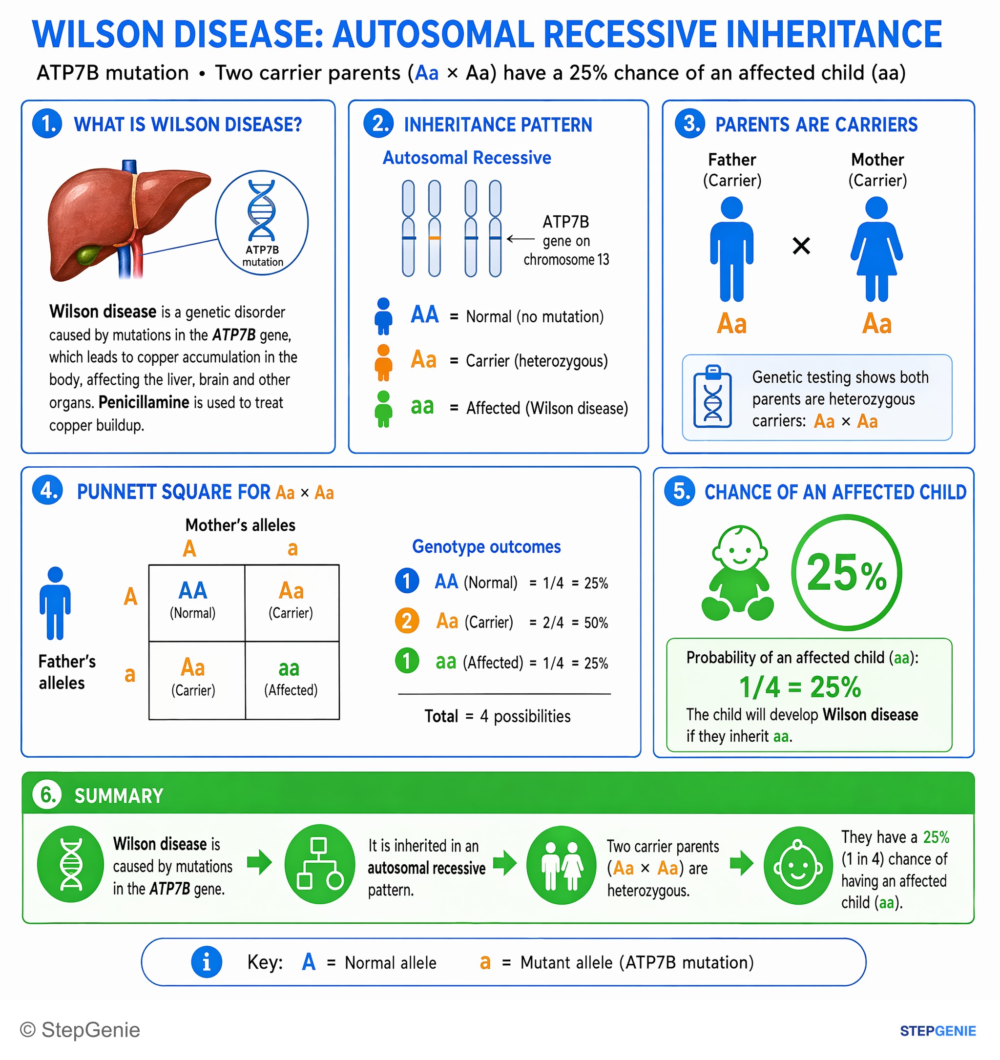
Wilson disease is autosomal recessive (ATP7B mutation); two heterozygous carriers have a 25% chance of having an affected child.
USMLE Step 1Frequently Asked Questions
Why does an unaffected sibling have a 2/3 chance of being a carrier?
When both parents are carriers (Aa × Aa), each child has four equally likely outcomes: AA, Aa, Aa and aa. Knowing the sibling is unaffected removes aa, leaving three equally likely outcomes, two of which are carriers, so the probability is 2/3. As a conditional probability, (1/2) ÷ (3/4) = 2/3. The rule applies only to a sibling whose parents are both carriers and whose unaffected status is certain; once the sibling has a carrier test, the result replaces the 2/3 estimate.
What is the chance of an affected child if both parents are carriers of an autosomal recessive disease?
Each pregnancy has a 1/4 (25%) chance of an affected child, a 1/2 (50%) chance of an unaffected carrier and a 1/4 chance of a child who is neither affected nor a carrier. Pregnancies are independent, so having one affected child does not change the risk for the next one. The chance that two specific children are both affected is 1/4 × 1/4, or 1/16.
How do you recognize autosomal recessive inheritance on a pedigree?
Look for affected siblings whose parents are unaffected, a trait that seems to skip generations, and males and females affected in equal numbers. Consanguinity makes autosomal recessive inheritance more likely, and unaffected parents of an affected child are obligate carriers. If nearly all affected people are male and no father passes the trait to a son, think X-linked recessive. If an affected parent has an affected child, consider pseudodominance (an affected person with a carrier partner) before calling it dominant.
If my brother has cystic fibrosis, what is the chance my child will have it?
If you are unaffected, your chance of being a carrier is 2/3. If your partner is White with no family history, Hardy-Weinberg applied to a cystic fibrosis incidence of about 1 in 2,500 gives a carrier frequency of about 1 in 25, so each child's risk is 2/3 × 1/25 × 1/4, or about 1 in 150. If your partner is a known carrier, the risk is 2/3 × 1/4, or 1 in 6. Carrier testing that includes your family's variant replaces these estimates.
What is a good mnemonic for autosomal recessive inheritance?
A RARE works well: Affected siblings with unaffected parents, Related parents (consanguinity), All sexes affected equally, Risk math (1/4 affected and 1/2 carriers for two carrier parents; 2/3 carrier for an unaffected sibling) and Enzyme deficiencies, since most inborn errors of metabolism are autosomal recessive. Remember the exceptions: ornithine transcarbamylase deficiency, Hunter syndrome and Fabry disease are X-linked, and acute intermittent porphyria is autosomal dominant.
What are examples of autosomal recessive diseases?
High-yield examples include cystic fibrosis, sickle cell disease, beta-thalassemia, phenylketonuria, Tay-Sachs disease, Gaucher disease, Hurler syndrome, von Gierke disease, classic galactosemia, Wilson disease, HFE hemochromatosis, 21-hydroxylase deficiency, spinal muscular atrophy, Friedreich ataxia, autosomal recessive polycystic kidney disease and oculocutaneous albinism. Common traps that are not autosomal recessive include Hunter syndrome, Fabry disease, OTC deficiency, Lesch-Nyhan syndrome and G6PD deficiency (X-linked), acute intermittent porphyria and ADPKD (autosomal dominant), and alpha-1 antitrypsin deficiency (codominant).