Locus heterogeneity means variants in different genes cause the same disease: oculocutaneous albinism can come from TYR, OCA2, TYRP1 or SLC45A2. Allelic heterogeneity means different variants in the same gene cause the same disease: more than 1,000 CFTR variants cause cystic fibrosis, and hundreds of HBB variants cause beta-thalassemia.
Both are forms of genetic heterogeneity, the National Cancer Institute's umbrella term for different genetic mechanisms that produce the same or similar phenotypes. Short version: different genes = locus; same gene, different variants = allelic.
Two stem clues settle most questions. "Negative for the common variant" of a known disease gene points to allelic heterogeneity. Two parents with the same recessive disorder who have unaffected children, or one disease that is X-linked in one family and autosomal in another, points to locus heterogeneity.
Locus vs allelic heterogeneity: the difference
| Locus heterogeneity | Allelic heterogeneity | |
|---|---|---|
| NCI definition | Variants at different gene loci cause the same or similar phenotype | Different variants at a single gene locus cause the same or similar phenotype |
| What differs between families | The gene | The variant within one gene |
| Classic examples | Albinism, retinitis pigmentosa, hearing loss, tuberous sclerosis (TSC1 or TSC2) | Cystic fibrosis (CFTR), beta-thalassemia (HBB), Marfan syndrome (FBN1) |
| Inheritance between families | Can differ: retinitis pigmentosa can be autosomal dominant, autosomal recessive or X-linked | X-linked stays X-linked and autosomal stays autosomal, because the locus is the same |
| Stem clue | Variant found in a different gene; affected recessive parents with unaffected children | "Negative for the most common variant"; two different variants in one patient |
| Testing logic | Multigene panel | Read the whole gene |
Genetic heterogeneity vs locus or allelic heterogeneity: genetic heterogeneity is the parent term. In NCI's words, "There are two types of genetic heterogeneity: allelic heterogeneity and locus heterogeneity," and MeSH files both under Genetic Heterogeneity. One disease can show both: albinism has four main genes (locus heterogeneity), and more than 100 TYR variants and more than 80 OCA2 variants have been found in people with types 1 and 2 (allelic heterogeneity).
The Q-bank question
A woman and a man who both have oculocutaneous albinism ask for genetic counseling before a fourth pregnancy. Each was born to unaffected parents. The woman has had white hair and very pale skin since birth; the man has light brown hair and pale skin. Both have nystagmus and reduced visual acuity. All three of their children have normal skin, hair and eye pigmentation, and paternity is confirmed. In both families, albinism follows an autosomal recessive pattern.
Which of the following best explains the children's normal pigmentation?
A. Allelic heterogeneity B. Incomplete penetrance C. Locus heterogeneity D. Phenotypic heterogeneity E. Pleiotropy F. Variable expressivity
Answer: C. Locus heterogeneity.
Why C is right: the two defects complement each other
Oculocutaneous albinism (OCA) is one phenotype with several autosomal recessive types, each from a different gene: OCA1 (TYR), OCA2 (OCA2), OCA3 (TYRP1) and OCA4 (SLC45A2), plus rarer types. Hair color is only a hint: type 1 typically gives white hair, type 2 light yellow, blond or light brown hair, and type 4 looks like type 2. The proof is the children. If she carries two TYR variants and he carries two OCA2 variants, every child gets one variant copy at each locus plus a working copy from the other parent:
| TYR locus | OCA2 locus | Pigmentation | |
|---|---|---|---|
| Mother (OCA1) | Two variant copies | Two normal copies | Albinism |
| Father (OCA2) | Two normal copies | Two variant copies | Albinism |
| Every child | One variant, one normal | One variant, one normal | Normal (carrier at both loci) |
Each parent supplies the working gene the other lacks, so the defects complement each other. NCI calls the children's genotype double heterozygosity: "two different mutated alleles at two separate genetic loci." If both parents had variants in the same gene, every child would inherit two variant copies of it and would be affected, so three pigmented children (paternity confirmed) place the parents' variants in different genes. Testing TYR and OCA2 would confirm it; the autosomal recessive inheritance review covers the carrier math.
This is the family version of the laboratory complementation test: MeSH defines it as checking whether a cell with a mutant phenotype regains function when another mutant genome with the same phenotype is introduced. A 1952 British Journal of Ophthalmology report described such a family: two albino parents, three normally pigmented children.
Why every other choice is wrong
A. Allelic heterogeneity. Different variants in the same gene cause one disease, as with the hundreds of HBB variants behind beta-thalassemia. Why wrong: if both parents had different TYR variants, each child would be a compound heterozygote (one variant from each parent) and would still have albinism.
B. Incomplete penetrance. Some people with a disease genotype show no signs; MedlinePlus's example is BRCA1 and BRCA2 carriers who never develop cancer. Why wrong: it would require all three children to carry an albinism genotype yet stay fully pigmented. Different genes explain the result without that assumption.
D. Phenotypic heterogeneity. Variants in one gene cause different disorders. RET variants that make a nonfunctional protein cause Hirschsprung disease, while overactivating ones cause multiple endocrine neoplasia type 2 (MEN syndromes); in HBB, Glu6Val gives sickle cell disease and other variants give beta-thalassemia. Why wrong: both parents have the same disorder, and the term says nothing about why the children escaped it.
E. Pleiotropy. One gene shapes "multiple and diverse phenotypic outcomes" (MeSH) in the same person. Untreated phenylketonuria causes intellectual disability, seizures, a musty odor, eczema and lighter skin and hair. Why wrong: pleiotropy describes how widely one genotype acts, not why a trait vanishes in the next generation.
F. Variable expressivity. People with the same genetic condition differ in severity or features: in Marfan syndrome, some are only tall and thin with long fingers, while others have life-threatening heart and blood vessel complications; NF1 is the other classic. Why wrong: expressivity varies only among people who have the condition; these children do not have it.
Locus heterogeneity examples
| Disease | Genes | Inheritance | Exam point |
|---|---|---|---|
| Oculocutaneous albinism | TYR, OCA2, TYRP1, SLC45A2 (types 1-4); rarer types include SLC24A5, LRMDA, DCT | Autosomal recessive (AR) | Affected parents can have unaffected children |
| Retinitis pigmentosa | More than 130 genes; RHO, USH2A and RPGR are among the most common | Autosomal dominant (AD; RHO most often), AR (USH2A), X-linked (RPGR); rarely digenic | One disease, three inheritance patterns |
| Nonsyndromic hearing loss | More than 155 genes; GJB2 causes about half of recessive cases, STRC is second | About 80% AR, 19% AD, 1% X-linked or mitochondrial | Even the commonest gene explains only part |
| Osteogenesis imperfecta | COL1A1 or COL1A2 in about 90%; CRTAP, P3H1, PPIB, SERPINF1 and others; MBTPS2 | AD (COL1A1/COL1A2, type V); AR (types VI-XVIII); X-linked (type XIX) | Unaffected parents can have a child with a recessive form (OI review) |
| Familial hypercholesterolemia | LDLR (most common), APOB, PCSK9 (gain of function); LDLRAP1 | AD; LDLRAP1 AR | Different genes, one pathway: LDL receptor clearance |
| Tuberous sclerosis complex | TSC1 (hamartin) or TSC2 (tuberin) | AD; about two-thirds de novo | The two proteins work together, so either gene gives TSC |
| Autosomal dominant PKD | PKD1 or PKD2; rarely ALG5, ALG9, DNAJB11, GANAB, IFT140 | AD | PKD2 disease is usually milder and later (ADPKD vs ARPKD) |
| Alport syndrome | COL4A5; COL4A3 or COL4A4 | X-linked (COL4A5, most families); AR or AD (COL4A3, COL4A4) | One kidney-ear-eye disease, three patterns |
| Chronic granulomatous disease | CYBB; CYBA, CYBC1, NCF1, NCF2, NCF4 | X-linked (CYBB); AR (the other five) | NADPH oxidase genes; girls get CGD too (CGD guide) |
| Xeroderma pigmentosum | Eight complementation groups, XP-A to XP-G plus XP-V (XPA, XPC, ERCC2, POLH and others) | AR | Groups XP-A to XP-G were defined by complementation |
| Fanconi anemia | 21 AR genes (FANCA, FANCC or FANCG in 80-90%); RAD51; FANCB | Mostly AR; RAD51 AD; FANCB X-linked | BRCA1, BRCA2 and PALB2 are FA genes too |
| Limb-girdle muscular dystrophy | Over 32 genes; CAPN3 most common | Mostly AR; some AD | Proximal weakness that overlaps other muscle disorders |
Exam point: XP complementation groups are locus heterogeneity by another name: fusing fibroblasts from different XP patients corrected the repair defect because the nuclei "supplied what the other was lacking" (DiGiovanna and Kraemer, 2012).
Allelic heterogeneity examples
Same gene, same disease, many variants. The practical consequence: a test for one variant, or a short panel, can miss the patient's variants.
| Disease | Gene | Variants | Exam point |
|---|---|---|---|
| Cystic fibrosis | CFTR | More than 2,000 reported; CFTR2 classed 1,085 as CF-causing by September 2024 | F508del is the most common; in UK Biobank it made up about 90% of CF-causing variants in Europeans but less elsewhere (V520F led in East Asians) |
| Beta-thalassemia | HBB | Hundreds | β0 variants abolish beta-globin, β+ variants reduce it; biallelic β0 typically means thalassemia major |
| Marfan syndrome | FBN1 | Over a thousand pathogenic variants | Most patients share the gene, not the severity |
| Duchenne and Becker muscular dystrophy | DMD | More than 2,000 | Duchenne variants typically leave no functional dystrophin; Becker variants leave an abnormal dystrophin with some function |
So allelic heterogeneity often explains severity, not just missed tests: the variant type helps separate thalassemia major from milder forms, and Duchenne from Becker. The alpha vs beta thalassemia table shows the beta genotypes.
Allelic heterogeneity vs compound heterozygosity
They work at different levels.
- Allelic heterogeneity describes a gene across patients: many different variants in one gene cause the same disease.
- Compound heterozygosity describes one person: "The presence of two different mutated alleles at a particular gene locus" (NCI), typically one from each parent in a recessive disease.
- Double heterozygosity is the two-gene version: "two different mutated alleles at two separate genetic loci" (NCI).
Allelic heterogeneity is what makes compound heterozygosity possible: when a gene has many disease-causing variants, an affected child can inherit a different one from each parent.
| Genotype | Meaning | Example |
|---|---|---|
| Homozygous | Two identical variants in one gene | Sickle cell anemia (HbSS): Glu6Val on both HBB copies |
| Compound heterozygous | Two different variants in one gene | HbS-beta thalassemia; congenital bilateral absence of the vas deferens from one mild and one CF-causing CFTR variant |
| Double heterozygous | One variant in each of two genes | The albinism couple's children: carriers, unaffected |
The terms work in dominant disease too. GeneReviews calls biallelic (homozygous or compound heterozygous) LDLR, APOB or PCSK9 variants homozygous FH, which starts earlier and is more severe; people heterozygous in two different FH genes are double heterozygotes, which "may have an additive effect". And in rare digenic retinitis pigmentosa, a variant must be present in two different genes to cause the disease (MedlinePlus).
Lookalike terms: phenotypic heterogeneity, pleiotropy, expressivity, penetrance
| Term | Gene vs phenotype | Classic example | Stem clue |
|---|---|---|---|
| Phenotypic heterogeneity | Same gene, different disorders | RET (Hirschsprung vs MEN2); HBB (sickle cell vs beta-thalassemia); FGFR3 (achondroplasia vs thanatophoric dysplasia) | "Same gene, different disease" |
| Pleiotropy | One gene, many systems in one person | Untreated PKU; Marfan syndrome (eye, skeleton, heart) | One variant explains several organs |
| Variable expressivity | Same condition, different severity | Marfan syndrome, NF1 | Relatives with the same variant, different severity |
| Incomplete penetrance | Genotype present, no signs in some carriers | BRCA1/BRCA2 | Unaffected obligate carrier; "skipped" generation |
| Anticipation | Earlier or more severe in each generation | Huntington disease, myotonic dystrophy, fragile X | Trinucleotide repeat expansion (anticipation question) |
| Mosaicism | Two or more cell lines in one person | See the mosaicism table | Variant in only some cells |
Naming varies. NCI limits allelic heterogeneity to variants causing "the same or similar" phenotype, as here. Others use it for any variants in one gene: a 2026 review names it the leading cause of phenotypic heterogeneity, where variants in one gene "do not invariably produce the same clinical phenotype". So HBB's sickle cell and thalassemia variants may get either label. On the exam, ask: one disease or two? Expressivity and penetrance are compared in penetrance vs expressivity.
What heterogeneity means for genetic testing
The kind of heterogeneity decides the test (MedlinePlus):
- Targeted variant test: looks for one known variant, such as the HBB sickle variant. Useful for relatives of a known case; weak when a gene has many disease-causing variants.
- Single-gene test: reads the whole gene, "particularly when there are many variants in the gene that can cause the suspected condition": the allelic-heterogeneity test. For beta-thalassemia, GeneReviews recommends HBB sequencing first, then deletion/duplication analysis if fewer than two variants are found.
- Gene panel: many genes at once, for a condition that "can be caused by variants in many genes": the locus-heterogeneity test (retinitis pigmentosa, hearing loss, or epilepsy, where a 2026 review counts around 2,000 genes linked to seizures or epilepsy).
- Exome or genome sequencing: when single-gene or panel testing has not given a diagnosis, or the cause is unclear.
Two cautions. A negative result does not rule out a heterogeneous disease: a genetic cause is found in only 50 to 80% of people with retinitis pigmentosa. And a panel built from one population can miss others: most of the common CF-causing variants in non-European populations are not on the American College of Medical Genetics' original 23-variant carrier-screening panel (Ideozu, 2024), which ACMG expanded to a 100-variant pan-ethnic panel in 2023 (GeneReviews).
USMLE traps
- A negative targeted test is not a different gene. CF has one gene; a negative F508del result means another CFTR variant. Read the whole gene, and remember that symptoms plus a sweat chloride of 60 mmol/L or more establish CF on their own (GeneReviews).
- Different inheritance, same disease = locus heterogeneity. Alport syndrome and CGD are often remembered as X-linked, but both have autosomal forms (COL4A3/COL4A4 Alport syndrome; five autosomal recessive CGD genes), so girls can be severely affected.
- Compound vs double heterozygote. Two different variants in one gene cause recessive disease; one variant in each of two genes usually does not. That contrast is the whole albinism question.
- "Albinism = four types" is out of date. MedlinePlus describes additional rare types, with testing entries up to type 8 and genes such as SLC24A5, LRMDA and DCT.
- Pleiotropy vs variable expressivity. Pleiotropy is breadth in one person (PKU: brain, skin, odor); expressivity is a range of severity across people with the same condition.
- Not anticipation. Earlier or worse disease in each generation points to a repeat expansion, not heterogeneity.
Three more USMLE-style questions
For more practice, try the free biochemistry and genetics questions.
1. The negative CF test
A 6-year-old boy has a chronic productive cough, recurrent sinus and lung infections, greasy stools and poor weight gain. Sweat chloride is 88 mmol/L on two occasions. A targeted test for F508del, the most common CFTR variant, is negative. His parents ask how he can have cystic fibrosis if "the gene test was negative." Which of the following best explains this result?
A. Allelic heterogeneity B. Anticipation C. Locus heterogeneity D. Variable expressivity E. Incomplete penetrance
Answer: A. Allelic heterogeneity. Symptoms plus a sweat chloride of 60 mmol/L or more establish CF. CF has one gene, CFTR, but more than 2,000 CFTR variants have been reported and CFTR2 had classed 1,085 as CF-causing by September 2024, so a one-variant test can miss his. Next step: sequence the whole gene; the result also shows whether he has CFTR modulator-responsive variants.
- B: anticipation comes from repeat expansions (Huntington disease, myotonic dystrophy, fragile X); CF is not a repeat disorder.
- C: tempting, because "gene test negative" sounds like "different gene," but no second gene causes CF.
- D: variable expressivity means different severity among people with the same condition; it does not explain a negative test.
- E: incomplete penetrance describes an unaffected carrier; this boy has the disease.
2. Alport syndrome in two families
Two families have Alport syndrome. In family 1, a 19-year-old man has hematuria, proteinuria, sensorineural hearing loss and anterior lenticonus; his maternal uncle started dialysis at 30, and his mother has only microscopic hematuria. In family 2, a 16-year-old girl has the same kidney, ear and eye findings; her parents, who are first cousins, both have isolated microscopic hematuria. Which of the following best explains why the disease looks X-linked in one family and autosomal recessive in the other?
A. Allelic heterogeneity B. Anticipation C. Locus heterogeneity D. Skewed X-inactivation in the girl E. Variable expressivity
Answer: C. Locus heterogeneity. Family 1 fits COL4A5 (X-linked: affected males through mildly affected females). Family 2 fits biallelic COL4A3 or COL4A4 variants (autosomal recessive): consanguinity, a severely affected girl and parents with only hematuria, as heterozygotes may have. Without treatment, recessive Alport syndrome leads to kidney failure in most males and females alike (GeneReviews). More kidney detail is in the Alport syndrome question.
- A: different COL4A5 variants would keep the disease X-linked; the locus, not the allele, sets the pattern.
- B: nothing worsens across generations, and Alport syndrome is not a repeat disorder.
- D: skewed X-inactivation may affect severity in females with X-linked Alport syndrome, but it explains neither the consanguinity nor her father's isolated hematuria: all males with the X-linked form develop proteinuria (GeneReviews).
- E: expressivity changes severity, not the inheritance pattern.
3. One gene, two variants
A 10-year-old boy has sickle cell disease with painful crises since early childhood. His mother has sickle cell trait. His father has beta-thalassemia trait, and testing confirms a β0 HBB variant in the father. Which of the following best describes the boy's genotype?
A. Allelic heterogeneity B. Compound heterozygosity C. Double heterozygosity D. Homozygosity E. Locus heterogeneity
Answer: B. Compound heterozygosity. He has two different variants in one gene, HBB: the sickle variant (Glu6Val) from his mother and a β0 variant from his father. This is HbS-beta thalassemia, which is severe with β0 and generally milder with β+ (MedlinePlus).
- A: allelic heterogeneity describes HBB across patients (hundreds of variants), not one person's genotype; it is why his compound genotype is possible.
- C: double heterozygosity needs variants at two different loci; both of his are in HBB.
- D: homozygosity means two identical variants, as in HbSS (see the sickle cell disease question).
- E: locus heterogeneity needs different genes; only HBB is involved.
Key takeaways
- Locus heterogeneity: different genes, same disease. Know albinism, retinitis pigmentosa, hearing loss, tuberous sclerosis, Alport syndrome and CGD.
- Allelic heterogeneity: same gene, different variants, same disease. Know cystic fibrosis (CFTR) and beta-thalassemia (HBB).
- Both are types of genetic heterogeneity. Compound heterozygosity is one person with two different variants in one gene; double heterozygosity is one variant in each of two genes.
- Affected recessive parents with unaffected children, or one disease with different inheritance in different families, means locus heterogeneity.
- "Negative for the common variant" means allelic heterogeneity: read the whole gene. Many candidate genes: use a panel.
- Phenotypic heterogeneity, pleiotropy, expressivity, penetrance and anticipation are the usual distractors.
Sources
- NCI Dictionary of Genetics Terms - National Cancer Institute: locus heterogeneity, allelic heterogeneity, genetic heterogeneity, compound heterozygosity, double heterozygosity
- MeSH - National Library of Medicine: Genetic Heterogeneity, Genetic Complementation Test, Genetic Pleiotropy
- MedlinePlus Genetics conditions - National Library of Medicine: oculocutaneous albinism (2024), retinitis pigmentosa (2026), nonsyndromic hearing loss (2026), osteogenesis imperfecta (2020), familial hypercholesterolemia (2020), tuberous sclerosis complex (2022), polycystic kidney disease (2014), Alport syndrome (2023), chronic granulomatous disease (2016), xeroderma pigmentosum (2023), Fanconi anemia (2025), limb-girdle muscular dystrophy (2025), cystic fibrosis (2021), sickle cell disease (2024), phenylketonuria (2023), neurofibromatosis type 1 (2020)
- MedlinePlus Genetics genes and explainers - National Library of Medicine: TYR (2007), OCA2 (2022), HBB (2024), CFTR (2008), RET (2018), FGFR3 (2023), FBN1 (2026), DMD (2017), reduced penetrance and variable expressivity (2021), anticipation (2021), types of genetic tests (2021)
- Cystic Fibrosis (Savant, Bojanowski, Upadia) - GeneReviews, NCBI Bookshelf (2026)
- Beta-Thalassemia (Langer) - GeneReviews, NCBI Bookshelf (2026)
- Alport Syndrome (Nozu, Yamamura, Horinouchi) - GeneReviews, NCBI Bookshelf (2025)
- Chronic Granulomatous Disease (Leiding, Holland) - GeneReviews, NCBI Bookshelf (2026)
- Tuberous Sclerosis Complex (Northrup, Koenig, Pearson, Au) - GeneReviews, NCBI Bookshelf (2024)
- Polycystic Kidney Disease, Autosomal Dominant (Harris, Torres) - GeneReviews, NCBI Bookshelf (2022)
- Familial Hypercholesterolemia (Ison, Clarke, Knowles) - GeneReviews, NCBI Bookshelf (2025)
- Fanconi Anemia (Mehta, Ebens) - GeneReviews, NCBI Bookshelf (2026)
- FBN1-Related Marfan Syndrome (Dietz) - GeneReviews, NCBI Bookshelf (2022)
- Ideozu JE, et al. Diversity of CFTR variants across ancestries characterized using 454,727 UK Biobank whole exome sequences - Genome Medicine (2024)
- McGarry ME, Raraigh KS, et al. Cystic Fibrosis Newborn Screening: A Systematic Review-Driven Consensus Guideline from the United States Cystic Fibrosis Foundation - International Journal of Neonatal Screening (2025)
- DiGiovanna JJ, Kraemer KH. Shining a light on xeroderma pigmentosum - Journal of Investigative Dermatology (2012)
- Lehmann AR, McGibbon D, Stefanini M. Xeroderma pigmentosum - Orphanet Journal of Rare Diseases (2011)
- Saad AK, Akawi N. Heterogeneity of monogenic epilepsy in loci, phenotypes, and treatment approaches - Neurological Sciences (2026)
- Trevor-Roper PD. Marriage of two complete albinos with normally pigmented offspring - British Journal of Ophthalmology (1952)
Visual explanations from the StepGenie QBank
Infographics StepGenie drew to explain questions in its own QBank, matched to this article's topic. Each caption is the takeaway of the question it explains.
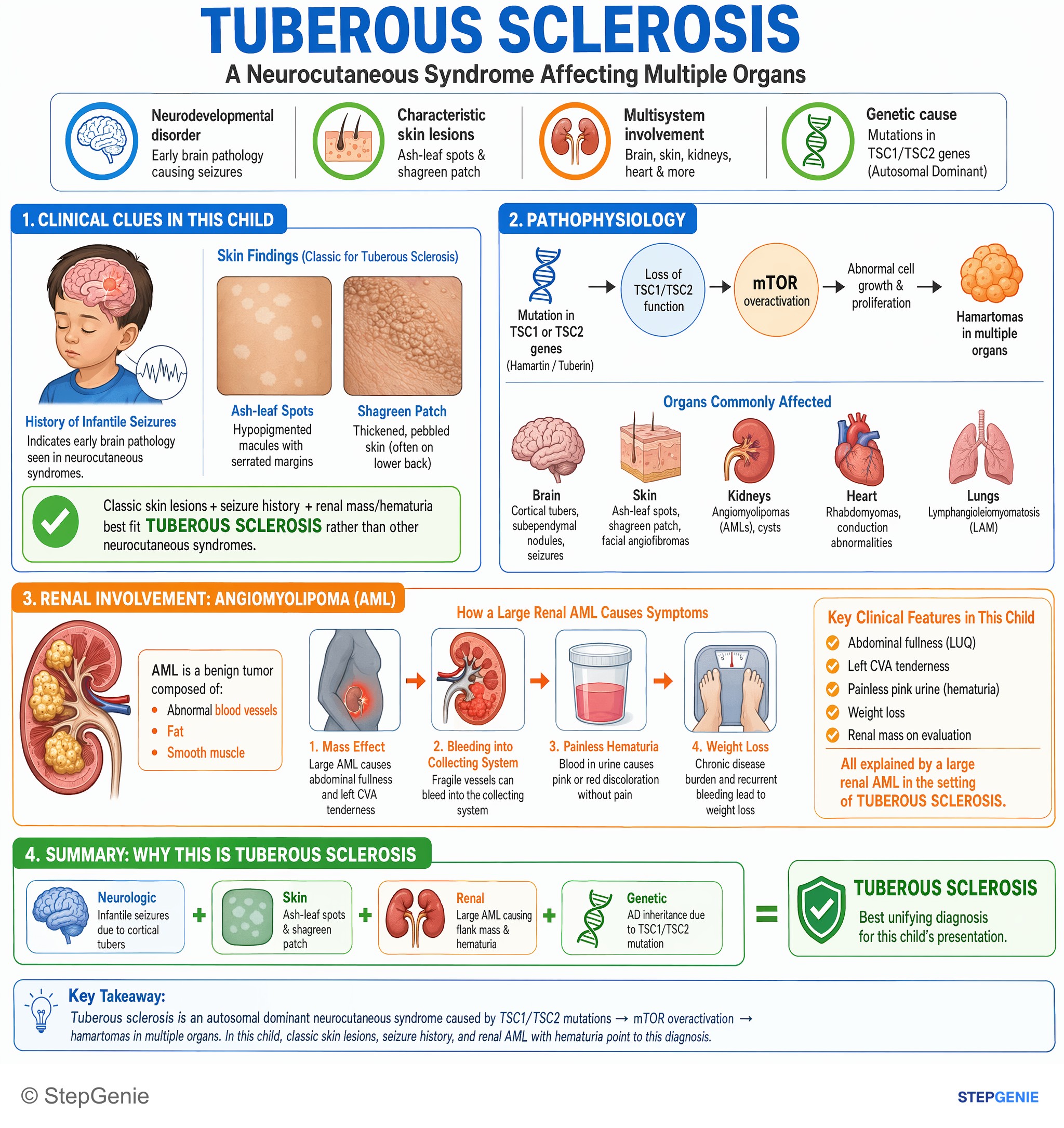
Tuberous sclerosis: AD TSC1/TSC2 mutation → cortical tubers, infantile seizures, ash-leaf spots, shagreen patch, and risk of renal…
USMLE Step 1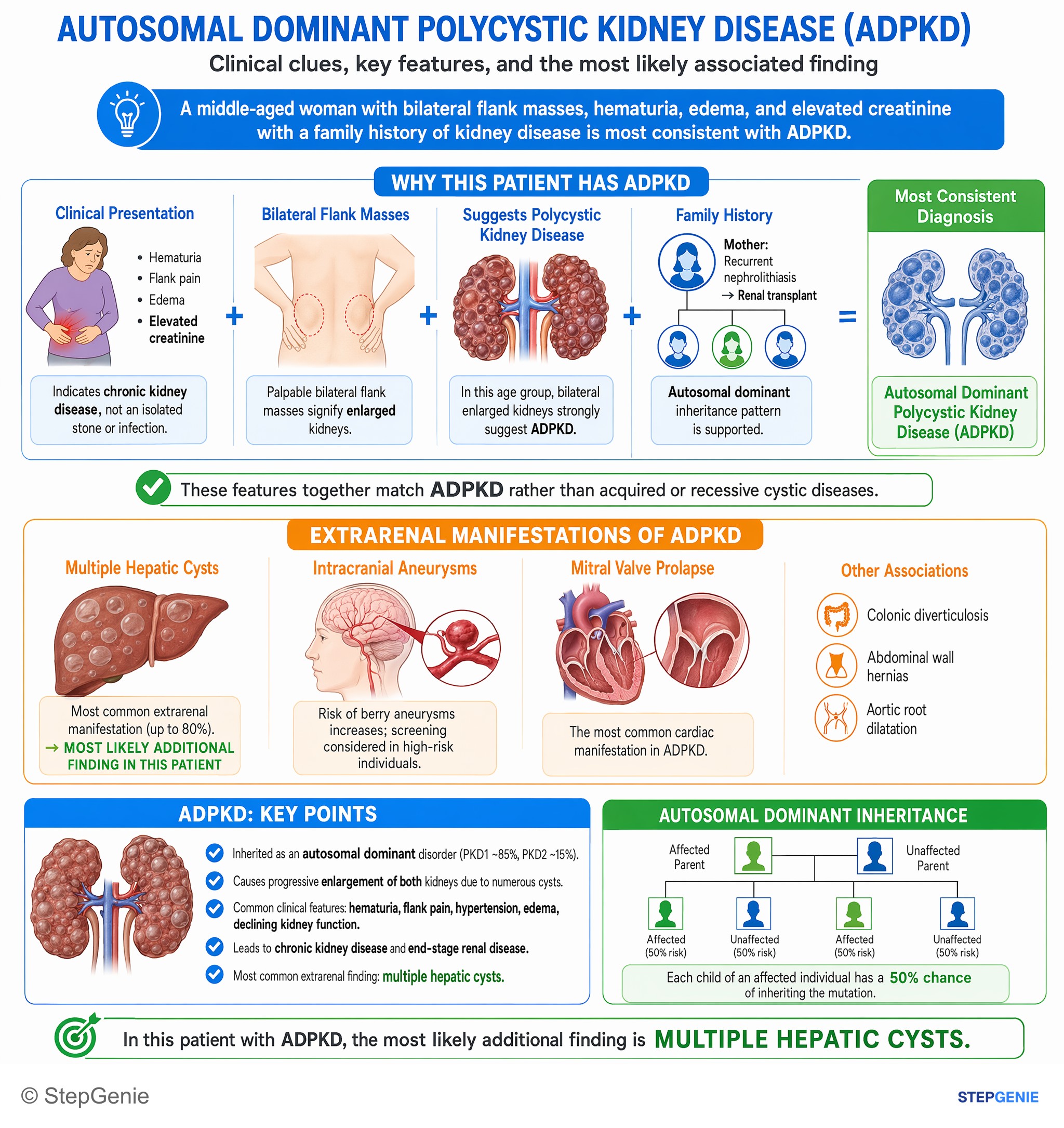
Autosomal dominant polycystic kidney disease causes bilateral renal enlargement, hematuria, renal failure, and is commonly associated with…
USMLE Step 1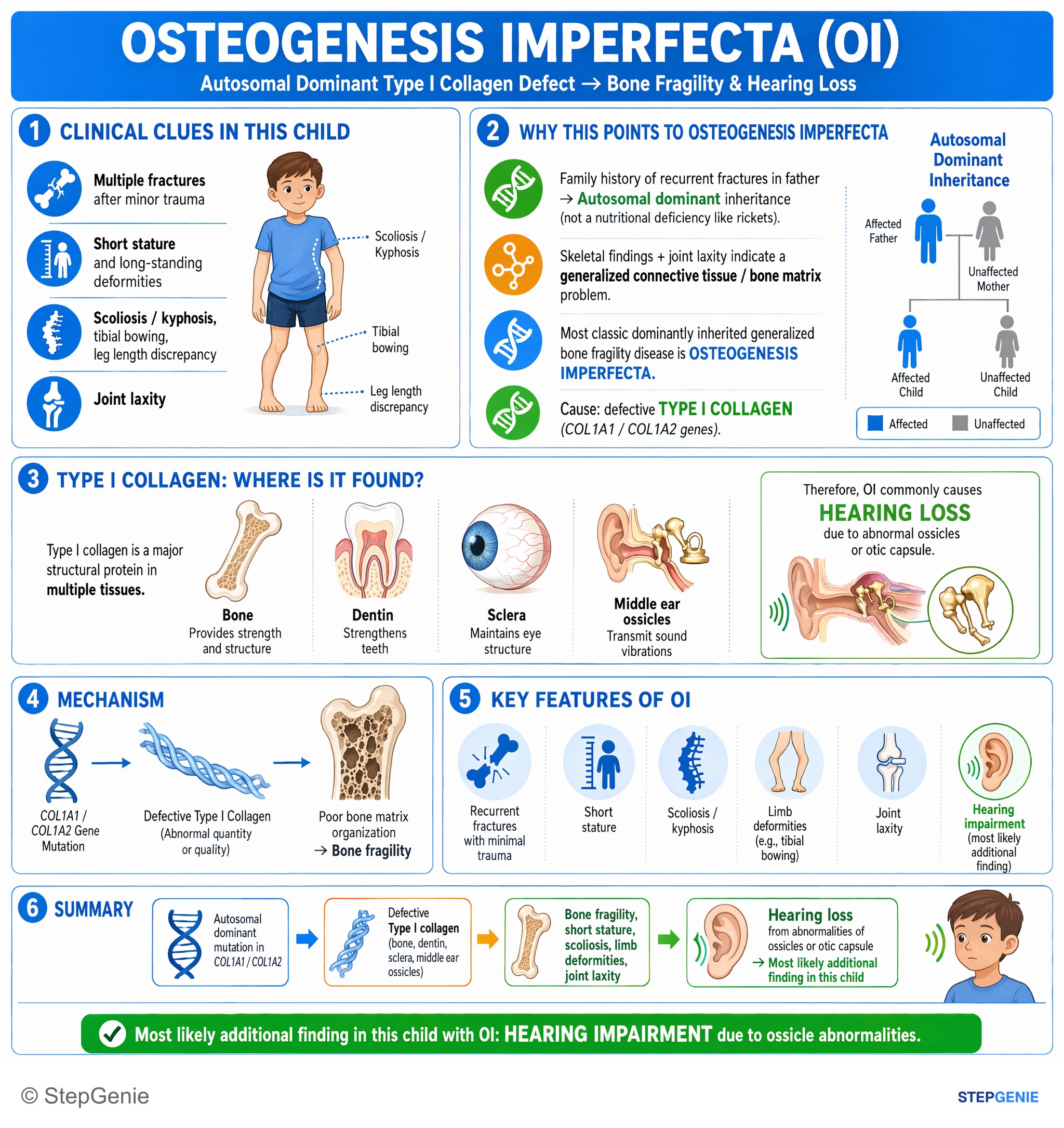
Osteogenesis imperfecta: autosomal dominant collagen I defect → bone fragility, short stature, scoliosis, limb deformities, and hearing…
USMLE Step 1Frequently Asked Questions
What is the difference between locus heterogeneity and allelic heterogeneity?
The difference is how many genes are involved. In locus heterogeneity, variants in different genes cause the same disease, as when oculocutaneous albinism comes from TYR in one family and OCA2 in another. In allelic heterogeneity, different variants in one gene cause the same disease, as with the more than 1,000 CFTR variants behind cystic fibrosis or the hundreds of HBB variants behind beta-thalassemia. A negative test for the common variant of a known gene points to allelic heterogeneity.
What are examples of locus heterogeneity?
Locus heterogeneity means variants at different gene loci cause the same or a similar disease. Classic examples are oculocutaneous albinism (TYR, OCA2, TYRP1, SLC45A2), retinitis pigmentosa (more than 130 genes), nonsyndromic hearing loss (more than 155 genes), tuberous sclerosis (TSC1 or TSC2), Alport syndrome (COL4A5, COL4A3, COL4A4) and chronic granulomatous disease (CYBB and five autosomal recessive genes). Because the genes differ, inheritance can differ between families with the same diagnosis.
What are examples of allelic heterogeneity?
Allelic heterogeneity means different variants in a single gene cause the same disease. Cystic fibrosis is the classic: more than 2,000 CFTR variants have been reported, and by September 2024 the CFTR2 project had classified 1,085 as CF-causing. Beta-thalassemia has hundreds of HBB variants, Marfan syndrome over a thousand FBN1 variants, and Duchenne and Becker muscular dystrophy more than 2,000 DMD variants. That is why a test for one common variant can miss the diagnosis.
What is the difference between allelic heterogeneity and compound heterozygosity?
Allelic heterogeneity describes a gene across many patients: different variants in that gene cause the same disease. Compound heterozygosity describes one person who carries two different disease-causing variants in the same gene, usually one from each parent, as in HbS-beta thalassemia. Allelic heterogeneity makes compound heterozygosity possible. Double heterozygosity is different again: one variant in each of two separate genes, which usually does not cause a recessive disease.
Is genetic heterogeneity the same as locus heterogeneity?
No. Genetic heterogeneity is the umbrella term for different genetic mechanisms that produce the same or similar phenotype. The National Cancer Institute divides it into two types: locus heterogeneity, where variants in different genes cause the disease, and allelic heterogeneity, where different variants in one gene cause it. Retinitis pigmentosa shows locus heterogeneity across more than 130 genes; cystic fibrosis shows allelic heterogeneity within CFTR.
How can two parents with albinism have children with normal pigmentation?
If the parents' albinism comes from different genes, for example TYR in one parent and OCA2 in the other, each child inherits one variant and one working copy of each gene. The children are carriers at both loci, called double heterozygotes, and are normally pigmented because the working copies complement each other. This is locus heterogeneity. If both parents had variants in the same gene, every child would have albinism.