Down syndrome is caused by an extra copy of chromosome 21 material. About 95% of cases are free trisomy 21 (47,XX,+21 or 47,XY,+21) from meiotic nondisjunction, usually in the mother's egg and mostly in meiosis I, which is why risk climbs with maternal age; about 3-4% are Robertsonian translocations (46 chromosomes, most often der(14;21)) that can be inherited from a balanced-carrier parent; and 1-2% are mosaic. Newborns show hypotonia, upslanting palpebral fissures, epicanthal folds, a flat facial profile, a single transverse palmar crease and a sandal gap. About half have congenital heart disease, with atrioventricular septal defect (endocardial cushion defect) the classic lesion. Other favorites: duodenal atresia, Hirschsprung disease, transient abnormal myelopoiesis and acute megakaryoblastic leukemia, hypothyroidism, atlantoaxial instability and early Alzheimer disease from an extra APP gene.
The vignette
A 39-year-old woman, gravida 2, para 1, delivers a boy at 38 weeks' gestation; she declined prenatal screening. The newborn is floppy, with upslanting palpebral fissures, epicanthal folds, a flat facial profile, small rounded ears, a single transverse palmar crease on both hands and a wide gap between the first and second toes. A systolic murmur is heard, and echocardiography shows a complete atrioventricular septal defect. A peripheral blood karyotype shows 47,XY,+21 in every cell examined. Which mechanism most likely produced this karyotype?
A. Nondisjunction during maternal meiosis B. Nondisjunction during paternal meiosis C. Unbalanced Robertsonian translocation inherited from a carrier parent D. Mitotic nondisjunction after fertilization (mosaicism) E. Microdeletion of chromosome 22q11.2 F. Loss of paternally expressed genes at 15q11.2-q13
Answer: A. Nondisjunction during maternal meiosis
A whole, free extra chromosome 21 in every cell is free trisomy 21, the cause of about 95% of Down syndrome. Nondisjunction is the failure of homologous chromosomes (meiosis I) or sister chromatids (meiosis II) to separate, so one gamete carries two copies of chromosome 21 and fertilization adds a third. The extra chromosome is typically maternal, and the error arises predominantly in meiosis I of oocyte formation.
That is why age matters. Merck puts the risk at about 1 in 1,476 births at maternal age 20, 1 in 352 at 35 and 1 in 85 at 40. Even so, CDC notes that most babies with Down syndrome are born to mothers younger than 35, simply because more babies are born to younger women.
Why every other choice is wrong
B. Nondisjunction during paternal meiosis. It happens, and a routine karyotype doesn't show which parent contributed the extra chromosome. But the extra 21 is typically maternal, and this stem adds a 39-year-old mother. Choose paternal origin only if the stem says DNA studies traced the extra chromosome to the father.
C. Robertsonian translocation. A real cause (about 3-4% of cases), but it changes the count. The extra 21q is fused to another acrocentric chromosome, usually 14, so the child has two normal chromosomes 21 plus the fused one: 46 chromosomes, written for example as 46,XY,der(14;21)(q10;q10),+21. In about half of cases the translocation is new; in the other half a parent, almost always the mother, is a healthy balanced carrier with 45 chromosomes. Choose translocation when the karyotype shows 46 chromosomes, or when a young mother has a second affected child. Recurrence risk (Merck) is about 1 in 10 if the mother carries t(14;21), 1 in 20 if the father does, and 100% if a parent carries t(21;21), so both parents should be karyotyped.
D. Mosaicism. Nondisjunction in the embryo after fertilization leaves two cell lines, 46 and 47,+21 (1-2% of cases). The phenotype varies widely, and some people with mosaicism have only subtle signs and normal intelligence; mosaic males may be fertile, whereas most men with Down syndrome are not. Every cell here is 47,+21, so this is not mosaicism. See the mosaicism comparison table.
E. 22q11.2 microdeletion. A microdeletion removes a small segment and leaves 46 chromosomes; chromosomal microarray or FISH finds it. The 22q11.2 deletion syndrome brings conotruncal heart defects (tetralogy of Fallot, interrupted aortic arch, truncus arteriosus, VSD), palatal anomalies, immune deficiency and hypocalcemia from hypoparathyroidism; over 90% of deletions are new. Compare this DiGeorge case.
F. Loss of paternal 15q11.2-q13 expression (Prader-Willi syndrome). Severe neonatal hypotonia and feeding difficulty overlap with Down syndrome, which is why this distractor works. But Prader-Willi comes from a paternal deletion (about 60-70%), maternal uniparental disomy (roughly 30-40% across its subtypes) or an imprinting defect (about 2-4%), all with 46 chromosomes; DNA methylation analysis makes the diagnosis, and hyperphagia and obesity develop in childhood, along with short stature and hypogonadism. See uniparental disomy.
The three karyotypes of Down syndrome
| Type | Share | Karyotype | Mechanism | Recurrence clue |
|---|---|---|---|---|
| Free trisomy 21 | About 95% | 47,XX,+21 or 47,XY,+21 | Meiotic nondisjunction, usually maternal meiosis I | Not inherited; risk rises with maternal age |
| Robertsonian translocation | About 3-4% | 46 chromosomes: der(14;21) plus two normal 21s | Extra 21q fused to chromosome 14 (sometimes 21 or 22) | Half are inherited from a balanced carrier (45 chromosomes): karyotype the parents |
| Mosaic | About 1-2% | Two cell lines: 47,+21 and normal 46 | Nondisjunction after fertilization | Not inherited; phenotype varies, sometimes very mild |
Findings by system
| System | Findings | Exam hook |
|---|---|---|
| Face and limbs | Upslanting palpebral fissures, epicanthal folds, Brushfield spots, flat facial profile, protruding tongue, small rounded ears; short broad hands with a single transverse palmar crease; sandal gap | Recognize the newborn |
| Neuro | Hypotonia in nearly all infants; intellectual disability (mean IQ about 50) | Hypotonia alone also fits Prader-Willi |
| Heart | Congenital heart disease in about 50%; AVSD is about 40% of these, and VSD is also common | Echocardiogram for every newborn |
| GI | Duodenal atresia (30-40% of babies with duodenal atresia have Down syndrome); Hirschsprung disease (about 2%); celiac disease | Double bubble, polyhydramnios, vomiting (bilious if the block is below the ampulla) |
| Blood | Transient abnormal myelopoiesis in about 10% of newborns, and 20-30% of those develop acute megakaryoblastic leukemia (GATA1) before age 4; ALL; neonatal polycythemia | Leukemia overall about 10 times more likely |
| Endocrine | Hypothyroidism in about 15% | Repeated TSH checks |
| Neck | Atlantoaxial and atlanto-occipital instability | No routine x-rays; image if symptoms suggest it |
| Senses and sleep | Hearing loss (mostly conductive), cataracts, glaucoma, strabismus, refractive errors; obstructive sleep apnea | Sleep study by age 4 |
| Aging brain | Early Alzheimer disease: APP sits on chromosome 21; about half of adults develop it | Dementia in a 50-year-old with Down syndrome |
| Other | Most men are infertile; median life expectancy about 60 years | Mosaic males may be fertile |
Hirschsprung disease (aganglionic megacolon) affects about 2% of people with Down syndrome; review it in Hirschsprung disease.
Prenatal screening and diagnosis
- Offer everyone both. ACOG (Practice Bulletin 226, 2020): all patients should be offered both screening and diagnostic tests, and may accept or decline after counseling, whatever their age.
- Cell-free DNA (from 10 weeks) is far more sensitive than serum screening, about 99% for trisomy 21 in pooled data, but it is still a screen: confirm a positive result with diagnostic testing.
- First-trimester screen (11-14 weeks): increased nuchal translucency, low PAPP-A, high hCG; detects about 85%.
- Quad screen (15-20 weeks): low AFP, low unconjugated estriol, high hCG, high inhibin A; detects about 80%. Hook: in Down syndrome the "HI" markers (hCG and inhibin) are high. All four low suggests trisomy 18; a high AFP points to an open neural tube defect.
- Diagnostic tests: chorionic villus sampling from 10 weeks to the end of the first trimester (about 1% of samples show confined placental mosaicism) or amniocentesis after 14 weeks, each with a procedure-related loss of about 0.2% or less. A karyotype confirms the diagnosis and shows the type, which drives recurrence counseling.
After birth: what to check
Merck's surveillance schedule: an echocardiogram prenatally or at birth; TSH at birth, 6 months, 12 months and then yearly; hearing at birth and every 6 months until normal hearing is established (about age 4), then yearly; an eye exam by 6 months; a sleep study by age 4; and a yearly CBC from age 1. Routine screening for atlantoaxial instability and celiac disease is not recommended; test when symptoms suggest them.
Don't confuse it with trisomy 18 or 13
| Trisomy 18 (Edwards) | Trisomy 13 (Patau) | |
|---|---|---|
| Classic clues | Clenched fists with overlapping fingers, micrognathia, low-set malformed ears, low birth weight, heart and kidney defects | Holoprosencephaly, cleft lip and palate, postaxial polydactyly, microphthalmia, scalp cutis aplasia |
| Extra clue | All four quad-screen markers low | About 50% die in the first month and up to 90% in the first year |
| Deep dive | Edwards syndrome | Patau syndrome |
How USMLE tests it
- 47 chromosomes = nondisjunction; 46 with Down syndrome = translocation; 46/47 = mosaic.
- Young mother, second affected child: think translocation and karyotype both parents. A t(21;21) carrier parent means every liveborn child has Down syndrome.
- Bilious vomiting and a double bubble in a newborn: duodenal atresia.
- Newborn with a transient myeloproliferative disorder: 20-30% progress to acute megakaryoblastic leukemia (GATA1) before age 4.
- Quad screen "HI": high hCG and inhibin A; low AFP and estriol.
- Most babies with Down syndrome are born to mothers under 35, even though risk per pregnancy rises with age.
Key takeaways
- Free trisomy 21 (about 95%) comes from nondisjunction, typically maternal and mostly in meiosis I; risk rises with maternal age.
- Robertsonian translocation (about 3-4%): 46 chromosomes in the child, 45 in a balanced-carrier parent; recurrence about 1 in 10 (carrier mother), 1 in 20 (carrier father), 100% for t(21;21).
- Mosaicism (about 1-2%): post-fertilization nondisjunction, two cell lines.
- AVSD, duodenal atresia, Hirschsprung, TAM/AMKL, hypothyroidism, atlantoaxial instability and early Alzheimer are the high-yield associations.
- Screening: cfDNA is the most sensitive screen; quad screen shows high hCG and inhibin A; confirm with CVS or amniocentesis.
- Distractors with 46 chromosomes: 22q11.2 deletion (conotruncal defects, hypocalcemia) and Prader-Willi (paternal 15q loss; hypotonia then hyperphagia).
Sources
- Down Syndrome (Trisomy 21) — Merck Manual Professional Edition (2025)
- Down syndrome — MedlinePlus Genetics, NLM (2020)
- Down Syndrome — StatPearls, NCBI Bookshelf (2026)
- About Down Syndrome — Centers for Disease Control and Prevention (2026)
- Genetics, Chromosome Abnormalities — StatPearls, NCBI Bookshelf (2026)
- Screening for Fetal Chromosomal Abnormalities, Practice Bulletin 226 — American College of Obstetricians and Gynecologists (2020)
- Noninvasive Prenatal Fetal Screening Tests — Merck Manual Professional Edition (2024)
- Procedures for Prenatal Genetic Diagnosis — Merck Manual Professional Edition (2024)
- Duodenal Atresia and Stenosis — StatPearls, NCBI Bookshelf (2023)
- 22q11.2 Deletion Syndrome — GeneReviews, NCBI Bookshelf (2025)
- Prader-Willi Syndrome — GeneReviews, NCBI Bookshelf (2026)
- Trisomy 13 (Patau Syndrome) — StatPearls, NCBI Bookshelf (2026)
Visual explanations from the StepGenie QBank
Infographics StepGenie drew to explain questions in its own QBank, matched to this article's topic. Each caption is the takeaway of the question it explains.
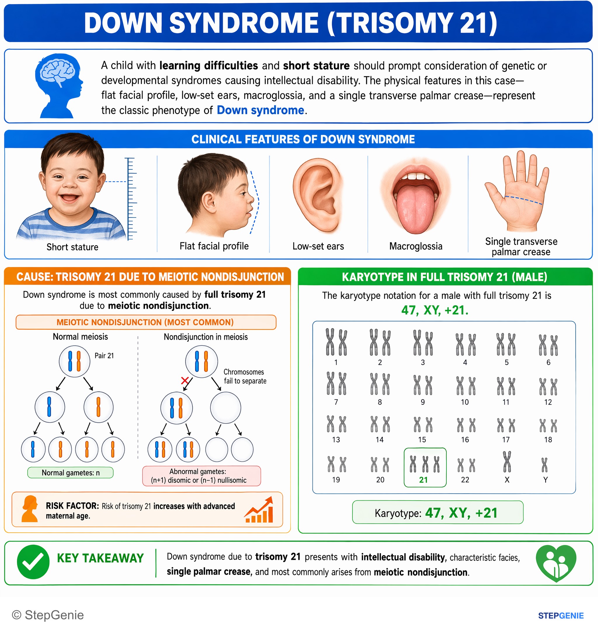
Down syndrome due to trisomy 21 presents with intellectual disability, characteristic facies, single palmar crease, and most commonly…
USMLE Step 1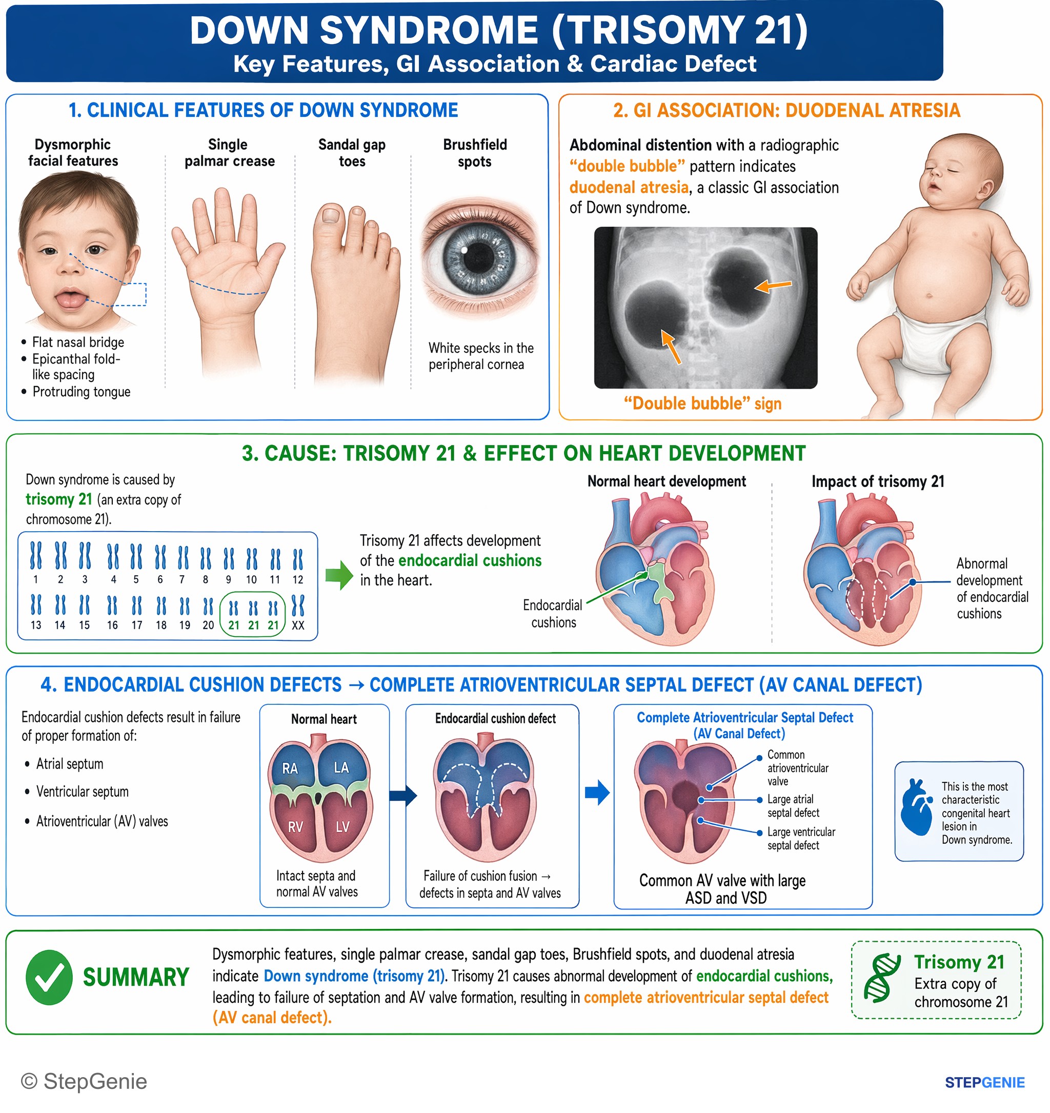
Down syndrome (trisomy 21) with duodenal atresia is strongly associated with complete atrioventricular septal defect due to endocardial…
USMLE Step 1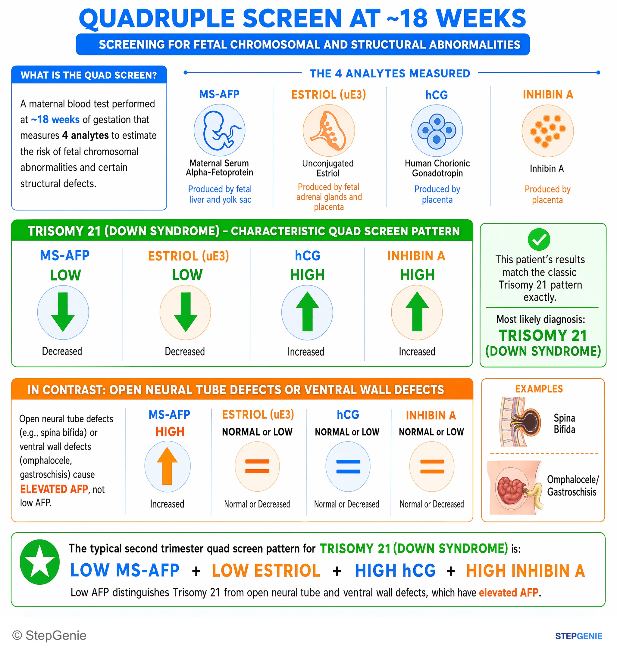
Trisomy 21 (Down syndrome) in 2nd trimester: low MS-AFP, low estriol, high hCG, high inhibin A on quadruple screen.
USMLE Step 1Frequently Asked Questions
What is the most common cause of Down syndrome?
About 95% of Down syndrome is free trisomy 21 caused by meiotic nondisjunction, the failure of chromosomes to separate during gamete formation. The extra chromosome 21 is typically maternal, and the error happens mostly in meiosis I of the egg, which is why risk rises with maternal age: roughly 1 in 352 births at age 35 and 1 in 85 at age 40. The karyotype is 47,XX,+21 or 47,XY,+21, and this form is not inherited.
How is translocation Down syndrome different from trisomy 21?
In Robertsonian translocation Down syndrome (about 3-4% of cases), extra chromosome 21 material is fused to another acrocentric chromosome, usually 14, so the child has 46 chromosomes instead of 47. About half of these translocations are new and half are inherited from a healthy balanced-carrier parent, who has 45 chromosomes. Recurrence risk is about 1 in 10 if the mother carries t(14;21), 1 in 20 if the father does, and 100% if a parent carries t(21;21), so both parents are karyotyped.
What heart defect is classically associated with Down syndrome?
About half of babies with Down syndrome have congenital heart disease. The classic lesion is an atrioventricular septal defect, also called an endocardial cushion or AV canal defect, which makes up about 40% of these heart defects; ventricular septal defects are also common. That is why every newborn with Down syndrome gets an echocardiogram, either prenatally or at birth, as part of routine surveillance.
What does the quad screen show in Down syndrome?
The second-trimester quad screen (15 to 20 weeks) shows low alpha-fetoprotein, low unconjugated estriol, high hCG and high inhibin A in Down syndrome, and detects about 80% of cases. A memory hook is that the HI markers, hCG and inhibin, are high. In trisomy 18 all four markers are low. The first-trimester screen shows increased nuchal translucency, low PAPP-A and high hCG. Cell-free DNA is more sensitive, but any positive screen needs CVS or amniocentesis to confirm.
Which blood cancers are linked to Down syndrome?
Leukemia is about 10 times more likely in Down syndrome. About 10% of newborns have transient abnormal myelopoiesis (a transient myeloproliferative disorder), and 20-30% of those go on to develop acute megakaryoblastic leukemia before age 4, a form linked to GATA1 mutations. Acute lymphoblastic leukemia is also more common. Neonates can also have polycythemia or thrombocytopenia, and children get a yearly blood count from age 1.
Why do people with Down syndrome get early Alzheimer disease?
The amyloid precursor protein gene, APP, sits on chromosome 21, so an extra copy of chromosome 21 means an extra copy of APP, which raises the risk of Alzheimer disease at a younger age. About half of adults with Down syndrome develop Alzheimer disease, and StatPearls reports dementia in 50% to 70% by age 60. On exams, new memory loss or functional decline in a middle-aged adult with Down syndrome points to early-onset Alzheimer disease.