Patau syndrome (trisomy 13) is caused by an extra chromosome 13, usually from maternal meiotic nondisjunction, and the classic newborn has midline defects (holoprosencephaly, cleft lip and palate, microphthalmia), postaxial polydactyly, punched-out scalp defects (cutis aplasia) and severe congenital heart disease. About 75-80% of cases are complete (free) trisomy 13, about 5% are mosaic, and most of the rest are translocation forms, typically a Robertsonian 13;14 translocation. It occurs in roughly 1 in 10,000 to 20,000 live births, and survival is poor: about half of affected infants die in the first month and only about 1 in 10 lives past the first birthday.
The Q-bank vignette
A newborn girl is born at 38 weeks to a 41-year-old woman who had no prenatal care. Her weight is below the 3rd percentile. She has bilateral cleft lip and palate, small eyes, a 2-cm hairless, ulcerated defect over the vertex of the scalp and an extra digit on the ulnar side of each hand. A holosystolic murmur is heard at the left lower sternal border. She has several apneic spells on her first day. Cranial ultrasound shows a single midline ventricle with no falx.
Which karyotype is most likely?
- A. 47,XX,+21
- B. 47,XX,+18
- C. 47,XX,+13
- D. 46,XX,del(5p)
- E. 46,XX with a 22q11.2 microdeletion
Answer: C, trisomy 13 (Patau syndrome). A single ventricle with no falx is alobar holoprosencephaly: the forebrain (prosencephalon) failed to divide into two hemispheres. Add midline clefting, microphthalmia, postaxial polydactyly, cutis aplasia of the scalp and a VSD-type murmur, and the pattern is trisomy 13. Advanced maternal age fits nondisjunction, and central apnea is one of the main causes of neonatal death in this condition, alongside heart disease, pulmonary hypertension, aspiration and airway obstruction.
Why every other answer choice is wrong
- A, trisomy 21 (Down syndrome): expect hypotonia, a flat facial profile, upslanting palpebral fissures, epicanthal folds, Brushfield spots and a sandal gap. The signature heart lesion is an atrioventricular septal defect, and duodenal atresia is the GI clue. Holoprosencephaly, polydactyly and scalp defects are not part of it. A single transverse palmar crease does not help: Merck lists it in trisomy 13 too. More in the Down syndrome breakdown.
- B, trisomy 18 (Edwards syndrome): the closest mimic (severe growth restriction, heart defects, early death, older mother). But the hands are clenched, with the index finger overlapping the third and fourth fingers, and the typical extras are rocker-bottom feet, a prominent occiput, micrognathia, a small mouth and a short sternum. Clefting and holoprosencephaly are not its signature. See the Edwards syndrome breakdown.
- D, cri-du-chat (5p deletion): a deletion, not a trisomy. The clue is a high-pitched, cat-like cry with microcephaly, hypotonia, widely spaced eyes and intellectual disability; some children have heart defects. Compare it in the cri-du-chat table.
- E, 22q11.2 deletion (DiGeorge syndrome): palate problems and a heart defect overlap, but the heart lesions are conotruncal (tetralogy of Fallot, interrupted aortic arch, truncus arteriosus) and the clues are hypocalcemia from low PTH and T-cell deficiency. Holoprosencephaly and cutis aplasia are not part of it, and polydactyly is only an occasional finding. Full picture in the DiGeorge syndrome breakdown.
Follow-up twist: the karyotype instead shows 46 chromosomes, with the extra chromosome 13 long arm fused to a chromosome 14 (a Robertsonian translocation). The next step is to karyotype both parents: a healthy balanced carrier has 45 chromosomes and, unlike after sporadic nondisjunction, a raised risk of another affected pregnancy.
Patau syndrome features: what to look for
| System | Findings (frequency where reported) |
|---|---|
| Brain | Holoprosencephaly spectrum (about 74%); trisomy 13 is its most common chromosomal cause. Severe intellectual disability, seizures, apneic spells |
| Face and eyes | Cleft lip and/or palate (about 45%); microphthalmia or anophthalmia (53-60%); iris coloboma, retinal dysplasia; hypotelorism or even cyclopia with severe holoprosencephaly; micrognathia; low-set, malformed ears; hearing loss |
| Skin | Cutis aplasia (focal scalp defects); loose skin folds on the neck |
| Hands | Postaxial polydactyly (63-67%); single transverse palmar crease; narrow, hyperconvex fingernails |
| Heart | Severe congenital heart disease in about 57-80%: VSD, ASD, PDA; dextrocardia is common |
| Abdomen and genitourinary | Omphalocele; renal anomalies (about 71%); cryptorchidism (about 78%); abnormal scrotum in boys, bicornuate uterus in girls |
| Growth | Small for gestational age |
Frequencies are from StatPearls and Merck; any infant can show only part of the list.
Why it happens: three genetic routes
| Form | Share | Mechanism | Counseling point |
|---|---|---|---|
| Complete trisomy (47,XX,+13 or 47,XY,+13) | About 75-80% | Meiotic nondisjunction; maternal in about 91%, mostly in meiosis I; risk rises with maternal age | Sporadic; recurrence risk is low |
| Translocation or partial trisomy | Most of the rest (Orphanet: about 20%) | Usually a Robertsonian translocation (typically 13;14) carrying extra 13q material; Robertsonian translocations join acrocentric chromosomes (13, 14, 15, 21, 22) | May be inherited from a balanced carrier parent: karyotype both parents |
| Mosaic trisomy 13 | About 5% | Postzygotic error: a mix of normal and trisomic cells | Milder, more variable features and longer survival are possible; severity does not reliably track the percentage of trisomic cells |
For how a second cell line arises after fertilization, see the mosaicism comparison table.
Prenatal screening and diagnosis
- First-trimester combined screen (11-14 weeks): nuchal translucency is often 3.5 mm or more, with low free beta-hCG and low PAPP-A. This pattern does not reliably separate trisomy 13 from trisomy 18. Down syndrome differs: high beta-hCG, low PAPP-A and a thick nuchal translucency.
- Cell-free DNA screening (from 10 weeks): the most sensitive screen, but less so for trisomy 13. In Merck's summary of meta-analysis data, sensitivity is about 91% for trisomy 13, versus 98% for trisomy 18 and 99% for trisomy 21. The "fetal" DNA actually comes from placental trophoblast, so a positive result is a screen, never a diagnosis.
- Ultrasound: structural anomalies such as holoprosencephaly, heart defects or facial clefts raise suspicion.
- Diagnosis: karyotype, FISH or chromosomal microarray on chorionic villus sampling (from 10 weeks to the end of the first trimester) or amniocentesis (safest after 14 weeks). Each carries a procedure-related loss risk of roughly 0.1-0.2% (Merck). Confined placental mosaicism turns up in about 1% of CVS samples and can mislead; rarely, amniocentesis is needed to settle it. After birth, a blood karyotype confirms the diagnosis, and FISH gives a rapid answer.
- Maternal risk: trisomy 13 pregnancies have higher rates of preeclampsia and preterm delivery (StatPearls).
Prognosis and management
- Most trisomy 13 pregnancies never reach a live birth: StatPearls cites antenatal mortality above 95%, partly reflecting elective termination. After birth, about 50% die within the first month and up to 90% within the first year; MedlinePlus puts survival past age 1 at 5-10%. Survivors have severe intellectual disability.
- There is no cure. Care is individualized after shared decision-making: comfort-focused palliative care, or selected interventions such as ventilatory support, gastrostomy feeding, seizure treatment and cardiac or cleft repair. Merck notes that survival has improved in children who undergo surgery.
- Mosaic and partial trisomy 13 can be milder, with longer survival.
Patau vs Edwards vs Down: comparison table
| Feature | Patau (trisomy 13) | Edwards (trisomy 18) | Down (trisomy 21) |
|---|---|---|---|
| Frequency | About 1 in 10,000-20,000 live births | About 1 in 5,000 live births | About 1 in 700 live births |
| One-line picture | Midline defects + polydactyly | Clenched fists + rocker-bottom feet | Hypotonia + flat facies |
| Head and face | Holoprosencephaly, cleft lip/palate, microphthalmia, cutis aplasia | Prominent occiput, micrognathia, small mouth, low-set malformed ears | Flat facial profile, upslanting palpebral fissures, epicanthal folds, Brushfield spots |
| Hands and feet | Postaxial polydactyly | Index finger overlapping third and fourth; rocker-bottom feet, clubfeet | Short broad hands, single palmar crease, sandal gap |
| Heart | VSD, ASD, PDA; dextrocardia | PDA, VSD | Congenital heart disease in about 50%; VSD and AV septal defect most common |
| Other clues | Omphalocele, renal anomalies | Short sternum, renal anomalies; more common in females | Duodenal atresia, Hirschsprung disease, hypothyroidism, leukemia, early-onset Alzheimer disease |
| Prenatal serum screen | First trimester: thick NT, low beta-hCG, low PAPP-A | Same first-trimester pattern; second trimester: low AFP, hCG and estriol | First trimester: thick NT, high beta-hCG, low PAPP-A |
| Cell-free DNA sensitivity | About 91% | About 98% | About 99% |
| Usual cause | Maternal nondisjunction; about 75-80% complete trisomy | Maternal nondisjunction; over 95% complete trisomy | Nondisjunction about 95%, translocation about 4%, mosaicism most of the rest |
| Survival | About 5-10% live past age 1 | Many die before birth or in the first month; about 5-10% live past age 1 | Median life expectancy about 60 years (high-resource settings) |
All three become more likely with advanced maternal age.
How USMLE tests trisomy 13
- Midline + extra digits = 13. Holoprosencephaly, cyclopia, cleft lip, a scalp defect and polydactyly: any two should make you think trisomy 13.
- The foot is a distractor. Rocker-bottom feet are the classic trisomy 18 foot (Merck and StatPearls list them under trisomy 18, not trisomy 13), but some review books and older Q-bank stems attach them to trisomy 13 as well. A stem that pairs them with clefting, polydactyly or holoprosencephaly still points to trisomy 13, so decide on the hands (polydactyly versus overlapping fingers) and the midline findings.
- The heart is not specific. VSD and PDA appear in both trisomy 13 and 18; an AV septal defect points to Down syndrome; a conotruncal lesion with hypocalcemia points to 22q11.2 deletion.
- Translocation twist. Recurrent affected pregnancies, or a karyotype with 46 chromosomes and extra chromosome 13 material, points to a Robertsonian translocation: karyotype the parents.
- Mosaic twist. A milder phenotype or longer-than-expected survival suggests mosaic trisomy 13.
- Screen versus diagnosis. An abnormal cell-free DNA or combined screen leads to CVS or amniocentesis, not straight to a diagnosis.
Key takeaways
- Trisomy 13 = holoprosencephaly, cleft lip/palate, microphthalmia, postaxial polydactyly, cutis aplasia and severe heart disease.
- About 75-80% complete trisomy from maternal meiotic nondisjunction (older mothers), about 5% mosaic, most of the rest Robertsonian translocations (typically 13;14): karyotype the parents.
- First-trimester screen: thick NT, low beta-hCG, low PAPP-A (same as trisomy 18); cell-free DNA detects about 91%; confirm with CVS or amniocentesis.
- About half die in the first month; only about 1 in 10 survives the first year; mosaic forms can live longer.
- 13 vs 18: polydactyly and midline defects vs clenched hands with overlapping fingers, a prominent occiput and a small jaw (rocker-bottom feet are classic for 18 but do not settle the question alone). 21: hypotonia, flat facies, AV septal defect, duodenal atresia.
Sources
- Trisomy 13 (Patau Syndrome) — StatPearls, NCBI Bookshelf (2026)
- Trisomy 13 (Patau Syndrome) — Merck Manual Professional Edition (2025)
- Trisomy 13 — MedlinePlus Genetics (2021)
- Trisomy 18 — Merck Manual Professional Edition (2025)
- Trisomy 18 — MedlinePlus Genetics (2021)
- Down Syndrome (Trisomy 21) — Merck Manual Professional Edition (2025)
- Noninvasive Prenatal Fetal Screening Tests — Merck Manual Professional Edition (2024)
- Procedures for Prenatal Genetic Diagnosis — Merck Manual Professional Edition (2024)
- Cri-du-chat Syndrome — MedlinePlus Genetics (2022)
- 22q11.2 Deletion Syndrome — GeneReviews, NCBI Bookshelf (2025)
- Trisomy 18 (Edwards Syndrome) — StatPearls, NCBI Bookshelf (2026)
- Trisomy 13 — Orphanet (2023)
Visual explanations from the StepGenie QBank
Infographics StepGenie drew to explain questions in its own QBank, matched to this article's topic. Each caption is the takeaway of the question it explains.
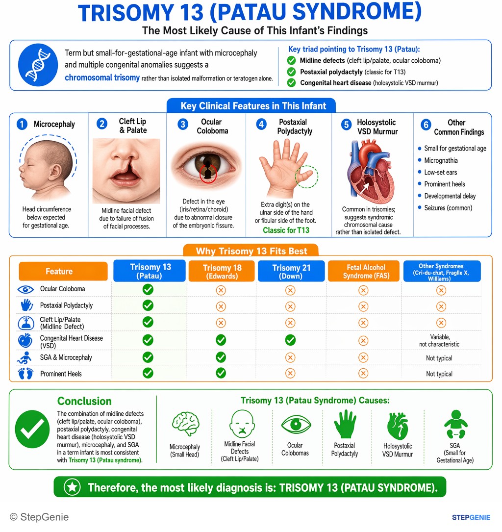
Trisomy 13 (Patau syndrome) causes microcephaly, midline facial defects, postaxial polydactyly, holosystolic VSD murmur, and ocular…
USMLE Step 1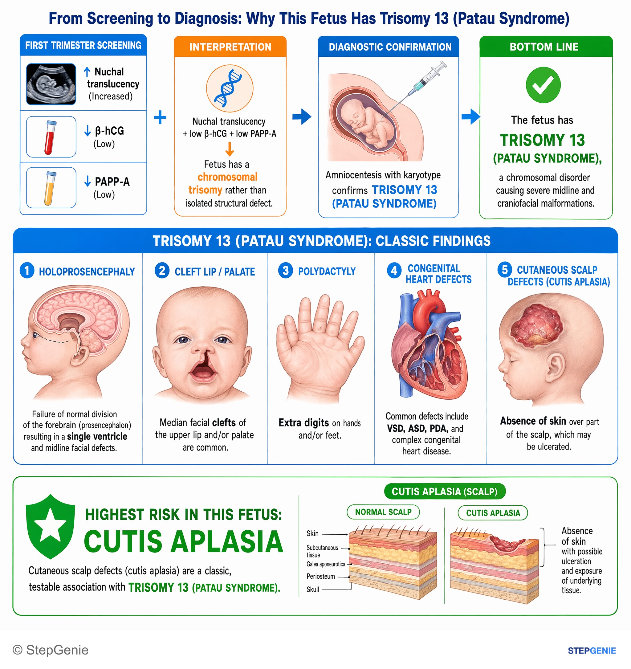
Trisomy 13 (Patau syndrome) causes severe midline defects: cleft lip/palate, holoprosencephaly, polydactyly, and cutaneous scalp defects…
USMLE Step 1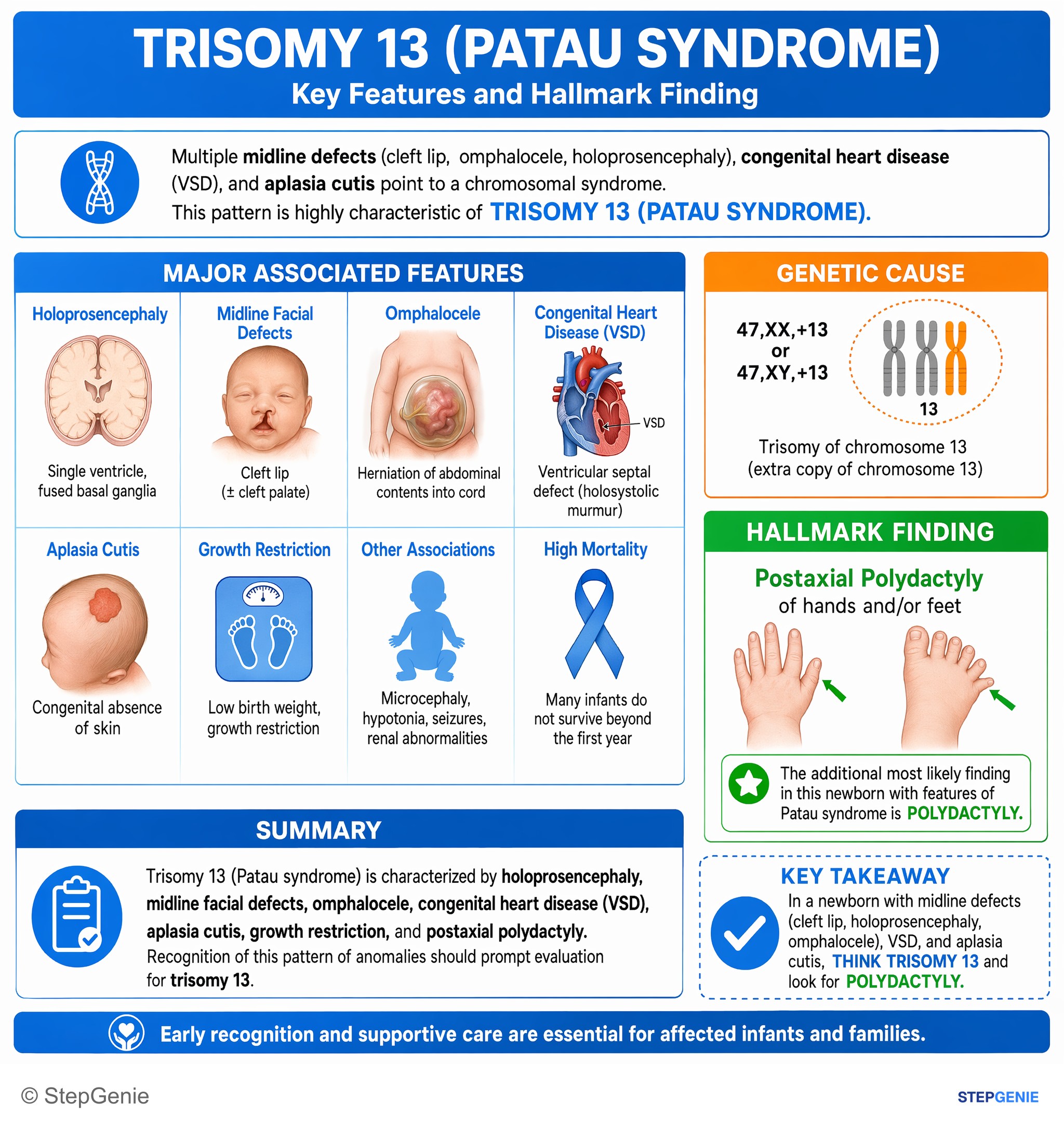
Trisomy 13 (Patau syndrome) causes holoprosencephaly, midline facial defects, omphalocele, VSD, growth restriction, and postaxial…
USMLE Step 1Frequently Asked Questions
What are the classic features of Patau syndrome?
The classic newborn with trisomy 13 has midline defects and extra digits: holoprosencephaly (the forebrain fails to divide into two hemispheres), cleft lip and/or palate, small or absent eyes, postaxial polydactyly, punched-out scalp defects called cutis aplasia, and severe congenital heart disease such as a VSD, ASD or PDA. Other findings include low-set malformed ears, omphalocele, kidney anomalies, small size for gestational age, apneic spells, seizures and severe intellectual disability.
What causes trisomy 13?
About 75 to 80% of cases are complete trisomy 13, caused by meiotic nondisjunction, usually in the mother's egg during meiosis I, so the risk rises with maternal age. About 5% are mosaic: the error happens after fertilization, so only some cells carry the extra chromosome. Most of the rest are translocation forms, typically a Robertsonian translocation between chromosomes 13 and 14, which can be inherited from a parent who carries a balanced translocation.
How long do babies with trisomy 13 live?
Survival is poor. About half of liveborn infants with trisomy 13 die within the first month and up to 90% within the first year; only about 5 to 10% live past their first birthday. Deaths usually come from central apnea, severe heart disease, pulmonary hypertension, aspiration or airway obstruction. Mosaic and partial forms can be milder, and survival has improved in some children who undergo surgery. Survivors have severe intellectual disability.
How is trisomy 13 detected before birth?
Screening raises suspicion and diagnostic testing confirms it. On the first-trimester combined screen, trisomy 13 typically shows a nuchal translucency of 3.5 mm or more with low free beta-hCG and low PAPP-A, the same pattern as trisomy 18. Cell-free DNA screening from 10 weeks detects about 91% of cases, fewer than for trisomy 21. Ultrasound may show holoprosencephaly, heart defects or clefts. Diagnosis needs chorionic villus sampling or amniocentesis with karyotype, FISH or microarray.
What is the difference between Patau syndrome and Edwards syndrome?
Both are severe autosomal trisomies linked to older maternal age, growth restriction, heart defects and early death. Patau syndrome (trisomy 13) is the midline syndrome: holoprosencephaly, cleft lip and palate, microphthalmia, cutis aplasia and postaxial polydactyly. Edwards syndrome (trisomy 18) brings clenched fists with the index finger overlapping the third and fourth fingers, rocker-bottom feet, a prominent occiput, a small jaw and mouth, and a short sternum. On second-trimester serum screening, trisomy 18 typically shows low AFP, hCG and unconjugated estriol.
Is trisomy 13 inherited, and can it happen again?
Most trisomy 13 is not inherited: it results from a random nondisjunction error during egg or sperm formation, and the recurrence risk is low. The exception is translocation trisomy 13. If the baby's karyotype shows a Robertsonian translocation, both parents should be karyotyped, because a healthy parent can carry a balanced translocation and then has an increased risk of another affected pregnancy. Mosaic trisomy 13 arises after fertilization and is not inherited.