Williams syndrome (Williams-Beuren syndrome) is a contiguous gene deletion syndrome: a heterozygous 1.5-1.8 Mb deletion at chromosome 7q11.23 that removes about 25-27 genes, including ELN (elastin). Losing one ELN copy causes an elastin arteriopathy, classically supravalvular aortic stenosis (SVAS) and peripheral pulmonary stenosis, while the wider deletion gives elfin facies, usually mild intellectual disability with relatively strong language but very weak visuospatial skills, and an overfriendly, anxious personality. Hypercalcemia (20-40%, most often symptomatic before age 2) is the classic lab clue. Most cases are de novo but the deletion is autosomal dominant, and a normal karyotype does not exclude it: chromosomal microarray or FISH confirms it.
The 5-second rule: in Williams, everything is "above"
When a stem gives you a chatty, overfriendly child with a murmur, say it in one breath: "7q11.23 deletion, ELN lost, everything is above."
| Above what? | The finding | How it shows up in a stem |
|---|---|---|
| Above the valve | Supravalvular aortic stenosis: narrowing at the sinotubular junction, just above the aortic sinuses | Harsh systolic ejection murmur at the upper sternal border, usually without an ejection click; right-arm systolic BP often higher than left |
| Calcium above normal | Hypercalcemia (20-40%) and hypercalciuria (about 30%) | Infant with irritability, vomiting and constipation; nephrocalcinosis in severe cases |
| Sociability above normal | Overfriendliness, social disinhibition and excessive empathy ("cocktail party" personality) | Hugs strangers and talks to everyone, yet anxiety and ADHD are common |
| Language above visuospatial | Relative strength in language and verbal short-term memory; extreme weakness in visuospatial construction | Fluent talker who cannot copy a simple shape, draw or finish a puzzle |
Then add the face: "elfin" facies, with a broad forehead, periorbital fullness, a stellate (lacy) iris, a short nose with a broad tip, a long philtrum, a wide mouth with full lips, a small jaw and, in young children, small widely spaced teeth. A hoarse voice (about 90%) is another connective tissue clue.
Flip it for DiGeorge. The other microdeletion classic, 22q11.2 deletion (DiGeorge) syndrome, brings calcium below normal (hypoparathyroidism), conotruncal heart defects instead of SVAS, and T-cell deficiency from thymic hypoplasia.
What is deleted, and why one deletion does so much
- The lesion: a heterozygous 1.5-1.8 Mb deletion at 7q11.23 containing about 25-27 genes. One event removes a row of neighboring genes, which is what contiguous gene deletion syndrome means, and it is why so many organ systems are involved.
- Why the same deletion recurs: the region is flanked by low-copy repeats with nearly identical sequence. When these repeats misalign, nonallelic homologous recombination deletes the segment between them, so unrelated patients carry nearly identical deletions.
- ELN haploinsufficiency: with one ELN copy, elastin production falls by about half. Arterial walls become thick, stiff and narrow (elastin arteriopathy), and reduced elastin also contributes to connective tissue problems in the skin and joints.
- The other genes: loss of GTF2I, GTF2IRD1 and LIMK1 is thought to shape the cognitive and behavioral profile (LIMK1 with visuospatial difficulty). Patients whose deletion includes NCF1 have less hypertension.
- Inheritance: autosomal dominant. Most cases are de novo, so parents are usually unaffected, but each child of an affected person has a 50% chance of inheriting the deletion.
- How common: about 1 in 7,500 to 1 in 18,000 people.
Heart and vessels: an elastin arteriopathy
Cardiovascular disease affects about 80% of patients and is the main cause of illness and death.
| Lesion | How often | Course and exam clues |
|---|---|---|
| Supravalvular aortic stenosis | About 75% | Hourglass narrowing at the sinotubular junction; may worsen, especially in the first 5 years; about one-third of children with SVAS need surgery |
| Peripheral pulmonary stenosis | 40-60% | Common in infancy; usually improves without intervention |
| Hypertension | About 50% | Stiff arteries or renal artery stenosis; measure BP in all four limbs |
| Other arterial stenoses (coronary, renal, mesenteric; any artery) | Variable | Coronary artery stenosis is implicated in sudden death (about 1 per 1,000 patient-years, 25-100 times the age-matched rate); sedation and anesthesia raise the risk of cardiac arrest |
Because the obstruction sits above the valve, there is usually no ejection click; in congenital valvular aortic stenosis, a click can be heard while the stiff leaflets are still mobile. The right arm reads higher than the left because of the Coanda effect: the jet clings to the aortic wall and streams into the innominate artery.
Calcium: the lab clue
- Who and when: hypercalcemia occurs in 20-40% and is most often symptomatic in the first 2 years (irritability, vomiting, constipation). It can cause dehydration and hypercalciuria (about 30%); nephrocalcinosis is uncommon (fewer than 5%).
- Why: unknown. Gut calcium absorption is increased, and vitamin D promotes that absorption.
- Management: avoid vitamin supplements that contain vitamin D, especially in young children; keep dietary calcium at no more than 100% of the recommended intake; encourage fluids; use oral corticosteroids for refractory cases and IV pamidronate for severe symptomatic hypercalcemia. Serum calcium is checked every 4-6 months before age 2.
- Exam framing: "Williams = hypercalcemia" is the classic pairing, but only a minority have it. For the other causes of high and low calcium, see hypocalcemia vs hypercalcemia.
Brain, behavior and the rest of the body
- Cognition: intellectual disability, usually mild, in about 75%. Adults may read at a high-school level, but writing, drawing and math are hard.
- Personality: the distinctive Williams personality (overfriendliness, social disinhibition, empathy) is present in about 95%. Non-social anxiety (about 80%, including specific phobias), ADHD (about 65% of children) and autism spectrum disorder (10-20%) are common. About 35% have hyperacusis (discomfort with ordinary sounds), and many do well at music.
- Endocrine: early puberty (about 50%), hypothyroidism (about 10%), and diabetes (about 20%, mostly in adults).
- GI and connective tissue: feeding difficulties in infancy (about 70%), constipation (about 50%), umbilical and inguinal hernias, colon diverticula (about 30%), soft lax skin, lax joints in infancy and contractures later, dental malocclusion and growth deficiency.
Diagnosis: why the karyotype is normal
- Confirm with chromosomal microarray (CMA) or targeted deletion testing such as FISH; both detect the recurrent deletion. In practice it is often found on CMA ordered for developmental delay, intellectual disability or autism.
- A routine G-banded karyotype cannot detect it: 1.5-1.8 Mb is below the resolution of conventional banding. On exams, "karyotype normal" never rules out a microdeletion.
- Workup after diagnosis: echocardiogram and ECG, blood pressure in all four limbs, serum calcium and urine calcium-to-creatinine ratio, thyroid function, and developmental, vision and hearing assessment.
Management in one minute
There is no cure; care is lifelong and directed at complications.
- Cardiology at diagnosis, then at least yearly until age 5 and every 2-3 years after that; surgical repair for significant SVAS.
- Anesthesia consultation before any sedation or surgery, because of the risk of cardiac arrest.
- Development: early intervention; speech, occupational, physical and feeding therapy; special education; treatment of anxiety and ADHD.
- Genetic counseling: 50% risk for each child of an affected person; prenatal and preimplantation testing are possible once the deletion is known.
Williams vs the look-alikes
| Syndrome | Genetics | Heart | Calcium | Other clues |
|---|---|---|---|---|
| Williams | 7q11.23 deletion (ELN); AD, mostly de novo | SVAS, peripheral pulmonary stenosis | High (20-40%) | Elfin face, overfriendly, language > visuospatial |
| 22q11.2 deletion (DiGeorge) | 22q11.2 deletion (TBX1); more than 90% de novo | Conotruncal: tetralogy of Fallot, truncus arteriosus, interrupted aortic arch, VSD | Low (hypoparathyroidism) | Palatal anomalies, T-cell immune deficiency, learning difficulties |
| Noonan | AD; PTPN11 in about half (RAS/MAPK pathway) | Valvar pulmonary stenosis, hypertrophic cardiomyopathy | Normal | Short stature, widely spaced eyes, low-set ears |
| Turner | 45,X or mosaic (seen on karyotype) | Bicuspid aortic valve, coarctation | Normal | Short stature, ovarian insufficiency; usually normal intelligence (Turner review) |
| Isolated (familial) SVAS | ELN variant; AD | SVAS | Normal | No Williams face, personality or cognitive profile |
How USMLE tests it (and the traps)
- Supra- vs valvular. Williams narrows the aorta above a normal valve, so there is usually no ejection click. Congenital valvular AS (often a bicuspid valve) can have one while the leaflets are stiff but not yet immobile.
- Elastin vs fibrillin. ELN loss narrows arteries; FBN1 variants (Marfan syndrome) dilate the aortic root, with aneurysm and dissection. See the Marfan syndrome review.
- Hyper vs hypo. High calcium points to Williams; low calcium with tetany or seizures points to 22q11.2.
- "Karyotype normal." Microdeletions sit below karyotype resolution, so pick CMA or FISH.
- Happy is not always Williams. Angelman syndrome (loss of maternal UBE3A on chromosome 15) also brings a happy, excitable demeanor with frequent laughter, but with severe speech impairment, seizures and ataxia. Williams children talk a lot.
Practice question
A 4-year-old girl is evaluated for a heart murmur. She greets the nurse with a hug and chats happily with everyone in the room, but her preschool teacher says she cannot copy simple shapes or finish puzzles. In infancy she had irritability, vomiting and constipation, and her serum calcium was elevated. She has a broad forehead, periorbital fullness, a lacy (stellate) iris pattern, a wide mouth with full lips and a hoarse voice. A harsh systolic ejection murmur is heard at the upper right sternal border without an ejection click, and systolic blood pressure is higher in the right arm than the left. Echocardiography shows narrowing of the ascending aorta at the sinotubular junction with a normal trileaflet aortic valve. A G-banded karyotype is 46,XX. Which of the following best explains her condition?
A. Heterozygous deletion at 7q11.23 that includes ELN B. Heterozygous deletion at 22q11.2 that includes TBX1 C. Pathogenic variant in FBN1 D. Gain-of-function variant in PTPN11 E. Monosomy X
Answer: A. Overfriendliness, weak visuospatial construction, infantile hypercalcemia, elfin facies, a hoarse voice and supravalvular aortic stenosis add up to Williams syndrome. Losing one copy of ELN halves elastin production and narrows the aorta just above the valve. The karyotype is normal because a 1.5-1.8 Mb deletion is below its resolution; chromosomal microarray or FISH would confirm it.
- B: a 22q11.2 deletion causes hypocalcemia (hypoparathyroidism), conotruncal defects such as tetralogy of Fallot or truncus arteriosus, palatal anomalies and T-cell deficiency. A routine karyotype misses it too, so the calcium level and the heart lesion are what separate the two.
- C: FBN1 variants cause Marfan syndrome, which dilates the aortic root rather than narrowing the aorta above the valve; tall stature and lens subluxation are its clues.
- D: PTPN11 gain-of-function variants are the most common cause of Noonan syndrome, whose classic heart lesion is valvar pulmonary stenosis (or hypertrophic cardiomyopathy); calcium is normal.
- E: monosomy X (Turner syndrome) would show on the karyotype as 45,X and causes a bicuspid aortic valve and coarctation, not SVAS.
Key takeaways
- Williams = heterozygous 7q11.23 deletion (1.5-1.8 Mb, about 25-27 genes, including ELN): a contiguous gene deletion syndrome, autosomal dominant but usually de novo.
- Everything is above: stenosis above the valve (SVAS, about 75%), calcium above normal (hypercalcemia, 20-40%, most often symptomatic before age 2), sociability above normal, language above visuospatial skills.
- Elastin arteriopathy can narrow any artery: peripheral pulmonary stenosis (often improves), renal artery stenosis with hypertension, coronary stenosis with sudden death and anesthesia risk.
- Elfin face (broad forehead, periorbital fullness, stellate iris, wide mouth, full lips) and a hoarse voice.
- Diagnose with chromosomal microarray or FISH; the karyotype is normal.
- Avoid vitamin D-containing supplements, and get an anesthesia consultation before any sedation or surgery.
- DiGeorge is the mirror image: 22q11.2, hypocalcemia, conotruncal defects, T-cell deficiency.
Sources
- Williams Syndrome — GeneReviews, NCBI Bookshelf (2023)
- Williams syndrome — MedlinePlus Genetics, National Library of Medicine (2022)
- ELN gene — MedlinePlus Genetics, National Library of Medicine (2022)
- 7q11.23 Duplication Syndrome — GeneReviews, NCBI Bookshelf (2021)
- Williams Syndrome — StatPearls, NCBI Bookshelf (2025)
- Supravalvar Aortic Stenosis — StatPearls, NCBI Bookshelf (2023)
- Supravalvular aortic stenosis — MedlinePlus Genetics, National Library of Medicine (2012)
- Aortic Stenosis — Merck Manual Professional Edition (2025)
- 22q11.2 Deletion Syndrome — GeneReviews, NCBI Bookshelf (2025)
- Noonan syndrome — MedlinePlus Genetics, National Library of Medicine (2018)
- Turner syndrome — MedlinePlus Genetics, National Library of Medicine (2023)
- Angelman syndrome — MedlinePlus Genetics, National Library of Medicine (2022)
Visual explanations from the StepGenie QBank
Infographics StepGenie drew to explain questions in its own QBank, matched to this article's topic. Each caption is the takeaway of the question it explains.
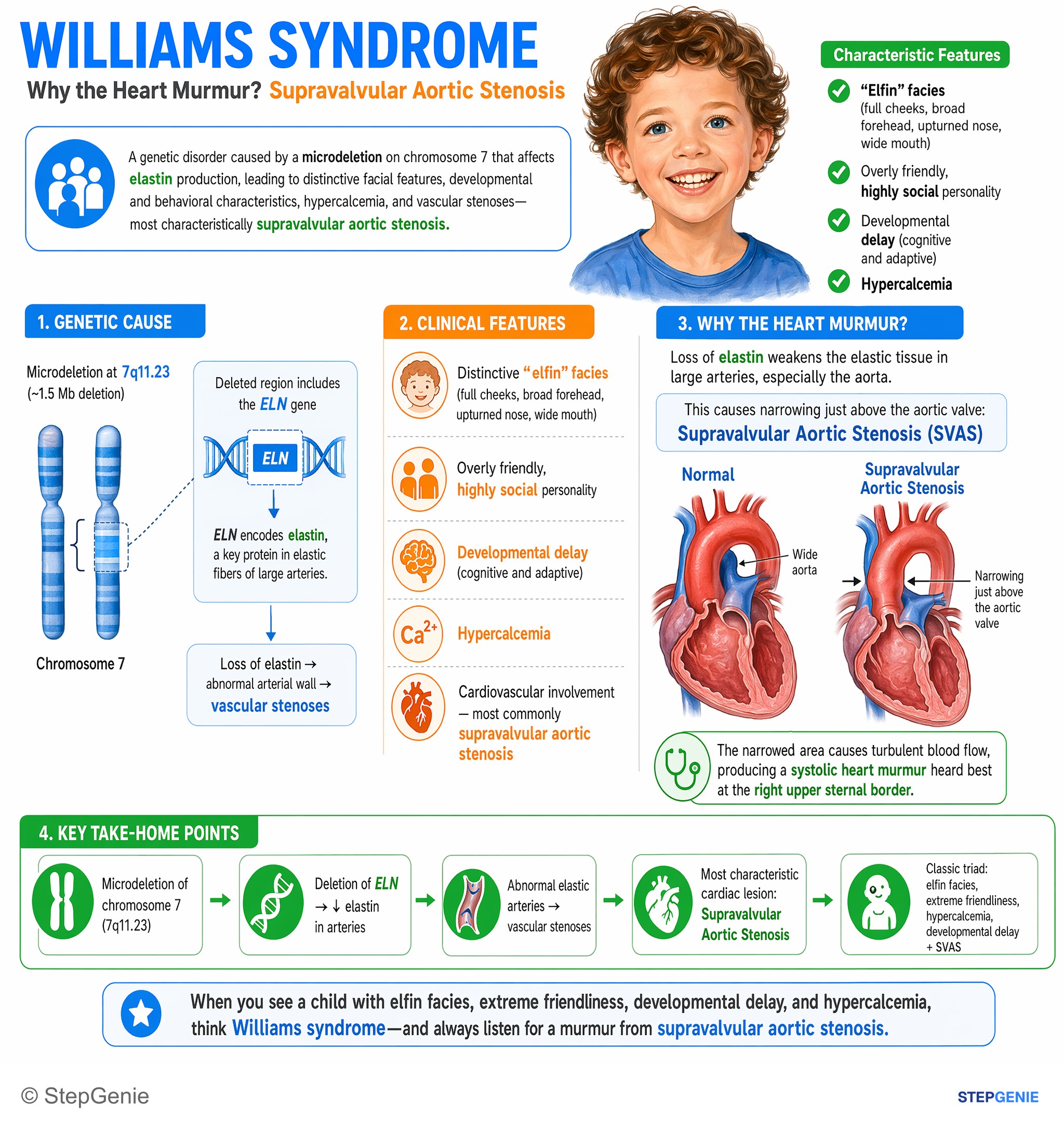
Williams syndrome: microdeletion of chromosome 7 → hypercalcemia, elfin facies, extreme friendliness, developmental delay, and…
USMLE Step 1Frequently Asked Questions
What chromosome deletion causes Williams syndrome?
Williams syndrome is caused by a heterozygous deletion of about 1.5 to 1.8 million base pairs at chromosome 7q11.23, which removes roughly 25 to 27 genes, including ELN, the elastin gene. Because one event removes several neighboring genes, it is a contiguous gene deletion syndrome. Most deletions arise de novo through nonallelic homologous recombination between low-copy repeats, but the condition is autosomal dominant, so an affected person has a 50% chance of passing it to each child.
Why does Williams syndrome cause supravalvular aortic stenosis?
The deletion removes one copy of ELN, so elastin production falls by about half. Arterial walls become thick, stiff and narrow, an elastin arteriopathy that is most obvious at the sinotubular junction just above the aortic valve. About 75% of patients have supravalvular aortic stenosis and 40-60% have peripheral pulmonary stenosis. Any artery can narrow, including the renal and coronary arteries, which explains the hypertension, the risk of sudden death and the need for careful anesthesia planning.
Why do children with Williams syndrome get hypercalcemia?
The exact cause is unknown, but calcium absorption from the gut is increased and vitamin D promotes that absorption. Hypercalcemia affects about 20-40% of patients and is most often symptomatic in the first two years of life, with irritability, vomiting and constipation; hypercalciuria and, rarely, nephrocalcinosis can follow. Management avoids vitamin supplements that contain vitamin D, especially in young children, keeps calcium intake no higher than the recommended amount, encourages fluids, and uses oral corticosteroids or IV pamidronate for refractory or severe cases.
What is the Williams syndrome personality and cognitive profile?
Most people with Williams syndrome are unusually friendly, socially disinhibited and empathic, often called a cocktail party personality, yet anxiety affects about 80% and ADHD about 65% of children. Intellectual disability is usually mild and occurs in about 75%. The profile is uneven: relative strengths in language and verbal short-term memory, with extreme weakness in visuospatial construction, such as copying shapes, drawing and puzzles. Many do well at music, and about a third have hyperacusis, an unusual sensitivity to sound.
How is Williams syndrome diagnosed, and can a karyotype detect it?
Diagnosis is confirmed by finding the 7q11.23 deletion with chromosomal microarray or a targeted test such as FISH; both detect the recurrent deletion. A routine G-banded karyotype cannot, because the 1.5 to 1.8 Mb deletion is below its resolution, so a normal karyotype does not exclude Williams syndrome. After diagnosis, patients need an echocardiogram and ECG, blood pressure in all four limbs, serum and urine calcium, thyroid tests, and developmental, vision and hearing assessment.
How do you tell Williams syndrome from DiGeorge syndrome?
Both are microdeletion syndromes that a routine karyotype misses, but they point in opposite directions. Williams syndrome (7q11.23, ELN) causes hypercalcemia, supravalvular aortic stenosis, elfin facies and an overfriendly personality. DiGeorge syndrome (22q11.2, TBX1) causes hypocalcemia from hypoparathyroidism, conotruncal heart defects such as tetralogy of Fallot or truncus arteriosus, palatal anomalies and T-cell deficiency from thymic hypoplasia. On a question, the calcium level and the type of heart lesion usually separate them within seconds.