Uniparental disomy (UPD) means both copies of a chromosome, or of part of one, come from the same parent; in heterodisomy they are that parent's two different homologs, and in isodisomy they are two identical copies of a single homolog. Heterodisomy classically starts with nondisjunction in meiosis I, and isodisomy with nondisjunction in meiosis II, monosomy rescue or a post-zygotic error. The chromosome count stays at 46, so a karyotype looks normal, and most UPD causes no disease. When it does, there are two routes. On the chromosomes with clinically important imprinted genes (6, 7, 11, 14, 15 and 20), it changes which parent's genes are active: maternal UPD 15 causes Prader-Willi syndrome and paternal UPD 15 causes Angelman syndrome. And isodisomy, but not pure heterodisomy, can make a child homozygous for a recessive variant that only one parent carries. UPD occurs in roughly 1 in 2,000 births.
Heterodisomy vs isodisomy
| Feature | Heterodisomy | Isodisomy |
|---|---|---|
| What the child inherits | Both of one parent's homologs: two different chromosomes | Two identical copies of one of that parent's homologs |
| Classic origin | Nondisjunction in meiosis I, then trisomy rescue | Nondisjunction in meiosis II, monosomy rescue or a post-zygotic error |
| Region around the centromere | Heterozygous (meiosis I error) | Homozygous (meiosis II error or monosomy rescue) |
| Can it unmask a recessive variant? | Not by itself; only within isodisomic segments left by crossing over | Yes: a carrier parent's variant becomes homozygous |
| Can it cause an imprinting disorder? | Yes, on chromosomes 6, 7, 11, 14, 15 and 20 | Yes, on the same chromosomes |
| SNP array of the child alone | Seen only where crossing over left runs of homozygosity; pure heterodisomy is invisible | A long run of homozygosity (ROH) with normal copy number |
| Classic example | Maternal UPD 15 after a maternal meiosis I error (Prader-Willi syndrome) | Maternal isodisomy 7 unmasking a CFTR variant (cystic fibrosis) |
Memory hook: iso = identical (one homolog, copied); hetero = different (both homologs). To place the error, remember what each division separates: meiosis I separates homologs and meiosis II separates sister chromatids. A meiosis I failure passes on both different homologs (heterodisomy); a meiosis II failure passes on two identical sisters (isodisomy).
Many real cases are mixed. Crossing over happens before either division, so sister chromatids are no longer identical along their whole length. A meiosis I error keeps heterozygosity around the centromere, where recombination is suppressed, but often leaves runs of homozygosity farther out; a meiosis II error always leaves homozygosity around the centromere. Monosomy rescue gives complete isodisomy, with no heterodisomic regions. The numbers reflect this:
- Prader-Willi syndrome (GeneReviews, 2026): complete isodisomy causes 4–5% of cases, UPD with isodisomic segments 17–23%, and heterodisomy without any isodisomic region about 8–11%.
- General population: among 205 UPD findings in 23andMe parent–child data, heterodisomy (76) and partial isodisomy (74) each outnumbered complete isodisomy (55), and maternal UPD outnumbered paternal UPD.
Isodisomy vs uniparental disomy. Isodisomy is a type of UPD, not an alternative to it: all uniparental isodisomy is UPD, but much UPD is heterodisomy. And not every run of homozygosity is UPD. Extended ROH on several chromosomes most often means the parents are related; ROH confined to one chromosome is the pattern that prompts UPD testing.
Meiosis I or meiosis II: how UPD arises
Nondisjunction is the failure of homologous chromosomes (meiosis I) or sister chromatids (meiosis II) to separate. After a meiosis I error, two of the four gametes carry both of the parent's homologs and two carry none. After a meiosis II error, one gamete carries two copies of the same homolog (its sister chromatids), one carries none and two are normal. Nondisjunction becomes more likely with maternal age, the same effect that raises the risk of trisomy 21, and most of it happens in maternal meiosis I.
UPD usually needs a second event that restores the count to 46:
- Trisomy rescue, the classic route. A disomic gamete meets a normal one, the zygote is trisomic, and early cells lose one of the three copies. If the lost copy is the only one from the other parent, which in theory happens in one-third of rescues, the embryo has UPD. Because most nondisjunction is maternal meiosis I, the usual product is maternal heterodisomy.
- Monosomy rescue. A gamete missing the chromosome meets a normal one, and the monosomic embryo duplicates its single copy. The result is complete isodisomy. This is rarer than trisomy rescue.
- Gamete complementation. A gamete missing a chromosome meets one carrying two copies of it. This is rare.
- Post-zygotic mitotic recombination. An exchange between chromatids in an early embryonic cell gives mosaic, segmental UPD of the end of a chromosome arm. This is how paternal UPD of 11p15 arises in Beckwith-Wiedemann syndrome.
- Structural rearrangements. In a fetus carrying a Robertsonian translocation between two different chromosomes, such as der(13;14), the UPD risk is about 0.6%; with a homologous rearrangement, mostly a de novo isochromosome, it is about 66%. Either one involving chromosome 14 or 15 is an indication for UPD testing.
The placenta keeps the evidence. After trisomy rescue, the trisomic cell line can persist in the placenta, so chorionic villus sampling (CVS) shows trisomy while the fetus is 46,XX or 46,XY: confined placental mosaicism. For trisomy 15 found this way, the chance of UPD has been estimated at 11–25%, which is why mosaicism for chromosome 6, 7, 11, 14, 15 or 20 on CVS prompts UPD testing even when amniocentesis is normal.
Why UPD causes disease, and why it usually doesn't
Most UPD is harmless. For most chromosomes it has no clinical consequence. In 23andMe data, no link between UPD and harmful traits survived correction for multiple testing, and UPD that presents with an imprinting or recessive disorder has an estimated prevalence of only 1 in 3,500 to 1 in 5,000. Disease comes from two mechanisms.
1. Imprinting. For a small share of genes, only the copy from one parent is active; the other is silenced by methylation marks laid down during egg or sperm formation. The clinically important imprinted regions lie on chromosomes 6, 7, 11, 14, 15 and 20. Two maternal copies of such a region leave none of its paternally expressed genes active, the functional equivalent of deleting the father's copy, and double the dose of its maternally expressed genes. The methylation mechanism is covered in genomic imprinting.
2. Unmasking a recessive variant. With isodisomy, a heterozygous carrier parent can pass the same variant twice. The first Mendelian disease shown to arise this way, reported in 1988, was cystic fibrosis in a child who had inherited two copies of a CFTR variant from a carrier mother and nothing from the father; CFTR lies on chromosome 7. This is the answer when a child has an autosomal recessive disease but only one parent is a carrier, once misattributed paternity and a deletion of the other allele are ruled out (the usual carrier math is in the autosomal recessive inheritance guide).
The sex chromosomes follow the same logic: maternal UPD of the X can give a girl an X-linked recessive disorder, and, rarely, paternal UPD of both sex chromosomes allows father-to-son transmission of an X-linked condition. More genetics guides are collected in the Biochemistry hub.
Uniparental disomy diseases: maternal vs paternal
Maternal and paternal UPD of the same imprinted region usually cause different syndromes. The table gives the share of each syndrome that UPD causes; paternal UPD is sometimes searched as "unipaternal disomy".
| Chromosome (region) | Maternal UPD causes | Paternal UPD causes |
|---|---|---|
| 6 (6q24) | — | Transient neonatal diabetes mellitus (about 40% of 6q24 cases) |
| 7 | Silver-Russell syndrome (7–10%) | — |
| 11 (11p15.5) | Silver-Russell syndrome (rare) | Beckwith-Wiedemann syndrome (about 20%), usually mosaic and segmental |
| 14 (14q32) | Temple syndrome | Kagami-Ogata syndrome |
| 15 (15q11.2–q13) | Prader-Willi syndrome (about 25–40%) | Angelman syndrome (3–7%) |
| 20 | Mulchandani-Bhoj-Conlin syndrome | Pseudohypoparathyroidism type 1B (about 10% of sporadic cases, paternal UPD of 20q) |
Mnemonic for chromosome 15: name the syndrome by the parent whose copy is missing. Prader-Willi = no Paternal copy, so maternal UPD; AngelMan = no Maternal UBE3A, so paternal UPD. In neurons only the maternal UBE3A allele is active, so two paternal copies leave none working.
Chromosome 15: Prader-Willi and Angelman syndromes
UPD is the second most common cause of Prader-Willi syndrome and a minor cause of Angelman syndrome:
| Feature | Prader-Willi syndrome | Angelman syndrome |
|---|---|---|
| What is lost | Paternally expressed genes of 15q11.2–q13, including the SNORD116 cluster | Maternal UBE3A, the only active copy in neurons |
| UPD form | Maternal UPD 15 | Paternal UPD 15 |
| Share caused by UPD | About 25% (MedlinePlus) to roughly 30–40% (GeneReviews 2026, all UPD subtypes) | About 3–7% |
| Main cause | Paternal deletion, about 60–70% | Maternal deletion, about 70–75% |
| Other causes | Imprinting defect by epimutation, about 2–4%; imprinting center deletion, under 0.5% | UBE3A variants about 11%; imprinting defects about 3%; about 10% unexplained |
| Clinical picture | Severe hypotonia and feeding difficulty in infancy, then hyperphagia and obesity; hypogonadism, short stature, small hands and feet | Severe developmental delay and speech impairment, ataxia, seizures, small head, happy demeanor with frequent laughter |
| What differs with UPD | More often advanced maternal age and post-term birth; less often the typical face or hypopigmentation; more psychosis (up to 64% vs 25% with a deletion) and autism | Usually milder than the deletion, which brings more microcephaly, seizures and motor and language problems, and a lower BMI |
The maternal age link follows from the mechanism: trisomy rescue after a maternal meiosis I error. Full clinical comparisons are in Prader-Willi vs Angelman, and a worked question with UPD as a distractor is in the genomic imprinting question breakdown.
Beckwith-Wiedemann, Silver-Russell and the rarer UPD syndromes
Beckwith-Wiedemann syndrome (paternal UPD 11p15, about 20%). BWS is an overgrowth spectrum: macroglossia, lateralized overgrowth, abdominal wall defects such as omphalocele, neonatal hypoglycemia, and embryonal tumors such as Wilms tumor and hepatoblastoma. The most common cause is loss of methylation at imprinting center 2 (about 50%). Paternal UPD arises by post-zygotic recombination, so it is mosaic; it is more often linked with lateralized overgrowth, and higher levels of mosaicism go with a more severe phenotype. Tumor risk depends on the mechanism:
| BWS mechanism | Any tumor | Wilms tumor | Hepatoblastoma |
|---|---|---|---|
| Gain of methylation at imprinting center 1 | 28.1% | 24% | Rare |
| Paternal UPD 11p15 | 16% | 7.9% | 3.5% |
| Loss of methylation at imprinting center 2 | 2.6% | 0.2% | 0.7% |
GeneReviews describes surveillance as abdominal ultrasound every 3 months until age 4, then kidney ultrasound every 3 months until age 7, with serum AFP every 3 months until age 4.
Silver-Russell syndrome (maternal UPD 7, 7–10%). The opposite growth picture: small for gestational age, postnatal growth failure, relative macrocephaly at birth, a prominent forehead, body asymmetry and feeding difficulties, the six Netchine-Harbison criteria. The largest group (35–67%) has hypomethylation of imprinting center 1 at 11p15. Compared with that group, children with maternal UPD 7 have developmental delay more often (65% vs 20% in one series) and asymmetry less often.
Transient neonatal diabetes mellitus (paternal UPD 6, about 40%). Overactivity of the paternally expressed PLAGL1 gene at 6q24 is thought to cause it. Diabetes usually starts in the first week of life, after severe intrauterine growth restriction, and generally resolves between 3 and 18 months of age, but up to half of patients develop permanent diabetes later in life.
Chromosome 14. Maternal UPD 14 causes Temple syndrome and paternal UPD 14 causes Kagami-Ogata syndrome, which can show a "coat-hanger" appearance of the ribs on prenatal imaging. A Robertsonian translocation involving chromosome 14 is a risk factor for both.
Chromosome 20. Paternal UPD of 20q causes about 10% of sporadic pseudohypoparathyroidism type 1B: resistance to parathyroid hormone, mainly in the kidneys, with low calcium, high phosphate and high PTH. Maternal UPD 20 causes Mulchandani-Bhoj-Conlin syndrome.
Diagnosis: why UPD hides on routine tests
| Test | What it shows in UPD | What it misses |
|---|---|---|
| Karyotype | Usually normal; can reveal a risk factor such as a Robertsonian translocation, isochromosome or marker chromosome | UPD itself |
| FISH or array CGH (copy number only) | Normal copy number, which excludes a deletion | UPD itself |
| SNP microarray (child only) | Whole-chromosome isodisomy as extensive ROH, and isodisomic segments | Heterodisomy without ROH; about one-third of confirmed UPD cases show no extended ROH |
| DNA methylation analysis | An abnormal imprint; first-line for suspected imprinting disorders | The cause: it can't tell UPD from an imprinting defect, and methylation-specific PCR can't separate either from a deletion (MS-MLPA can) |
| DNA markers (STR or SNP) compared with both parents | Proves UPD, including heterodisomy; needs at least two fully informative loci | Can miss low-level mosaic UPD |
| Exome or genome sequencing | ROH from isodisomy; trio analysis can detect UPD | Heterodisomy without parental data |
A practical order for chromosome 15. For suspected Prader-Willi syndrome, GeneReviews recommends DNA methylation analysis and an oligo-SNP array together as first-tier tests. Methylation establishes the diagnosis but not its cause; the array finds a deletion or isodisomy. If it finds neither, comparing the child's DNA polymorphisms with the parents' separates maternal heterodisomy from an imprinting defect. In Angelman syndrome, methylation identifies about 80% of patients; it misses UBE3A variants (about 11%).
When to test for UPD. The American College of Medical Genetics and Genomics (ACMG) lists the clues above (placental or fetal mosaicism for chromosome 6, 7, 11, 14, 15 or 20; a Robertsonian translocation or isochromosome involving 14 or 15; a recessive disorder with one carrier parent) plus an imprinting disorder with abnormal methylation and a normal microarray, ROH confined to one chromosome, and unexplained X-linked disease in a homozygous girl or passed from father to son.
Recurrence risk is usually low. UPD typically arises as a random event in egg or sperm formation or early development. GeneReviews puts the recurrence risk below 1% for the vast majority of Prader-Willi families, but some mechanisms carry much higher risks, so the proband's mechanism and parental testing guide counseling.
How USMLE tests uniparental disomy: traps and look-alikes
| Trap | What is actually true |
|---|---|
| A normal karyotype rules out a chromosomal cause | UPD has 46 normal-looking chromosomes |
| Maternal UPD 15 causes Angelman syndrome | Maternal UPD 15 = Prader-Willi (no paternal copy); paternal UPD 15 = Angelman |
| Heterodisomy unmasks recessive disease | Only isodisomy, or an isodisomic segment, makes a carrier's variant homozygous |
| A meiosis II error gives heterodisomy | Meiosis I (homologs) gives heterodisomy; meiosis II (sister chromatids) gives isodisomy |
| An abnormal methylation result means a deletion | Methylation shows the imprint is wrong, not why: on methylation-specific PCR, deletion, UPD and imprinting defects look the same |
| A run of homozygosity means UPD | ROH on many chromosomes points to consanguinity; ROH on one chromosome suggests UPD |
| A complete mole is the same thing as UPD | UPD usually involves a single chromosome pair or segment; a complete mole is androgenetic, a whole-genome event: every nuclear chromosome is paternal, usually 46,XX from one sperm that duplicates (about 10% are 46,XY from two sperm); a partial mole is triploid (69,XXY or 69,XXX) |
Worked vignettes
Vignette 1. A 4-year-old boy needed tube feeding as a hypotonic newborn and now eats constantly. His mother was 41 at his birth. Methylation analysis of 15q11.2–q13 shows only the maternal pattern, and a SNP microarray shows normal copy number with no run of homozygosity on chromosome 15. DNA markers show no paternal chromosome 15 alleles and two different maternal alleles next to the centromere. Which event most likely caused his condition?
Answer: Nondisjunction in maternal meiosis I followed by trisomy rescue, leaving maternal heterodisomy 15 (Prader-Willi syndrome).
Reasoning: Maternal-only methylation means Prader-Willi syndrome, and normal copy number excludes a paternal deletion. No paternal alleles at all means UPD, not an imprinting defect, which would show one allele from each parent. Two different maternal alleles at the centromere mean both maternal homologs were kept, a meiosis I error; a meiosis II error leaves homozygosity there. Heterodisomy leaves no long runs of homozygosity, so the array looked normal, and advanced maternal age fits.
Vignette 2. A 4-year-old girl with cystic fibrosis confirmed by sweat chloride testing is homozygous for F508del. Her mother is heterozygous for F508del, her father carries no CFTR variant, and paternity is confirmed. She was small for gestational age and remains short. A SNP microarray shows normal copy number and a run of homozygosity spanning all of chromosome 7, with no long runs elsewhere. What is the most likely mechanism?
Answer: Maternal uniparental isodisomy of chromosome 7.
Reasoning: She inherited two identical copies of one maternal chromosome 7, both carrying F508del, and none from her father. ROH confined to one chromosome points to UPD rather than consanguinity, and isodisomy along the whole chromosome fits monosomy rescue. Maternal UPD 7 also causes 7–10% of Silver-Russell syndrome, which fits her growth. Because her father is not a carrier, a sibling's risk is very low.
Vignette 3. A 38-year-old woman has chorionic villus sampling at 12 weeks, and cultured villi show trisomy 15 in a proportion of cells. The fetal ultrasound is normal, and amniocentesis at 16 weeks shows 46,XX in all cells. What is the most appropriate next step?
Answer: Test the amniotic fluid for UPD 15, by methylation analysis of 15q11.2–q13 or by DNA markers compared with both parents.
Reasoning: Trisomy confined to the placenta with a normal fetal karyotype is the footprint of trisomy rescue. If the fetus lost the only paternal chromosome 15, it has maternal UPD 15, and therefore Prader-Willi syndrome, despite a normal karyotype; the risk after trisomy 15 placental mosaicism has been estimated at 11–25%. Reassurance based on the normal karyotype is the trap.
Vignette 4. A 3-year-old girl has severe developmental delay, no words, a wide-based unsteady gait, seizures and a happy, excitable manner with frequent laughter. Methylation analysis of 15q11.2–q13 shows only the paternal pattern, a chromosomal microarray shows no deletion, and DNA markers show that both chromosome 15s came from her father. What is the mechanism, and how does it compare with the commonest cause?
Answer: Paternal UPD 15 causing Angelman syndrome; the maternal deletion, which causes about 70–75% of cases, usually produces a more severe picture.
Reasoning: Two paternal chromosome 15s leave no active UBE3A in neurons. The deletion is the most severe form, with more microcephaly, seizures and motor and language problems and a lower BMI. A UBE3A variant, by contrast, would have left methylation normal.
Key takeaways
- UPD = both copies of a chromosome (or segment) from one parent, with 46 chromosomes; most UPD is harmless.
- Heterodisomy = both of one parent's homologs (meiosis I error); isodisomy = one homolog twice (meiosis II error, monosomy rescue, post-zygotic event). Crossing over makes many cases mixed.
- Only isodisomy unmasks a recessive variant; both types can cause imprinting disorders on chromosomes 6, 7, 11, 14, 15 and 20.
- Chromosome 15: maternal UPD = Prader-Willi (about 25–40% of cases); paternal UPD = Angelman (3–7%).
- Other UPD syndromes: paternal 11p15 = Beckwith-Wiedemann (mosaic, 16% tumor risk); maternal 7 = Silver-Russell; paternal 6 = transient neonatal diabetes; paternal 20q = pseudohypoparathyroidism 1B.
- Clues: a recessive disease with one carrier parent; trisomy on CVS with a normal fetus; a Robertsonian translocation involving chromosome 14 or 15.
- Diagnosis: methylation first for imprinting syndromes; SNP array finds isodisomy as ROH; parental marker studies prove UPD, including heterodisomy.
Sources
- Diagnostic testing for uniparental disomy: a points to consider statement from the American College of Medical Genetics and Genomics (ACMG) — Genetics in Medicine (2020)
- Genetic Disorders Caused by Imprinting Errors and Uniparental Disomy Not Detectable by Sequence Analysis — GeneReviews, NCBI Bookshelf (2017)
- Prader-Willi Syndrome — GeneReviews, NCBI Bookshelf (2026)
- Angelman Syndrome — GeneReviews, NCBI Bookshelf (2025)
- Beckwith-Wiedemann Syndrome — GeneReviews, NCBI Bookshelf (2023)
- Silver-Russell Syndrome — GeneReviews, NCBI Bookshelf (2025)
- What are genomic imprinting and uniparental disomy? — MedlinePlus Genetics (2024)
- Prader-Willi syndrome — MedlinePlus Genetics (2022)
- Angelman syndrome — MedlinePlus Genetics (2022)
- 6q24-related transient neonatal diabetes mellitus — MedlinePlus Genetics (2011)
- CFTR gene — MedlinePlus Genetics (2008)
- Spence JE, et al. Uniparental disomy as a mechanism for human genetic disease — American Journal of Human Genetics (1988)
- Nakka P, et al. Characterization of Prevalence and Health Consequences of Uniparental Disomy in Four Million Individuals from the General Population — American Journal of Human Genetics (2019)
- 13.2 Chromosomal Basis of Inherited Disorders — OpenStax Biology 2e (2018)
- Hydatidiform Mole — StatPearls, NCBI Bookshelf (2024)
- Pseudohypoparathyroidism type 1B — Genetic and Rare Diseases Information Center (GARD), NIH (2026)
Visual explanations from the StepGenie QBank
Infographics StepGenie drew to explain questions in its own QBank, matched to this article's topic. Each caption is the takeaway of the question it explains.
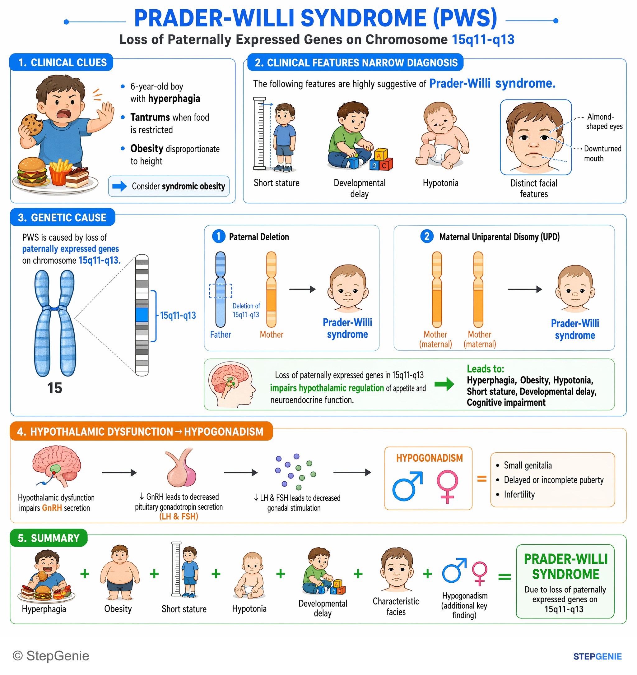
Prader-Willi syndrome due to paternal 15q11-q13 deletion → hyperphagia, obesity, short stature, hypotonia, developmental delay…
USMLE Step 1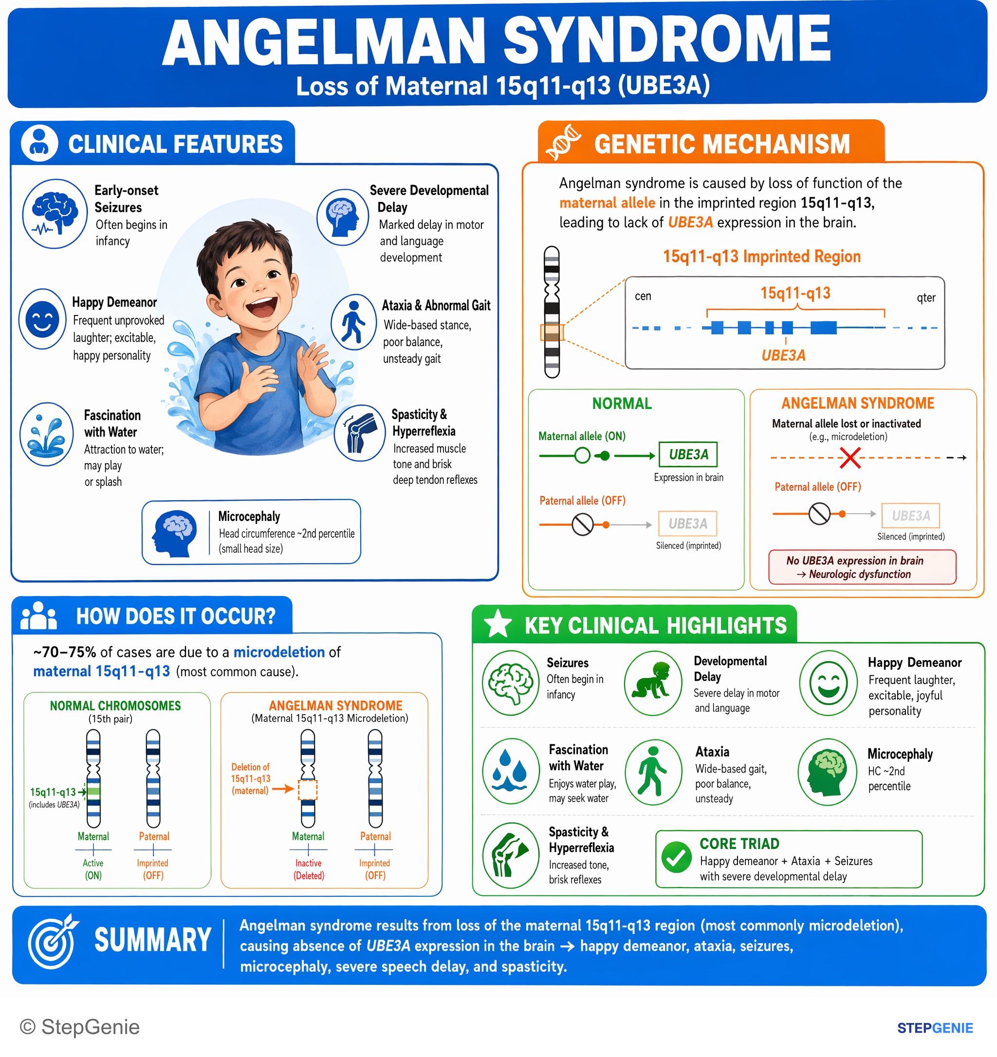
Angelman syndrome from loss of maternal 15q11-q13 (UBE3A) → happy demeanor, ataxia, seizures, microcephaly, severe speech delay, spasticity.
USMLE Step 1Frequently Asked Questions
What is the difference between heterodisomy and isodisomy?
Heterodisomy means a child inherited both of one parent's homologs of a chromosome pair, classically after nondisjunction in meiosis I and trisomy rescue. Isodisomy means the child has two copies of a single parental homolog, from a meiosis II error, monosomy rescue or a post-zygotic event. Both are forms of uniparental disomy and both can cause imprinting disorders, but only isodisomy can make a child homozygous for a recessive variant that one parent carries. Because of crossing over, many cases mix the two.
Is heterodisomy caused by an error in meiosis I or meiosis II?
Heterodisomy classically comes from meiosis I. Meiosis I separates homologous chromosomes, so a nondisjunction error there puts both of one parent's different homologs into the gamete; once trisomy rescue removes the other parent's copy, the child has heterodisomy. Meiosis II separates sister chromatids, so an error there passes on two copies of the same homolog and gives isodisomy. On testing, a meiosis I error leaves heterozygosity around the centromere, while a meiosis II error leaves homozygosity there.
What does uniparental disomy mean?
Uniparental disomy (UPD) means a person inherited both copies of a chromosome, or of part of one, from one parent and none from the other. The chromosome count stays at 46, so a karyotype usually looks normal. UPD occurs in roughly 1 in 2,000 births and is usually harmless. It causes disease when it involves imprinted genes, as in Prader-Willi and Angelman syndromes, or when isodisomy makes a child homozygous for a recessive variant.
What diseases are caused by uniparental disomy?
The classic UPD disorders involve imprinted regions. Maternal UPD 15 causes Prader-Willi syndrome and paternal UPD 15 causes Angelman syndrome. Paternal UPD of 11p15 causes about 20% of Beckwith-Wiedemann syndrome, maternal UPD 7 causes 7–10% of Silver-Russell syndrome, and paternal UPD 6 causes about 40% of 6q24 transient neonatal diabetes. Maternal and paternal UPD 14 cause Temple and Kagami-Ogata syndromes, and paternal UPD of 20q causes some pseudohypoparathyroidism type 1B. Isodisomy can also unmask an autosomal recessive disease such as cystic fibrosis.
How can a child have an autosomal recessive disease if only one parent is a carrier?
Once misattributed paternity and a deletion of the other allele are excluded, the classic explanation is uniparental isodisomy: the child received two copies of the carrier parent's variant-bearing chromosome and none from the other parent. The first such case, reported in 1988, was a child with cystic fibrosis who had inherited two copies of a CFTR variant from a carrier mother. A SNP array shows a long run of homozygosity on that chromosome, and parental DNA studies confirm it.
How is uniparental disomy diagnosed?
A karyotype is usually normal. For a suspected imprinting disorder such as Prader-Willi syndrome, DNA methylation analysis comes first, often together with a SNP microarray; methylation shows that the imprint is abnormal but not why. The array separates a deletion from isodisomy, which appears as a long run of homozygosity with normal copy number. Heterodisomy is proven only by comparing the child's DNA markers, such as STRs or SNPs, with both parents' DNA, showing that one parent contributed nothing.