Atopic dermatitis (eczema) is a chronic, relapsing, intensely itchy inflammatory skin disease that usually begins before age 5 (as early as 3 months) and affects up to 20% of children and 10% of adults in high-income countries. It arises from a leaky skin barrier, classically loss-of-function variants in the filaggrin gene (FLG), combined with Th2-skewed inflammation driven by IL-4 and IL-13, and it forms the atopic triad with asthma and allergic rhinitis. Infants get lesions on the face, scalp and extensor surfaces; older children and adults get them in the flexures. Treatment climbs from emollients and topical corticosteroids to nonsteroidal topicals (calcineurin, PDE4 and JAK inhibitors), then to biologics such as dupilumab or oral JAK inhibitors, which carry boxed warnings. Eczema herpeticum is an emergency that needs systemic acyclovir.
Pathophysiology: leaky barrier plus Th2 inflammation
1. The barrier defect
- Filaggrin (processed from profilaggrin) bundles structural proteins in the outermost skin cells into a strong barrier, and its breakdown products form part of the skin's natural moisturizing factor (MedlinePlus Genetics).
- FLG loss-of-function variants are found in 20-30% of people with atopic dermatitis vs 8-10% of the general population; about 10% of Europeans are heterozygous carriers (Merck). Carriers have more severe disease and higher IgE levels.
- The result is xerosis and increased transepidermal water loss, so irritants and allergens penetrate easily. Excessive washing and bathing are common triggers.
- FLG variants also cause ichthyosis vulgaris (dry, scaly skin), and ichthyosis is a recognized associated feature of atopic dermatitis.
2. Type 2 (Th2) inflammation
- Merck describes T-cell-mediated inflammation with a Th2-dominant component. IL-4 and IL-13 are the key cytokines (the targets of dupilumab), and IL-4 drives B cells to switch to IgE (see Th1 vs Th2 responses).
- The Th2-dominant inflammation downregulates antimicrobial peptides (Merck), which helps explain Staphylococcus aureus colonization and frequent skin infections.
- IL-31 is a neuroimmune cytokine that drives itch; nemolizumab blocks its receptor.
- Itch-scratch cycle: itch leads to scratching, which erodes the barrier and inflames the skin further; chronic rubbing produces lichenification.
3. The atopic march
Merck calls the triad of atopic dermatitis, allergic rhinoconjunctivitis and asthma atopy, or the atopic diathesis, and atopic dermatitis typically comes first, before rhinoconjunctivitis and asthma. FLG variants raise the risk of asthma only in people who also have atopic dermatitis (MedlinePlus).
Clinical picture by age
| Age | Typical sites | Look |
|---|---|---|
| Infants | Face (cheeks), scalp, neck, eyelids, extensor surfaces; groin and axillae (diaper area) usually spared | Red, scaly, eroded or crusted patches with excoriations |
| Older children and adults | Flexures: antecubital and popliteal fossae, neck; groin and axillae still spared | Dry, lichenified plaques from chronic scratching |
- Acute lesions: intensely itchy, red, thickened, scaly patches or plaques, eroded by scratching. Chronic lesions: dry and lichenified.
- Associated signs: xerosis, palmar hyperlinearity, keratosis pilaris, an infraorbital fold (Dennie-Morgan fold), thinning of the lateral eyebrows (Hertoghe sign), white dermographism and pityriasis alba.
- Course: childhood atopic dermatitis often abates by age 5, although flares are common through adolescence and into adulthood (Merck).
Diagnosis: clinical
There is no definitive laboratory test; diagnosis is clinical (Merck). The AAD criteria (Eichenfield, 2014) reproduced in Merck are:
- Essential: pruritus plus eczema (acute, subacute or chronic) with typical age-specific patterns and a chronic or relapsing history.
- Important: early onset, personal or family history of atopy, IgE reactivity, xerosis.
- Associated: the signs listed above, plus lichenification and facial pallor.
Total and allergen-specific IgE or prick testing can support an atopic diathesis but do not diagnose atopic dermatitis. For adults with treatment-resistant atopic dermatitis, AAD's first diagnostic-testing guideline (2026) covers when patch testing, skin biopsy and skin scrapings help. Biopsy, rarely needed, shows a nonspecific spongiotic (eczematous) pattern.
Complications
- S. aureus and streptococcal superinfection (impetigo, cellulitis): weeping, honey-colored crusts on a flaring patch. Treat with topical mupirocin or oral antistaphylococcal antibiotics.
- Eczema herpeticum (HSV): treat as an emergency. Grouped vesicles and punched-out erosions appear in areas of active or recent dermatitis, spread more widely than ordinary HSV, and can become systemic and occasionally fatal. Give systemic acyclovir or valacyclovir, and get an ophthalmology consult if the eye may be involved (Merck). Review the virus in the herpesviruses guide.
- Other viral skin infections: molluscum contagiosum and common warts.
- Allergic contact dermatitis: nickel allergy is twice as common as in people without atopy (see the contact dermatitis guide).
Treatment ladder (FDA status checked October 2026)
1. Everyone: skin care. Short, lukewarm baths, then moisturizers (ointments or creams; ceramide-containing products are useful). AAD makes a strong recommendation for moisturizers.
2. Flares: topical corticosteroids, twice daily, are effective for most mild to moderate disease. Use low potency on the face and skin folds; overuse causes atrophy, striae and telangiectasias.
3. Steroid-sparing topicals. AAD strongly recommends topical calcineurin, PDE4 and JAK inhibitors (2023) and tapinarof and roflumilast (2025 update); difamilast was approved after those reviews.
| Class | Drugs (FDA-labeled ages for atopic dermatitis) | Points to know |
|---|---|---|
| Calcineurin inhibitors | Tacrolimus ointment (0.03% from age 2; 0.1% from age 16), pimecrolimus 1% cream (from age 2) | Block calcineurin, so less IL-2 and T-cell activation; no skin atrophy, useful on face and eyelids; transient burning; boxed warning: long-term safety not established, rare skin cancer and lymphoma reports |
| PDE4 inhibitors (raise cAMP) | Crisaborole 2% ointment (from 3 months); roflumilast cream (0.15% from age 6, 0.05% for ages 2-5); difamilast 1% ointment (from age 2; FDA-approved February 2026) | Mild to moderate disease; crisaborole's most common adverse reaction is application-site pain (burning or stinging, about 4%) |
| Aryl hydrocarbon receptor agonist | Tapinarof cream (from age 2) | Also approved for plaque psoriasis |
| Topical JAK inhibitor | Ruxolitinib 1.5% cream (from age 2) | Mild to moderate disease in non-immunocompromised patients, short-term and non-continuous; carries the same boxed warning as oral JAK inhibitors |
4. Moderate to severe disease not controlled with topicals:
- Phototherapy (narrowband UVB).
- Biologics (none of the four below carries a boxed warning):
- Dupilumab: blocks IL-4 receptor alpha, so both IL-4 and IL-13 signaling stop; approved from 6 months of age. High-yield adverse effects: conjunctivitis and keratitis, injection-site reactions, oral herpes and eosinophilia; avoid live vaccines.
- Tralokinumab (from 12 years) and lebrikizumab (from 12 years and at least 40 kg): anti-IL-13.
- Nemolizumab: blocks the IL-31 receptor (itch); approved for atopic dermatitis in December 2024 for ages 12 and older, used with topical corticosteroids and/or calcineurin inhibitors.
- Oral JAK inhibitors: upadacitinib and abrocitinib, from 12 years, for refractory disease not controlled with other systemic drugs, including biologics, or when those are inadvisable. Boxed warning: serious infections, mortality, malignancy, major adverse cardiovascular events and thrombosis. Don't combine them with biologics or other immunosuppressants.
- Older immunosuppressants: cyclosporine, methotrexate, azathioprine, mycophenolate (conditional AAD recommendations).
- Systemic corticosteroids: AAD recommends against them; Merck allows short emergency use only.
Itch: oral antihistamines as needed (Merck prefers sedating hydroxyzine or diphenhydramine at bedtime); AAD's 2023 topical guideline recommends against topical antihistamines.
Guidelines in force: AAD's adult topical (2023) and phototherapy/systemic (2023) guidelines, a 2025 focused update with strong recommendations for tapinarof, roflumilast, lebrikizumab and nemolizumab, and AAD's first pediatric guidelines (2026), which strongly recommend against systemic corticosteroids.
Atopic vs contact vs seborrheic dermatitis vs psoriasis
| Feature | Atopic dermatitis | Allergic contact dermatitis | Seborrheic dermatitis | Psoriasis |
|---|---|---|---|---|
| Mechanism | Barrier defect (FLG) + Th2 (IL-4, IL-13) | Type IV T-cell reaction to a hapten (nickel, urushiol) | Malassezia yeast in sebum-rich skin | Th17 (IL-17, IL-22), TNF; keratinocyte hyperproliferation |
| Sites | Infants: face, extensors; later: flexures; spares groin and axillae | Wherever the allergen touched (watchband, streaks) | Scalp, eyebrows, nasolabial folds, behind ears, sternum; cradle cap and diaper area in infants | Extensor elbows and knees, scalp, sacrum, gluteal cleft |
| Look | Ill-defined, scaly, excoriated; lichenified when chronic | Vesicular, eczematous, geometric | Yellow, greasy scale | Well-demarcated plaques with silvery scale; nail pitting |
| Itch | Intense (hallmark) | Intense | Occasional | Variable: asymptomatic or itchy |
| Key test or clue | Clinical; atopic history | Patch test | Clinical | Clinical; Koebner phenomenon |
| First-line | Emollients, topical steroids | Avoid the allergen, topical steroids | Ketoconazole, selenium sulfide or zinc pyrithione shampoo | Topicals, then phototherapy or biologics |
More on plaques and nails in the psoriasis guide.
How USMLE tests it
- Infant with itchy cheeks and extensor rash, diaper area spared: atopic dermatitis; greasy yellow scalp scale with diaper involvement is seborrheic dermatitis.
- Child with flexural eczema plus asthma or allergic rhinitis: atopic march.
- Sudden painful, punched-out erosions and fever in a child with eczema: eczema herpeticum; start acyclovir.
- Eczema, thrombocytopenia with small platelets and recurrent infections in a boy: Wiskott-Aldrich syndrome. Eczema, cold staphylococcal abscesses, retained primary teeth and very high IgE: hyper-IgE (Job) syndrome.
- Mechanisms: dupilumab = IL-4Rα (blocks IL-4 and IL-13); tacrolimus = calcineurin, so less IL-2; crisaborole = PDE4, so more cAMP; JAK inhibitors = boxed warning.
- New red, irritated eyes on dupilumab: drug-associated conjunctivitis or keratitis.
Practice question
A 4-year-old boy with atopic dermatitis since infancy and mild asthma has had fever and a rapidly spreading, painful rash for 2 days. Temperature is 38.8°C (101.8°F). Over eczematous plaques on his face and neck are clusters of uniform, punched-out erosions with hemorrhagic crusts and a few intact vesicles. The eyes are not involved.
Which is the most appropriate next step in management?
- A. Switch to a high-potency topical corticosteroid
- B. Topical mupirocin alone
- C. Systemic acyclovir
- D. Start dupilumab
- E. Patch testing for contact allergens
Answer: C. Monomorphic punched-out erosions and vesicles over active eczema with fever are eczema herpeticum, a disseminated HSV infection of barrier-damaged skin. Start systemic acyclovir (or valacyclovir) promptly; HSV PCR of a lesion confirms it but should not delay treatment, and any eye involvement needs urgent ophthalmology.
- A: stronger corticosteroids do not treat HSV and can worsen a viral skin infection.
- B: mupirocin treats bacterial impetiginization (honey-colored crusts); bacterial superinfection can coexist, but the viral infection needs an antiviral.
- D: dupilumab is long-term control for moderate to severe disease, not therapy for an acute infection; herpes infections are among its listed adverse reactions.
- E: patch testing looks for allergic contact dermatitis and has no role in an acute febrile eruption.
Key takeaways
- Atopic dermatitis = barrier defect (FLG loss of function) + Th2 inflammation (IL-4, IL-13; IL-31 for itch); part of the atopic triad with asthma and allergic rhinitis.
- Infants: face, scalp, extensors (diaper area spared); older children and adults: flexures. Itch is the hallmark.
- Clinical diagnosis: pruritus, eczema in age-typical sites, chronic or relapsing course.
- Complications: S. aureus superinfection, eczema herpeticum (systemic acyclovir, emergency), molluscum, contact allergy.
- Ladder: emollients, topical corticosteroids, then calcineurin, PDE4 (crisaborole, roflumilast, difamilast), AhR (tapinarof) or JAK (ruxolitinib) topicals; then phototherapy, dupilumab (from 6 months), tralokinumab, lebrikizumab, nemolizumab, or oral JAK inhibitors (upadacitinib, abrocitinib; boxed warning).
- No long-term systemic corticosteroids (AAD recommends against).
Sources
- Atopic Dermatitis (Eczema) — Merck Manual Professional Edition (2025)
- Atopic dermatitis clinical guideline — American Academy of Dermatology (2023-2026)
- FLG gene — MedlinePlus Genetics, National Library of Medicine
- Tacrolimus ointment 0.1% (generic) label — DailyMed, NLM (2025)
- DUPIXENT (dupilumab) label — DailyMed, NLM (2026)
- OPZELURA (ruxolitinib) cream label — DailyMed, NLM (2026)
- EUCRISA (crisaborole) ointment label — DailyMed, NLM
- ZORYVE (roflumilast) cream label — DailyMed, NLM (2026)
- CIBINQO (abrocitinib) label — DailyMed, NLM (2026)
- EBGLYSS (lebrikizumab) label — DailyMed, NLM (2026)
- NEMLUVIO (nemolizumab-ilto) label — DailyMed, NLM (2025)
- ADQUEY (difamilast) ointment label — DailyMed, NLM (2026)
Visual explanations from the StepGenie QBank
Infographics StepGenie drew to explain questions in its own QBank, matched to this article's topic. Each caption is the takeaway of the question it explains.
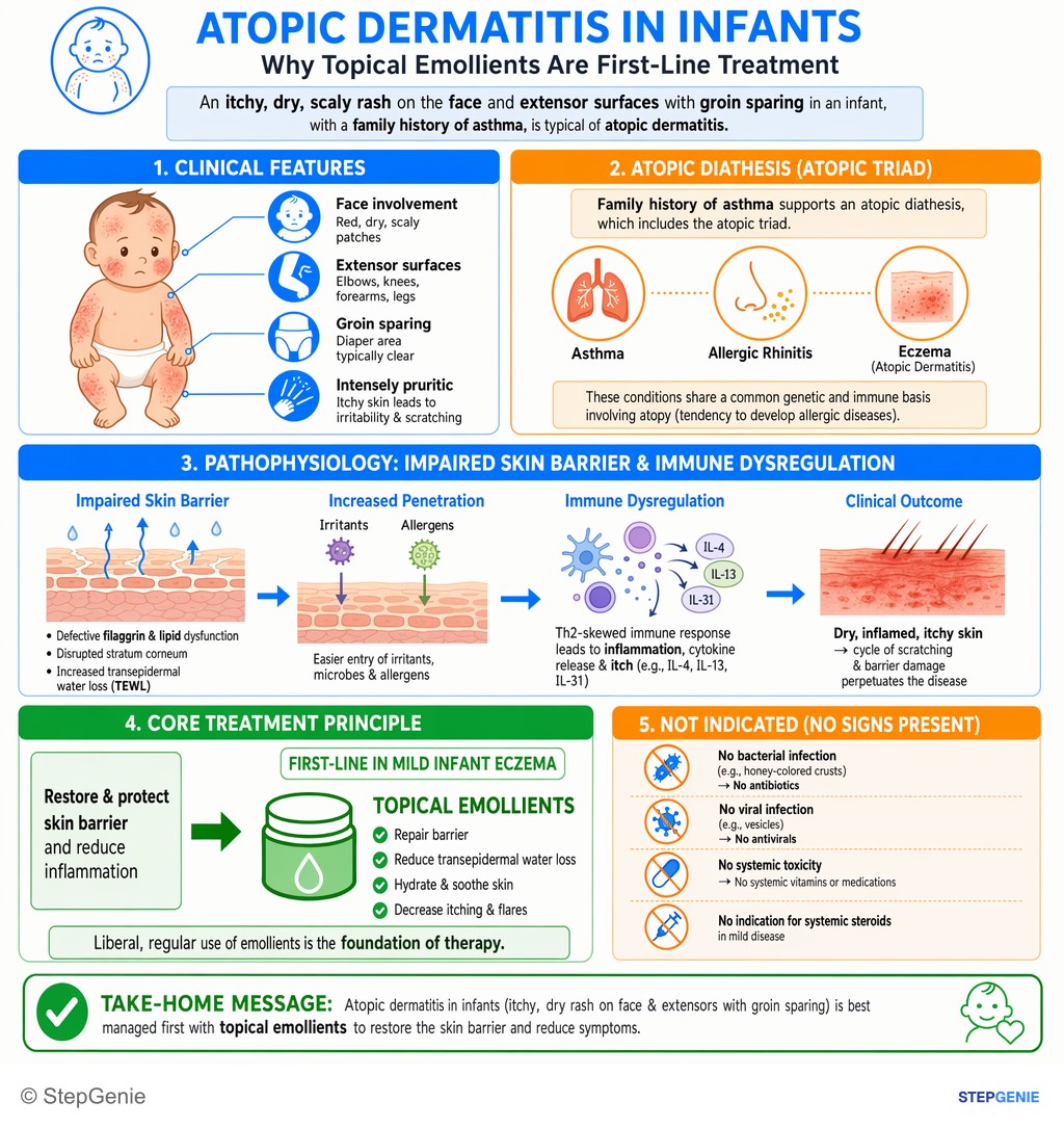
Atopic dermatitis in an infant (itchy, dry rash on face/extensors) is first-line treated with topical emollients to restore skin barrier.
USMLE Step 2Frequently Asked Questions
What causes atopic dermatitis?
Atopic dermatitis results from a defective skin barrier combined with Th2-skewed immune inflammation. Loss-of-function variants in the filaggrin gene (FLG), found in 20 to 30% of people with atopic dermatitis versus 8 to 10% of the general population, weaken the barrier and reduce the skin's natural moisturizing factor, so the skin dries out and irritants penetrate. The Th2 cytokines IL-4 and IL-13 drive inflammation and IgE, suppress antimicrobial peptides and favor Staphylococcus aureus colonization. Excessive washing and bathing are common triggers.
Where does atopic dermatitis appear in infants versus adults?
In infants, atopic dermatitis typically involves the face, especially the cheeks, plus the scalp, neck, eyelids and extensor surfaces, and it usually spares the groin and axillae, so the diaper area is often clear. In older children and adults it shifts to the flexures, especially the antecubital and popliteal fossae and the neck, where chronic scratching causes lichenification. Itch is the hallmark at every age; childhood disease often abates by age 5, although flares are common through adolescence and into adulthood.
How is atopic dermatitis diagnosed?
Diagnosis is clinical; there is no definitive laboratory test. The AAD criteria require pruritus plus eczema with typical age-specific patterns and a chronic or relapsing history. Supporting features include early onset, a personal or family history of atopy, IgE reactivity and dry skin, and associated signs include Dennie-Morgan folds, keratosis pilaris, palmar hyperlinearity and white dermographism. IgE or prick testing can support an atopic background but does not confirm atopic dermatitis; patch testing is used when allergic contact dermatitis is suspected.
What is eczema herpeticum and how is it treated?
Eczema herpeticum is herpes simplex virus infection spreading through skin damaged by atopic dermatitis. It presents with clusters of painful, uniform vesicles and punched-out erosions, often with hemorrhagic crusts and fever, in areas of active or recent eczema. It can become systemic and occasionally fatal, so it is treated as an emergency with systemic acyclovir or valacyclovir, without waiting for test results. Suspected eye involvement needs an ophthalmology consultation. It differs from bacterial impetiginization, which causes honey-colored crusts.
What is the treatment ladder for atopic dermatitis?
Everyone needs gentle skin care and regular moisturizers. Flares are treated with topical corticosteroids, using low potency on the face and folds. Steroid-sparing topicals include calcineurin inhibitors (tacrolimus, pimecrolimus), PDE4 inhibitors (crisaborole, roflumilast, difamilast), tapinarof and ruxolitinib cream. Moderate to severe disease uncontrolled by topicals moves to phototherapy, biologics (dupilumab, tralokinumab, lebrikizumab, nemolizumab) or oral JAK inhibitors (upadacitinib, abrocitinib), which carry boxed warnings for serious infections, mortality, malignancy, cardiovascular events and thrombosis. AAD recommends against systemic corticosteroids.
How does dupilumab work in atopic dermatitis?
Dupilumab is a monoclonal antibody against the IL-4 receptor alpha subunit, so it blocks signaling by both IL-4 and IL-13, the main Th2 cytokines in atopic dermatitis. It is FDA-approved for moderate to severe atopic dermatitis from 6 months of age when topical prescription therapies are inadequate or inadvisable, and it has no boxed warning. High-yield adverse effects are conjunctivitis and keratitis, injection-site reactions, oral herpes and eosinophilia, and live vaccines should be avoided during treatment.