Dermatitis herpetiformis (DH) is an intensely itchy, chronic autoimmune eruption of grouped papules and vesicles that is the skin manifestation of celiac disease: nearly all patients have celiac changes on small-bowel biopsy, even though most have no GI symptoms. The rash is symmetric on the extensor elbows and knees, buttocks, sacrum and scalp, and is often seen only as excoriations because patients scratch the vesicles off. The diagnostic test is a biopsy of perilesional skin for direct immunofluorescence (DIF), which shows granular IgA in the dermal papillae. Treatment is dapsone for rapid relief of itch (test G6PD first) plus a lifelong strict gluten-free diet, which treats the underlying disease and lets most patients stop dapsone over time.
Pathophysiology: from gluten to the skin
- Genetics. DH and celiac disease share HLA risk alleles, mainly HLA-DQ2 and, to a lesser extent, HLA-DQ8. More than 95% of patients with celiac disease carry one of them, but the alleles are not disease-specific, so a negative test helps rule celiac disease out while a positive one does not diagnose it. See the HLA associations question breakdown.
- Trigger. Gluten (its gliadin fraction) drives an immune response in the small bowel; in the gut the autoantigen is tissue transglutaminase.
- Skin autoantigen. DH is an IgA-dominant response to transglutaminases, and the key skin target is epidermal transglutaminase (TG3).
- Lesion. IgA deposits collect at the tips of the dermal papillae and recruit neutrophils, which form papillary microabscesses and a subepidermal blister.
- Reversibility. The IgA deposits can disappear with strict gluten avoidance.
- Flares. Iodine exposure can precipitate them.
- The name. "Herpetiformis" describes the clustered, herpes-like grouping of lesions; no virus is involved (contrast the true herpesviruses).
Clinical picture
| Feature | What to know |
|---|---|
| Who | Usually adults in their 30s-40s, but children and older adults can be affected; men somewhat more often (about 1.5-2:1); mostly people of Northern European descent, rare in Black and Asian people |
| Symptoms | Intense itching and burning; vesicles rupture quickly, so intact ones are often hard to find |
| Lesions | Clusters of erythematous papules, urticarial plaques and small vesicles, occasionally bullae; symmetric |
| Sites | Extensor elbows and knees, buttocks and sacrum, scalp and occiput |
| Gut | Most patients have no GI symptoms despite celiac changes on biopsy; some have diarrhea, bloating or weight loss |
| Clues from malabsorption | Iron deficiency anemia without bleeding, low folate, low bone density |
| Associations | Autoimmune thyroid disease, type 1 diabetes, pernicious anemia, Addison disease, vitiligo; dental enamel pits; small-bowel lymphoma risk |
DH and celiac disease travel together, but not one-for-one: only a minority of people with celiac disease develop DH (Merck: about 15-25%). Review the gut side in the celiac disease question breakdown.
Diagnosis
- Skin biopsy for DIF from perilesional skin (normal-looking skin next to a lesion). Expect granular IgA deposits in the dermal papillae (occasionally fibrillar). Merck gives DIF a sensitivity of 90-95% and a specificity of 95-100%.
- Routine histology (H&E) shows a subepidermal blister with neutrophils at the tips of the dermal papillae. It supports the diagnosis; DIF confirms it.
- Celiac serology: IgA anti-tissue transglutaminase (tTG) and IgA anti-endomysial antibodies; IgA anti-epidermal transglutaminase antibodies can also be measured. Always check total IgA, because IgA deficiency (which is associated with celiac disease) causes false negatives; IgA-deficient patients need IgG-based tests (IgG tTG or IgG deamidated gliadin peptide).
- Keep eating gluten until testing is done. A gluten-free diet started early can make serology and biopsies falsely reassuring.
- HLA-DQ2/DQ8 typing is useful mainly to rule out: without these variants, celiac disease is very unlikely.
- Small-bowel biopsy findings in celiac disease: villous atrophy, crypt hyperplasia and increased intraepithelial lymphocytes. Because almost all patients with DH have celiac disease on histology (Merck), a confirmed DH diagnosis means managing the patient for celiac disease too.
Immunofluorescence: DH vs the other blistering diseases
| Disease | Blister level | DIF pattern | Target | Classic clue |
|---|---|---|---|---|
| Dermatitis herpetiformis | Subepidermal | Granular IgA in dermal papillae | Epidermal transglutaminase (TG3) | Celiac disease; extensor surfaces; intense itch, excoriations |
| Linear IgA disease | Subepidermal | Linear IgA along the basement membrane zone | BP180 fragments (97-kDa antigen, LAD-1) | Drug-induced cases (vancomycin is the drug most often implicated); clustered lesions; mucosal involvement common; in children, annular "crown of jewels" or "string of pearls" blisters |
| Bullous pemphigoid | Subepidermal | Linear IgG and C3 along the basement membrane zone | BP180 and BP230 (hemidesmosomes) | Older than 60; tense bullae, urticarial plaques; mucosa rarely involved; Nikolsky negative; DPP-4 inhibitors, checkpoint inhibitors |
| Pemphigus vulgaris | Intraepidermal (suprabasal acantholysis) | Intercellular IgG, net-like | Desmoglein 3 (sometimes 1) | Middle age; flaccid bullae; painful oral erosions, often before skin lesions; Nikolsky positive |
The pemphigoid and pemphigus details are in pemphigus vulgaris vs bullous pemphigoid.
Treatment
Dapsone (fast control of the skin).
- FDA-labeled for DH and leprosy. The label starts adults at 50 mg daily and titrates (range 50-300 mg); Merck starts at 25-50 mg and notes most patients respond to 50-150 mg/day.
- Itching drops promptly, often within 1-3 days, followed by clearing of lesions. Its mechanism in DH has not been established.
- Test G6PD activity before starting. Hemolysis with Heinz bodies is dose-related and exaggerated in G6PD deficiency; see G6PD deficiency.
- Other toxicities: methemoglobinemia, hepatitis, peripheral neuropathy (mainly motor), severe skin reactions including toxic epidermal necrolysis, and agranulocytosis and aplastic anemia, which have caused deaths. The label cites an FDA advisory committee recommendation of a CBC weekly for the first month, monthly for 6 months, then every 6 months.
- If dapsone can't be used: a sulfonamide such as sulfasalazine (dapsone itself is a sulfone). Potent topical corticosteroids can ease itching in the short term.
Strict gluten-free diet (the long game).
- Treats the underlying gluten-sensitive enteropathy, corrects malabsorption and clears the IgA from the skin over time.
- Lets many patients reduce or stop dapsone; the dapsone label gives an average of 8 months before the dose can be lowered, and full control on diet alone can take months to years.
- Merck notes that keeping to it for 5-10 years lowers the risk of small-bowel lymphoma.
- It is lifelong, and patients who keep to it have excellent long-term survival.
How USMLE tests it
- "Itchy grouped vesicles on elbows, knees and buttocks" + iron deficiency or bloating = DH, a marker of celiac disease.
- Granular IgA = DH; linear IgA = linear IgA disease; linear IgG = bullous pemphigoid; net-like IgG = pemphigus vulgaris.
- Biopsy site: perilesional skin for DIF.
- Excoriations only: expect scratched-off papules rather than intact blisters. Location helps: atopic dermatitis in older children and adults favors flexures; see atopic dermatitis.
- Scabies also itches intensely, but look for burrows (finger webs, wrists) and itchy contacts; DIF is not part of its work-up.
- Dapsone + dark urine and fatigue = hemolysis (G6PD); dapsone + cyanosis = methemoglobinemia.
- Negative celiac serology in a patient with DH? Check total IgA and whether the patient had already cut out gluten.
Practice question
A 34-year-old man of Irish descent has a 4-month history of severely itchy, symmetric clusters of papules and tiny vesicles on the extensor surfaces of his elbows and knees, his buttocks and the back of his scalp. Most lesions are excoriated. He reports intermittent bloating. His hemoglobin is mildly low and his ferritin is low. A biopsy of perilesional skin is sent for direct immunofluorescence. Which finding is most likely?
A. Granular IgA deposits in the dermal papillae B. Linear IgA deposits along the basement membrane zone C. Linear IgG and C3 deposits along the basement membrane zone D. Intercellular IgG deposits in a net-like pattern E. No immunoglobulin deposits, with multinucleated giant cells on a Tzanck smear
Answer: A. Itchy, symmetric, grouped and excoriated papulovesicles on extensor surfaces, plus iron deficiency and bloating that hint at celiac disease, make this dermatitis herpetiformis; DIF shows granular IgA in the dermal papillae.
- B: linear IgA disease. Subepidermal blisters with linear IgA; it can be drug-induced (vancomycin is the drug most often implicated), and mucosal involvement is common.
- C: bullous pemphigoid. An older patient (usually over 60) with tense bullae, often after urticarial plaques; antibodies target BP180 and BP230.
- D: pemphigus vulgaris. Intraepidermal acantholysis with flaccid bullae, oral erosions and a positive Nikolsky sign.
- E: herpes simplex. A viral infection with grouped vesicles, but not symmetric extensor lesions with celiac clues; "herpetiformis" is only a description of the grouping.
Next steps: check G6PD before starting dapsone, send IgA tTG with total IgA while he is still eating gluten, and start a strict gluten-free diet.
Key takeaways
- DH = the skin form of celiac disease: intensely itchy, symmetric, grouped papulovesicles on elbows, knees, buttocks, sacrum and scalp; most patients have no GI symptoms.
- Mechanism: IgA against epidermal transglutaminase (TG3) deposits at the dermal papillary tips, draws in neutrophils and causes a subepidermal blister; HLA-DQ2/DQ8.
- Diagnosis: perilesional skin biopsy with DIF showing granular IgA in the dermal papillae, plus IgA tTG and total IgA while the patient is still eating gluten.
- Treatment: dapsone for rapid itch control after G6PD testing, with CBC monitoring for hemolysis, methemoglobinemia and agranulocytosis.
- A lifelong gluten-free diet treats the cause, allows dapsone to be tapered (often after about 8 months) and lowers lymphoma risk.
- Do not confuse it with linear IgA disease (linear IgA, vancomycin), bullous pemphigoid (linear IgG, tense bullae) or pemphigus vulgaris (net-like IgG, flaccid bullae, Nikolsky positive).
Sources
- Dermatitis Herpetiformis — Merck Manual Professional Edition (2024)
- Dermatitis Herpetiformis — StatPearls, NCBI Bookshelf (2023)
- Dapsone Tablets Label — DailyMed (2026)
- Celiac Disease — Merck Manual Professional Edition (2025)
- Diagnosis of Celiac Disease — NIDDK, NIH
- Bullous Pemphigoid — Merck Manual Professional Edition (2024)
- Pemphigus Vulgaris — Merck Manual Professional Edition (2024)
- Linear Immunoglobulin A Disease — Merck Manual Professional Edition
- Linear IgA Dermatosis — StatPearls, NCBI Bookshelf (2023)
- Selective IgA Deficiency — Merck Manual Professional Edition (2024)
Visual explanations from the StepGenie QBank
Infographics StepGenie drew to explain questions in its own QBank, matched to this article's topic. Each caption is the takeaway of the question it explains.
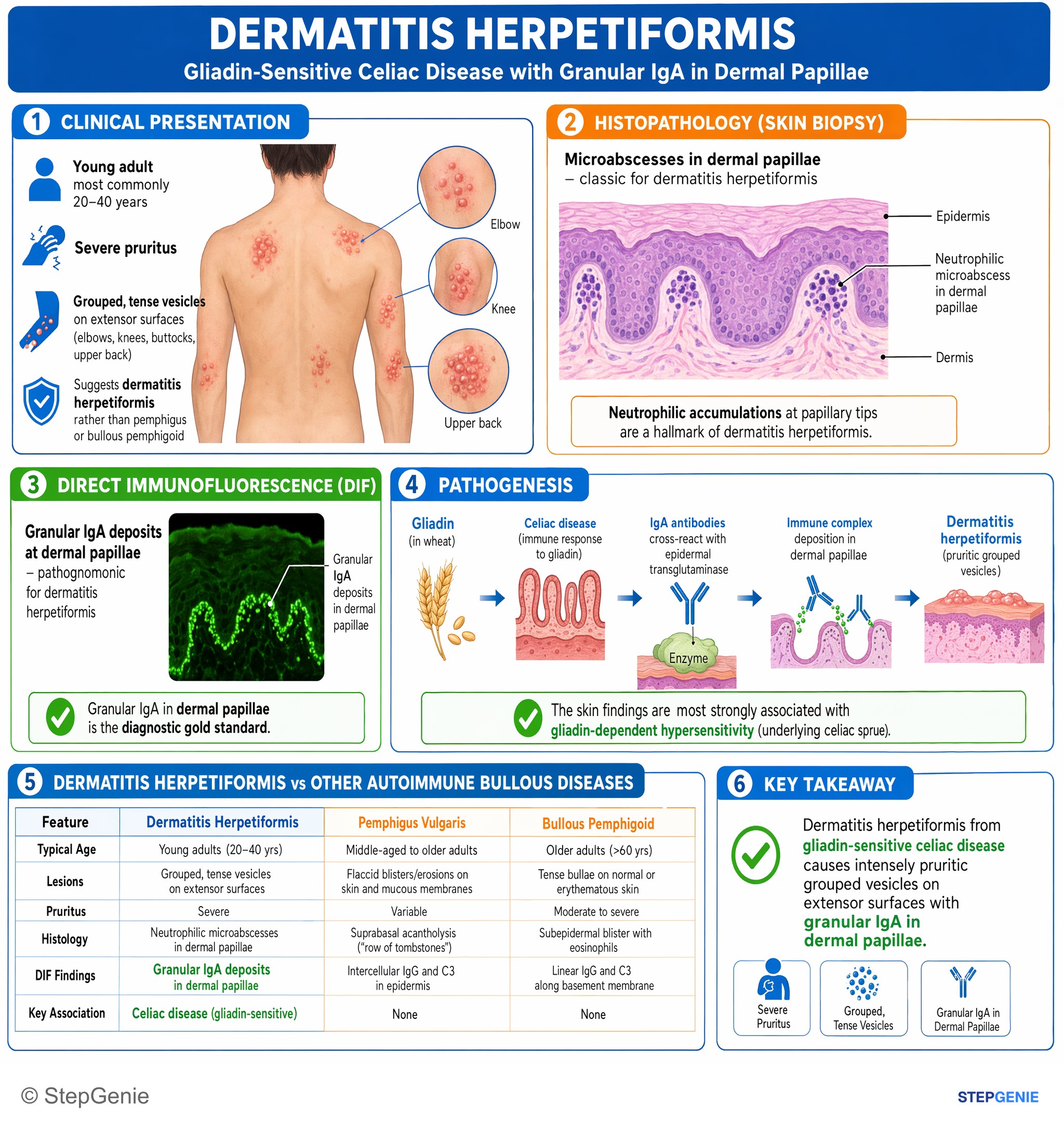
Dermatitis herpetiformis from gliadin-sensitive celiac disease causes intensely pruritic grouped vesicles on extensor surfaces with…
USMLE Step 1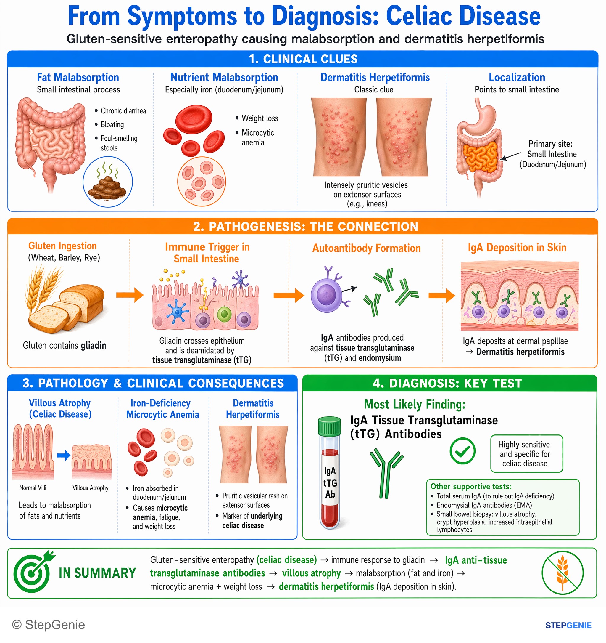
Celiac disease from gluten-sensitive enteropathy → villous atrophy, iron-deficiency microcytic anemia, and dermatitis herpetiformis with…
USMLE Step 1Frequently Asked Questions
What is dermatitis herpetiformis?
Dermatitis herpetiformis is a chronic, intensely itchy autoimmune skin disease and the skin manifestation of celiac disease. It causes symmetric clusters of red papules and small vesicles on the elbows, knees, buttocks, sacrum and scalp, which are usually scratched into excoriations. Nearly all patients have celiac changes in the small bowel, but most have no digestive symptoms. Despite the name, it has nothing to do with herpesviruses; herpetiformis describes the grouped look of the lesions.
What does direct immunofluorescence show in dermatitis herpetiformis?
A biopsy of perilesional skin, meaning normal-looking skin next to a lesion, shows granular IgA deposits in the dermal papillae. This separates it from linear IgA disease, which shows linear IgA along the basement membrane zone; bullous pemphigoid, which shows linear IgG and C3 along the basement membrane zone; and pemphigus vulgaris, which shows intercellular IgG in a net-like pattern. Routine histology shows neutrophils at the dermal papillary tips and a subepidermal blister.
How is dermatitis herpetiformis related to celiac disease?
Both come from an immune reaction to gluten in people who carry HLA-DQ2 or HLA-DQ8. In the gut the autoantigen is tissue transglutaminase; in the skin, IgA directed against epidermal transglutaminase (TG3) deposits in the dermal papillae and draws in neutrophils. Almost all patients with dermatitis herpetiformis have celiac disease on small-bowel histology, usually without digestive symptoms, so they should have celiac serology, including total IgA, and be managed for celiac disease.
What is the treatment for dermatitis herpetiformis?
Dapsone relieves the itching quickly, often within one to three days, and clears the lesions, but G6PD activity should be tested before starting it. The long-term treatment is a strict, lifelong gluten-free diet, which treats the underlying gluten sensitivity, heals the gut and gradually clears the IgA from the skin. The dapsone label gives an average of about 8 months before the dose can be reduced, and many patients eventually control the disease with diet alone.
What are the side effects of dapsone?
The key toxicities are dose-related hemolysis, which is worse in G6PD deficiency, and methemoglobinemia, which can cause cyanosis. Dapsone can also cause agranulocytosis and aplastic anemia, which have been fatal, so the label recommends blood counts weekly for the first month, monthly for six months and then every six months. Other risks include hepatitis, mainly motor peripheral neuropathy and severe skin reactions such as toxic epidermal necrolysis.
Can you test for dermatitis herpetiformis after starting a gluten-free diet?
Testing is most reliable while the patient is still eating gluten. A gluten-free diet started before testing can lower celiac antibodies and heal the intestine, and the IgA deposits in the skin can also fade with strict gluten avoidance, making results falsely reassuring. If serology is negative despite strong suspicion, check total IgA, because IgA deficiency causes false-negative IgA tests, and ask whether the patient had already cut out gluten.