Pemphigus vulgaris (PV) and bullous pemphigoid (BP) are two blistering disorders that USMLE loves because they look similar at first glance—but their level of skin split, antibody target, mucosal involvement, and Nikolsky sign cleanly separate them. If you can visualize where the “glue” fails (desmosomes vs hemidesmosomes), you can answer most Step questions in seconds.
Big-Picture: How to Tell Them Apart in 10 Seconds
The “Two Anchors” Mental Model
- Desmosomes = anchors between keratinocytes in the epidermis
- Hemidesmosomes = anchors between basal epidermis and basement membrane
If you attack:
- Desmosomes → PV → intraepidermal, flaccid bullae, + mucosa
- Hemidesmosomes → BP → subepidermal, tense bullae, usually no mucosa
High-Yield Comparison Table (Memorize This)
| Feature | Pemphigus vulgaris | Bullous pemphigoid |
|---|---|---|
| Typical patient | Middle-aged | Elderly |
| Level of split | Intraepidermal (suprabasal) | Subepidermal |
| Bullae | Flaccid, rupture easily | Tense, less likely to rupture |
| Mucosal involvement | Common (often first) | Uncommon/rare |
| Nikolsky sign | Positive | Negative (classically) |
| Pathogenesis | IgG vs desmoglein (desmosomes) | IgG vs hemidesmosomes (BP180/BP230) |
| Histology | Acantholysis, “row of tombstones” | Subepidermal split + eosinophils |
| DIF (direct IF) | Net-like (fishnet) IgG in epidermis | Linear IgG/C3 at BM |
| Treatment | Steroids + steroid-sparing immunosuppression | High-potency topical/systemic steroids; immunosuppression if severe |
Pemphigus Vulgaris (PV)
Definition (What it is)
An autoimmune blistering disease caused by antibodies against desmosomes, leading to loss of keratinocyte cohesion (acantholysis) and intraepidermal blisters.
Pathophysiology (Why it happens)
- Autoantibodies: IgG against desmoglein 1 and 3
- Desmogleins are cadherins in desmosomes
- The epidermis “unglues” from within → flaccid bullae
- Suprabasal acantholysis → basal cells remain attached to basement membrane → classic histology pattern
First Aid Cross-Reference
- FA Step 1 typically highlights:
- Anti-desmoglein
- Intraepidermal acantholysis
- Fishnet IF pattern
- Nikolsky positive
- Oral mucosa involvement
Clinical Presentation (How it looks on test day)
High-yield clues:
- Painful oral erosions often precede skin findings
- Flaccid bullae that rupture → erosions and crusting
- Positive Nikolsky sign: lateral pressure causes epidermal shearing
Exam stem vibes:
- Middle-aged patient with nonhealing oral ulcers + fragile blisters
- “Skin sloughs off with rubbing”
Diagnosis (What confirms it)
- Biopsy for H&E
- Suprabasal acantholysis
- “Row of tombstones” (basal layer attached)
- Direct immunofluorescence (DIF)
- Net-like / reticular IgG (and often C3) in epidermis
- Think: antibodies are between keratinocytes → “fishnet”
Treatment (What you do)
- Systemic corticosteroids are the backbone (often needed due to severity)
- Add steroid-sparing agents:
- Azathioprine, mycophenolate mofetil
- Rituximab (anti-CD20) is increasingly common in modern management
- Supportive care:
- Wound care, infection prevention, pain control, fluids/electrolytes if extensive
High-Yield Associations & Pitfalls
- PV can be life-threatening due to skin barrier loss → infection, fluid loss
- Mucosal involvement strongly favors PV over BP
- Flaccid bullae = think intraepidermal (roof is thin)
Bullous Pemphigoid (BP)
Definition (What it is)
An autoimmune blistering disorder caused by antibodies against hemidesmosomes, producing subepidermal blisters.
Pathophysiology (Why it happens)
- Autoantibodies: IgG against hemidesmosomal proteins:
- BP180 (type XVII collagen) and BP230
- Separation occurs at the dermal–epidermal junction → thicker roof → tense bullae
- Inflammatory response commonly features eosinophils
First Aid Cross-Reference
- FA Step 1 classic points:
- Anti-hemidesmosome
- Subepidermal blister
- Linear deposition on IF
- Tense bullae
- Eosinophils
Clinical Presentation (How it looks on test day)
High-yield clues:
- Elderly patient with pruritus and tense bullae
- Often on trunk, flexor surfaces
- Mucosal involvement uncommon
- Nikolsky sign usually negative
Exam stem vibes:
- Older patient with itchy urticarial plaques that evolve into tense blisters
- “Bullae don’t rupture easily”
Diagnosis (What confirms it)
- Biopsy for H&E
- Subepidermal split
- Eosinophils in blister cavity/dermis
- Direct immunofluorescence (DIF)
- Linear IgG and/or C3 along basement membrane zone
- Think: antibodies are at the basement membrane → “straight line”
Treatment (What you do)
- Mild/moderate disease:
- High-potency topical corticosteroids can be very effective
- More extensive disease:
- Systemic corticosteroids
- Steroid-sparing agents (depending on severity): doxycycline/niacinamide, azathioprine, mycophenolate, etc.
High-Yield Associations & Pitfalls
- BP is generally less severe than PV
- Tense bullae + pruritus + elderly is the classic triad
- Linear IF is the classic testable discriminator
Nikolsky Sign: What It Actually Means (and Why Step Cares)
- Positive Nikolsky = epidermal cohesion is weak → superficial layers shear off with pressure
- Classic in PV
- Negative Nikolsky (classically) = split is deeper and roof is sturdier
- Classic in BP
USMLE tip: If they explicitly mention Nikolsky sign and oral lesions, they’re practically handing you PV.
Rapid-Fire USMLE Differentiation Prompts
If the vignette says…
- “Painful oral ulcers” → PV
- “Tense bullae in an elderly patient with itching” → BP
- “Fishnet pattern on immunofluorescence” → PV
- “Linear deposition at the basement membrane” → BP
- “Acantholysis” → PV
- “Eosinophils + subepidermal split” → BP
- “Flaccid blisters that rupture” → PV
- “Nikolsky positive” → PV
Mini-Table: Pathology Words You Must Recognize
| Keyword | Points to | Why |
|---|---|---|
| Acantholysis | PV | Loss of keratinocyte adhesion (desmosomes) |
| Row of tombstones | PV | Basal cells remain attached |
| Eosinophils | BP | Common inflammatory infiltrate |
| Subepidermal split | BP | Hemidesmosomal separation |
| Suprabasal blister | PV | Desmosomal failure above basal layer |
| Fishnet | PV | Intercellular IgG |
| Linear | BP | BM zone IgG/C3 |
How First Aid Wants You to Think About It (Classic “Anchor” Points)
When you’re stuck between PV and BP, force yourself to answer these four questions:
- Is there mucosal involvement?
- Yes → PV
- Are the bullae flaccid or tense?
- Flaccid → PV
- Tense → BP
- Where is the split?
- Intraepidermal/suprabasal → PV
- Subepidermal → BP
- What’s the IF pattern?
- Fishnet → PV
- Linear → BP
Quick Practice Vignettes (Test Yourself)
Vignette 1
A 52-year-old with weeks of painful mouth sores now has easily ruptured blisters on the chest. Gentle rubbing causes skin to slough. DIF shows a net-like pattern.
Answer: Pemphigus vulgaris (anti-desmoglein; intraepidermal acantholysis)
Vignette 2
A 78-year-old with intense itching develops large, tense bullae on the abdomen and arms. Biopsy shows subepidermal splitting with eosinophils. DIF shows linear C3 along the basement membrane.
Answer: Bullous pemphigoid (anti-hemidesmosomes; subepidermal blister)
Take-Home Summary (What to Memorize)
- Pemphigus vulgaris: anti-desmoglein, intraepidermal (suprabasal) blister, flaccid bullae, + mucosa, + Nikolsky, fishnet IF.
- Bullous pemphigoid: anti-hemidesmosomes (BP180/BP230), subepidermal blister, tense bullae, pruritus, mucosa uncommon, linear IF, eosinophils.
Visual explanations from the StepGenie QBank
Infographics StepGenie drew to explain questions in its own QBank, matched to this article's topic. Each caption is the takeaway of the question it explains.
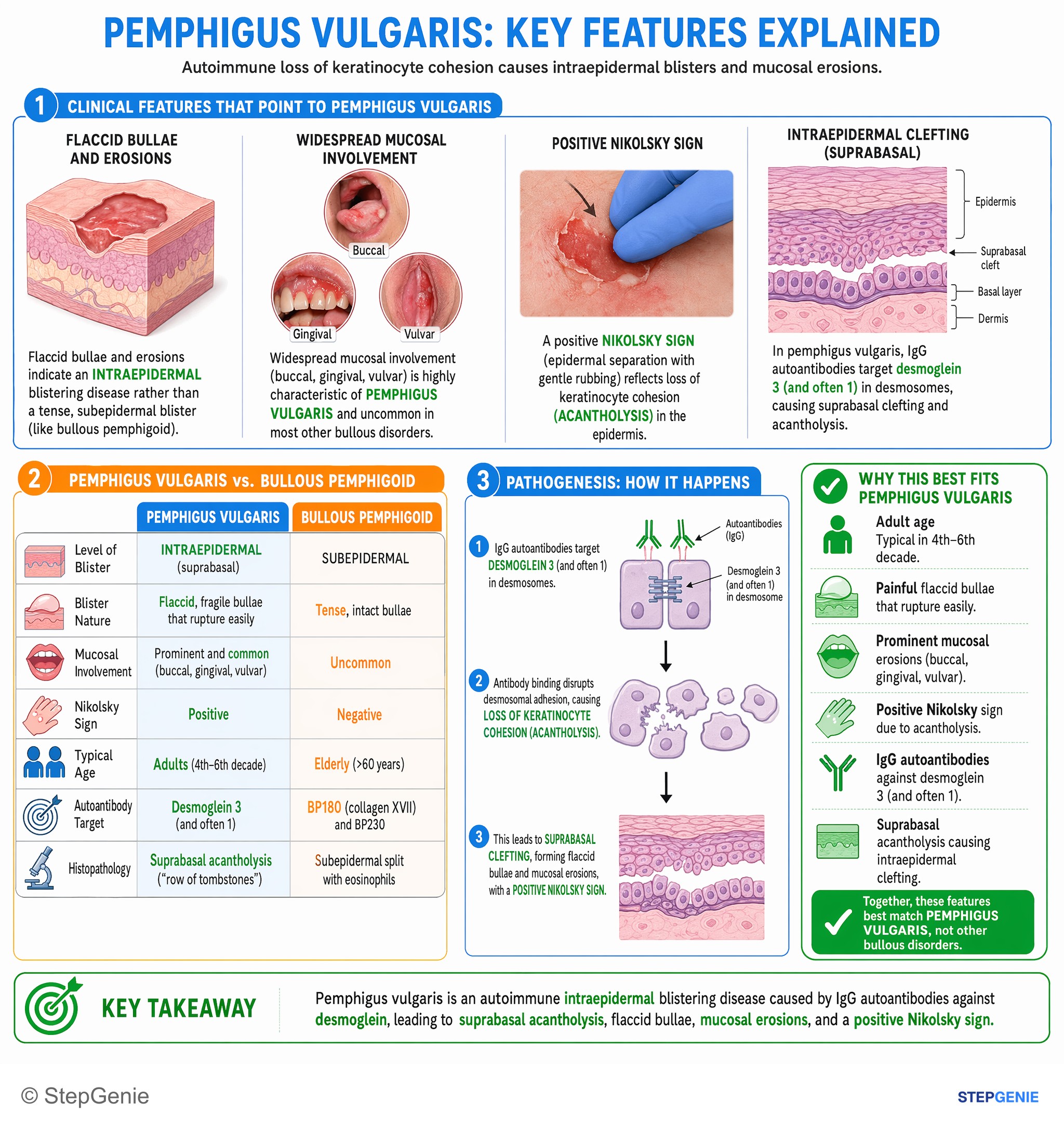
Pemphigus vulgaris: autoantibodies against desmoglein cause suprabasal acantholysis, flaccid bullae, mucosal erosions, and positive…
USMLE Step 1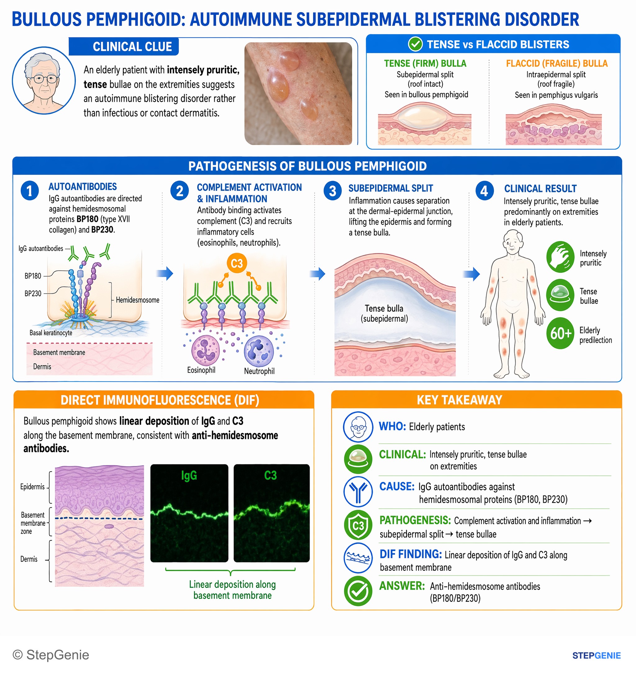
Bullous pemphigoid: elderly with tense, intensely pruritic bullae; IgG/C3 along basement membrane; due to anti-hemidesmosome antibodies…
USMLE Step 1Frequently Asked Questions
How do you tell pemphigus vulgaris from bullous pemphigoid quickly on Step 1?
The fastest way is to compare the level of skin split, antibody target, mucosal involvement, and Nikolsky sign. Pemphigus vulgaris attacks desmosomes, causing an intraepidermal suprabasal split with flaccid bullae, common oral mucosal involvement, and a positive Nikolsky sign. Bullous pemphigoid attacks hemidesmosomes, producing a subepidermal split with tense bullae, usually no mucosal involvement, and a classically negative Nikolsky sign.
Why does pemphigus vulgaris cause flaccid bullae and oral lesions?
Pemphigus vulgaris causes flaccid bullae because IgG antibodies attack desmoglein 1 and 3 in desmosomes, so keratinocytes lose cohesion within the epidermis. This intraepidermal suprabasal acantholysis leaves a thin blister roof that ruptures easily. Oral mucosal involvement is common and often appears first, so painful oral erosions or nonhealing oral ulcers are a classic clue favoring pemphigus vulgaris.
Why are bullae tense in bullous pemphigoid?
Bullae are tense in bullous pemphigoid because the split occurs below the epidermis at the dermal-epidermal junction. IgG antibodies target hemidesmosomal proteins BP180 and BP230, creating a subepidermal blister with a thicker roof that is less likely to rupture. That is why an elderly patient with pruritus and tense blisters, especially without mucosal disease, points toward bullous pemphigoid.
What biopsy and immunofluorescence findings confirm pemphigus vulgaris vs bullous pemphigoid?
Pemphigus vulgaris is confirmed by suprabasal acantholysis on biopsy, often described as a row of tombstones, with direct immunofluorescence showing net-like or fishnet IgG in the epidermis. Bullous pemphigoid shows a subepidermal split with eosinophils on biopsy, and direct immunofluorescence reveals linear IgG and/or C3 along the basement membrane zone. These patterns are classic testable discriminators.
What is the first-line treatment for pemphigus vulgaris and bullous pemphigoid?
Pemphigus vulgaris is treated with systemic corticosteroids as the backbone of therapy, often with steroid-sparing immunosuppression such as azathioprine, mycophenolate mofetil, or rituximab. Bullous pemphigoid is treated with high-potency topical corticosteroids for mild to moderate disease, while more extensive disease may need systemic corticosteroids and additional steroid-sparing agents depending on severity.