Vitiligo is an acquired, T-cell–mediated autoimmune loss of epidermal melanocytes that leaves sharply demarcated, chalk-white (depigmented) macules and patches, typically symmetric around the eyes and mouth and on the hands, elbows, knees and genitals, and bright blue-white under a Wood lamp. It affects about 0.5-2% of people, usually starts before age 30, and travels with other autoimmune diseases, above all autoimmune thyroid disease, so check thyroid function. Unlike albinism, where melanocytes are present but make little or no melanin, vitiligo skin has no melanocytes at all. Treatment starts with a topical corticosteroid or calcineurin inhibitor, ruxolitinib 1.5% cream (FDA-approved for nonsegmental vitiligo from age 12) and narrowband UVB for widespread disease; as of October 2026, no oral JAK inhibitor is FDA-approved for vitiligo.
The mnemonic: spell VITILIGO
The name carries the eight facts exams ask about.
| Letter | Stands for | What to remember |
|---|---|---|
| V | Vanished melanocytes | Biopsy shows complete absence of functioning melanocytes: the cells are destroyed, so the skin is depigmented, not just lighter |
| I | Immune attack | Melanocyte-specific CD8+ cytotoxic T cells release IFN-γ, which signals through JAK1/JAK2-STAT1 in keratinocytes; their CXCL9 and CXCL10 recruit more T cells |
| T | Thyroid (and other autoimmunity) | Strongest link: Hashimoto thyroiditis and Graves disease; also type 1 diabetes, pernicious anemia, Addison disease, alopecia areata |
| I | Illuminates under Wood lamp | Chalk-white, bright blue-white under Wood light (365 nm), sharply contrasting with normal skin |
| L | Leukotrichia | White hairs in a patch mean the follicular melanocyte reservoir is gone: a poor sign for repigmentation |
| I | Isomorphic (Koebner) response | New patches at sites of trauma, friction or sunburn, in about 20-60% of patients |
| G | Generalized and symmetric | Nonsegmental vitiligo is 85-90% of cases; segmental vitiligo is unilateral, starts early and stabilizes |
| O | Ointments, then light | Topical steroid or tacrolimus, ruxolitinib cream, then narrowband UVB; sunscreen for all |
One-liner: chalk-white, symmetric, scale-free patches + Wood lamp glow + thyroid disease = vitiligo; melanocytes are gone.
Why melanocytes disappear
- Autoimmunity drives it. Vitiligo is now recognized as a T-cell–mediated autoimmune disease (cell-mediated, type IV-like), and the IFN-γ → JAK-STAT → CXCL10 loop explains why JAK inhibitors repigment skin.
- Genetics: more than 30 genes contribute, including NLRP1 and PTPN22, and about one-fifth of patients have an affected close relative.
- Triggers: physical injury such as sunburn, emotional stress, some household chemicals and dyes (chemical leukoderma), and melanoma immunotherapy such as PD-1 inhibitors, which can trigger vitiligo as an immune adverse effect.
Presentation: nonsegmental vs segmental
| Nonsegmental (generalized) | Segmental | |
|---|---|---|
| Share of cases | 85-90% | About 10% |
| Pattern | Bilateral, symmetric: periorificial face, fingers, dorsal hands, wrists, elbows, knees, shins, armpits, groin, genitals, nipples | One side or one body segment |
| Course | Unpredictable, flares and spreads | Early onset, rapid spread, then stabilizes within 1-2 years |
| Autoimmune disease | Associated | Less often associated |
| Best extra option | Ruxolitinib cream (the FDA indication is nonsegmental) | Surgical grafting once stable |
Lesions are asymptomatic, with no scale or atrophy. Facial and neck lesions respond best; acral lesions (hands and feet) respond poorly.
Diagnosis and workup
- Clinical diagnosis in most patients; the Wood lamp confirms depigmentation and shows subtle patches on fair skin, and dermoscopy helps.
- Biopsy only if unsure: no melanocytes in the involved epidermis.
- Screen for autoimmunity. Up to 30% of patients have other autoimmune antibodies (thyroglobulin, adrenal or parietal cell) or an autoimmune endocrinopathy. Merck suggests a CBC, fasting glucose, thyroid function and anti-TPO antibodies. See the thyroid function test guide, Addison disease, type 1 diabetes and pernicious anemia within B12 deficiency.
Vitiligo vs albinism vs piebaldism
The highest-yield contrast: are the melanocytes there?
| Vitiligo | Albinism (oculocutaneous) | Piebaldism | |
|---|---|---|---|
| Onset | Acquired, usually before 30 | Congenital | Present at birth |
| Melanocytes | Absent (destroyed) | Normal number, absent or greatly reduced melanin (TYR/tyrosinase in type 1) | Absent in patches (KIT defect disrupts melanocyte migration) |
| Distribution | Patchy, progressive | Whole skin, hair and eyes | Fixed patches that don't grow; white forelock in about 90% |
| Inheritance | Polygenic | Autosomal recessive | Autosomal dominant |
| Eyes | Normal vision | Nystagmus, photophobia, strabismus, reduced acuity, foveal hypoplasia | Normal |
| Skin cancer | No increased risk shown | High, especially squamous cell carcinoma | — |
Other white spots
| Condition | Pigment | Discriminator |
|---|---|---|
| Tinea versicolor (Malassezia) | Hypo- or hyperpigmented, fine scale, trunk and neck | KOH: hyphae and budding yeast ("spaghetti and meatballs") |
| Pityriasis alba | Hypopigmented, ill-defined, faint scale | Children's faces, with atopic dermatitis |
| Postinflammatory hypopigmentation | Hypopigmented | A rash or injury came first, in the same place |
| Tuberculoid leprosy | Hypopigmented | Anesthetic patches, thickened nerves |
| Ash-leaf spots (tuberous sclerosis) | Hypopigmented | Present at birth; seizures, facial angiofibromas |
| Melanoma-associated leukoderma | Depigmented | Melanoma or checkpoint inhibitor therapy; examine the whole skin (see melanoma ABCDE) |
Treatment (as of October 2026)
| Situation | Treatment | Notes |
|---|---|---|
| Limited disease | Topical corticosteroid; topical calcineurin inhibitor (tacrolimus, pimecrolimus) | Calcineurin inhibitors suit the face and groin, where steroid atrophy is a worry |
| Nonsegmental, age 12+ | Ruxolitinib 1.5% cream (JAK1/JAK2 inhibitor), twice daily, up to 10% of body surface area | FDA-approved for vitiligo in July 2022; response may take more than 24 weeks; carries the JAK class boxed warning |
| Widespread | Narrowband UVB (NB-UVB) | Often the preferred initial treatment |
| Localized | Excimer laser (308 nm) | Targets small areas |
| Rapidly spreading | Short course of low-dose oral corticosteroid ("mini-pulse") | Stabilizes disease |
| Stable disease (segmental vitiligo stabilizes early) | Autologous micrografting, suction blister grafting, tattooing | Surgical options need stable disease |
| Extensive, refractory | Depigment the remaining skin with 20% monobenzone (monobenzyl ether of hydroquinone) | Permanent; evens out skin tone |
| Everyone | Sun protection, camouflage cosmetics | Depigmented skin burns easily |
Oral JAK inhibitors: none is FDA-approved for vitiligo as of October 2026. In the European Union, oral upadacitinib (15 mg once daily) was approved in July 2026, after a positive CHMP opinion in June 2026, for nonsegmental vitiligo in patients 12 and older who are candidates for systemic therapy; AbbVie reports that it is under FDA review. Ritlecitinib is FDA-approved only for severe alopecia areata. Oral JAK inhibitors such as tofacitinib, baricitinib, upadacitinib and ritlecitinib have shown benefit in refractory, extensive disease.
Sun and skin cancer: depigmented skin burns easily and needs protection with clothing or sunscreen. Merck advises discussing a theoretical increase in skin cancer risk, but epidemiologic studies show no increased risk of melanoma or nonmelanoma skin cancer in vitiligo (StatPearls), unlike albinism.
How USMLE tests it
- "Depigmented" vs "hypopigmented": chalk-white, sharp borders, no scale = vitiligo; lighter skin with scale suggests tinea versicolor or pityriasis alba.
- Biopsy question: absent melanocytes = vitiligo (or piebaldism); normal melanocytes without melanin = albinism.
- Next step after diagnosis: thyroid function tests (TSH, anti-TPO).
- New patch along a scratch: Koebner phenomenon.
- Congenital white forelock that never changes: piebaldism, not vitiligo.
- New depigmentation in a melanoma patient on a PD-1 inhibitor: immune-related vitiligo-like leukoderma.
Practice question
A 26-year-old woman has 6 months of spreading white patches around her eyes and mouth and on the backs of both hands, elbows and knees. Some eyelashes inside the patches are white, and a new patch appeared along a scratch on her forearm. The macules are sharply demarcated and chalk-white, with no scale or itch, and they glow bright blue-white under a Wood lamp. Sensation over the patches is normal. She takes levothyroxine for hypothyroidism. A punch biopsy from a patch would most likely show which of the following?
A. Absence of melanocytes in the basal epidermis B. A normal number of melanocytes with little or no melanin C. Hyphae and budding yeast in the stratum corneum D. Granulomas surrounding dermal nerves E. Increased melanin in basal keratinocytes
Answer: A. Symmetric periorificial and acral chalk-white patches, leukotrichia, a Koebner lesion, Wood lamp accentuation and autoimmune thyroid disease make this nonsegmental vitiligo, the autoimmune destruction of melanocytes. Next: check her thyroid function and screen for other autoimmune disease (CBC, fasting glucose), then start a topical corticosteroid, tacrolimus or ruxolitinib cream; white eyelashes predict a poorer response.
- B: normal melanocyte numbers with absent or greatly reduced melanin is albinism, which is congenital and generalized, with nystagmus and photophobia.
- C: hyphae and budding yeast ("spaghetti and meatballs") are tinea versicolor, which has fine scale and favors the trunk and neck.
- D: granulomas around nerves suggest tuberculoid leprosy, whose hypopigmented patches are anesthetic.
- E: increased basal melanin causes hyperpigmentation (for example, melasma or lentigines), the opposite problem.
Key takeaways
- VITILIGO: Vanished melanocytes, Immune (CD8+ T cells, IFN-γ, JAK-STAT), Thyroid, Illuminates under Wood lamp, Leukotrichia, Isomorphic (Koebner), Generalized and symmetric, Ointments then light.
- Depigmented, not hypopigmented: chalk-white, sharp borders, no scale.
- Melanocytes absent in vitiligo; present but not making melanin in albinism.
- Screen with thyroid function and anti-TPO (plus CBC and fasting glucose).
- Treat: topical steroid or calcineurin inhibitor, ruxolitinib 1.5% cream (FDA-approved, nonsegmental, age 12+), NB-UVB for widespread disease; grafting for stable segmental disease; monobenzone for extensive disease.
- Oct 2026: no oral JAK inhibitor is FDA-approved for vitiligo; upadacitinib is EU-approved (July 2026) and, per AbbVie, under FDA review.
Sources
- Vitiligo — Merck Manual Professional Edition (2024)
- Vitiligo — StatPearls, NCBI Bookshelf (2026)
- Vitiligo — MedlinePlus Genetics (2022)
- OPZELURA (ruxolitinib) cream label — DailyMed, NLM (2026)
- Drugs@FDA: Opzelura (NDA 215309) approval history — US Food and Drug Administration
- AbbVie Presents Long-Term RINVOQ (upadacitinib) Data in Non-Segmental Vitiligo — AbbVie (2026)
- AbbVie Announces European Commission Approval of RINVOQ for Non-Segmental Vitiligo — AbbVie (2026)
- LITFULO (ritlecitinib) capsules label — DailyMed, NLM (2026)
- Albinism — Merck Manual Professional Edition (2024)
- CHMP Post-Authorisation Summary of Positive Opinion for Rinvoq (non-segmental vitiligo) — European Medicines Agency (2026)
- Piebaldism — MedlinePlus Genetics (2013)
- Tinea Versicolor — Merck Manual Professional Edition (2025)
Visual explanations from the StepGenie QBank
Infographics StepGenie drew to explain questions in its own QBank, matched to this article's topic. Each caption is the takeaway of the question it explains.
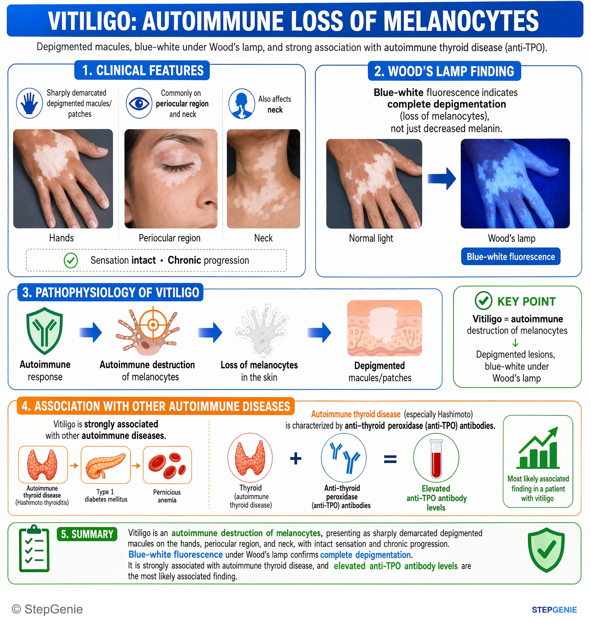
Vitiligo = autoimmune destruction of melanocytes → depigmented macules, blue-white under Wood’s lamp, strongly associated with autoimmune…
USMLE Step 1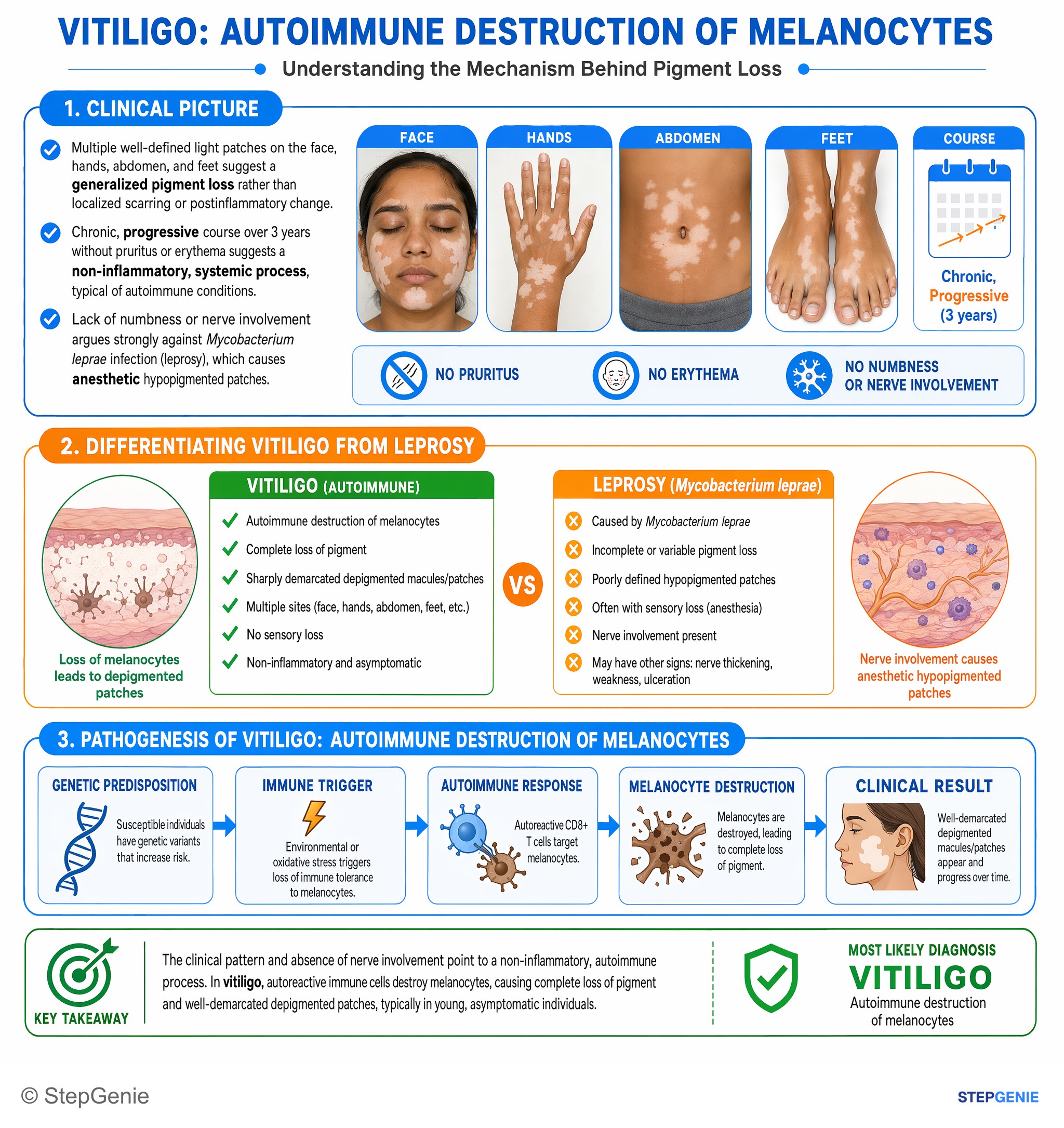
Vitiligo causes well-demarcated depigmented macules/patches due to autoimmune melanocyte destruction, often in asymptomatic young patients…
USMLE Step 1Frequently Asked Questions
What causes vitiligo?
Vitiligo is a T-cell–mediated autoimmune disease. Melanocyte-specific CD8+ cytotoxic T cells enter the epidermis and release interferon-gamma, which signals through JAK1, JAK2 and STAT1 in keratinocytes; the chemokines CXCL9 and CXCL10 then recruit more T cells and melanocytes are destroyed. More than 30 genes, including NLRP1 and PTPN22, raise the risk, and about one in five patients has an affected close relative. Sunburn or other skin injury, emotional stress, some chemicals and melanoma immunotherapy can trigger it.
What is the difference between vitiligo and albinism?
In vitiligo, melanocytes are destroyed, so acquired, patchy, chalk-white areas have no melanocytes on biopsy. In albinism, a normal number of melanocytes is present, but they make little or no melanin, most classically because of a TYR (tyrosinase) defect in type 1 oculocutaneous albinism. Oculocutaneous albinism is congenital, usually autosomal recessive and generalized, affects the eyes with nystagmus, photophobia and reduced vision, and carries a high skin cancer risk, especially squamous cell carcinoma.
What does vitiligo look like under a Wood lamp?
Under a Wood lamp (ultraviolet light at about 365 nm), depigmented vitiligo patches look chalk-white with a bright blue-white glow that contrasts sharply with the surrounding normal skin. The lamp is most useful on fair skin, where subtle patches are hard to see in normal light, and it helps separate true depigmentation from conditions that only lighten the skin, such as pityriasis alba or postinflammatory hypopigmentation.
Which autoimmune diseases are associated with vitiligo?
Autoimmune thyroid disease is the strongest association, both Hashimoto thyroiditis and Graves disease. Others include type 1 diabetes, pernicious anemia, Addison disease and alopecia areata, and up to 30% of patients have other autoimmune antibodies or an autoimmune endocrine disease. Merck suggests checking a complete blood count, fasting glucose, thyroid function and anti-thyroid peroxidase antibodies in patients with vitiligo. Segmental vitiligo is less often linked to other autoimmune disease.
What is the treatment for vitiligo?
Limited disease is treated with a topical corticosteroid or a topical calcineurin inhibitor such as tacrolimus, which suits the face and groin. Ruxolitinib 1.5% cream, a JAK inhibitor, is FDA-approved for nonsegmental vitiligo in patients 12 and older, applied twice daily to up to 10% of body surface area. Narrowband UVB phototherapy is often first for widespread disease, short oral steroid courses can halt rapid spread, grafting suits stable disease, and monobenzone can depigment the remaining skin in extensive disease.
Is there an oral JAK inhibitor approved for vitiligo?
Not in the United States as of October 2026. No oral JAK inhibitor is FDA-approved for vitiligo; the only FDA-approved JAK inhibitor for it is ruxolitinib 1.5% cream. In the European Union, oral upadacitinib 15 mg once daily was approved in July 2026, after a positive opinion from the European Medicines Agency's CHMP in June 2026, for nonsegmental vitiligo in patients 12 and older who are candidates for systemic therapy, and AbbVie reports that it is under FDA review. Ritlecitinib is FDA-approved only for severe alopecia areata.