Psoriasis is one of those “classic derm” diagnoses that shows up everywhere on Step 1—vignette stems, image-based questions, immunology tie-ins, and pharm. If you can explain why the plaques look the way they do (and what the immune system is doing underneath), you’ll stop memorizing random facts and start racking up easy points.
Big Picture (What Psoriasis Is)
Psoriasis is a chronic, immune-mediated inflammatory skin disease characterized by well-demarcated erythematous plaques with silvery scale, driven largely by T-cell cytokines that accelerate keratinocyte proliferation.
- Core idea: epidermis turns over way too fast → thickened stratum corneum + scale
- Pattern recognition: extensor surfaces (elbows, knees), scalp, sacrum, nails
Pathophysiology (The Step 1 “Why”)
Psoriasis is a T-cell–mediated disease with a prominent Th17/Th1 signature.
Immune pathway (high yield)
- Dendritic cells activate T cells → secretion of cytokines:
- IL-23 promotes Th17 maintenance
- Th17 produces IL-17 and IL-22
- TNF-α amplifies inflammation
- These cytokines drive:
- Keratinocyte hyperproliferation
- Angiogenesis and increased dermal blood flow (→ erythema)
- Neutrophil recruitment (→ microabscesses)
What this means histologically
- Acanthosis: epidermal thickening (↑ stratum spinosum)
- Parakeratosis: retained nuclei in stratum corneum (rapid turnover)
- Hypogranulosis: diminished granular layer
- Dilated capillaries in dermal papillae
- Neutrophils in stratum corneum = Munro microabscesses (classic buzzword)
First Aid cross-reference: Dermatology (psoriasis) + Immunology (Th17/IL-17, TNF-α) + Pharm (biologics targeting TNF-α, IL-17, IL-23).
Clinical Presentation (How It Appears on Exams)
Classic lesions
- Well-demarcated erythematous plaques with silvery scale
- Usually on extensor surfaces: elbows, knees
- Also common: scalp, lumbosacral area, gluteal cleft
Key high-yield signs
- Auspitz sign: scraping scale causes pinpoint bleeding
- Mechanism: dilated capillaries close to surface
- Koebner phenomenon: lesions appear at sites of skin trauma
- Think: scratches, surgical scars, sunburn
Nail findings (Step loves these)
- Nail pitting
- Onycholysis (nail plate separates)
- “Oil drop” discoloration (yellow-brown)
Psoriatic arthritis (don’t miss)
- Inflammatory arthritis associated with psoriasis
- Can involve:
- DIP joints
- Dactylitis (“sausage digits”)
- Enthesitis
- Typically seronegative (RF negative); belongs to seronegative spondyloarthropathies
Diagnosis (Mostly Clinical)
Psoriasis is primarily a clinical diagnosis, especially with classic plaques + distribution.
When biopsy shows up
If Step gives histology, look for:
- Parakeratosis
- Acanthosis
- Munro microabscesses
- Thinning of suprapapillary plates + dilated dermal capillaries (ties to Auspitz)
Helpful differentiation (rapid table)
| Condition | Key Clue | Distribution | Scale |
|---|---|---|---|
| Psoriasis | Well-demarcated plaques, Auspitz, Koebner, nail pitting | Extensor surfaces | Silvery |
| Atopic dermatitis | Pruritus, allergies/asthma, lichenification | Flexural in older kids/adults | Variable |
| Seborrheic dermatitis | Greasy scale, dandruff | Scalp, nasolabial folds | Greasy, yellow |
| Tinea corporis | Annular lesion with central clearing | Anywhere | Scaly border; KOH + |
| Lichen planus | “6 P’s” (pruritic, purple, polygonal, planar, papules, plaques) | Wrists/ankles | Fine white lines (Wickham striae) |
Triggers & Associations (High-Yield Integrations)
Psoriasis can flare with:
- Infections (notably streptococcal pharyngitis)
- Stress
- Skin trauma (Koebner)
- Medications:
- Lithium
- β-blockers
- Antimalarials (e.g., hydroxychloroquine)
- Interferons
- Withdrawal of systemic corticosteroids (can precipitate severe flare)
Special presentation: guttate psoriasis
- “Raindrop” small scaly papules on trunk/proximal extremities
- Often after strep throat
- Exam phrasing: teenager with sore throat → sudden eruption of many small lesions
Comorbidities to remember
- Psoriatic arthritis
- Metabolic syndrome (obesity, insulin resistance)
- Increased cardiovascular risk (in severe disease)
Treatment (Step-Style Approach)
Treatment is typically framed by severity and body surface area (BSA), but Step 1 focuses on mechanisms and drug targets.
Mild–moderate (localized plaques)
- Topical corticosteroids
- MOA: anti-inflammatory; decrease cytokine transcription
- Pearl: avoid high-potency chronic use on face/groin (atrophy)
- Vitamin D analogs (e.g., calcipotriene)
- ↓ keratinocyte proliferation and modulates differentiation
- Topical retinoids (e.g., tazarotene)
- Coal tar or salicylic acid (less Step-heavy)
Moderate–severe (widespread) / refractory
- Phototherapy (UVB)
- Slows keratinocyte proliferation; immunomodulatory
Systemic agents (know the big ones + toxicities)
- Methotrexate
- Inhibits dihydrofolate reductase (↓ DNA synthesis) + anti-inflammatory
- Toxicities: hepatotoxicity, bone marrow suppression, pulmonary toxicity
- Antidote/support: folinic acid (leucovorin)
- Cyclosporine
- Calcineurin inhibitor → ↓ IL-2 transcription → ↓ T-cell activation
- Toxicities: nephrotoxicity, hypertension, neurotoxicity
- Acitretin (oral retinoid)
- Teratogenic; mucocutaneous dryness; dyslipidemia
Biologics (very high yield)
These map directly onto the Th17/TNF pathway:
- Anti–TNF-α: infliximab, adalimumab, etanercept
- Anti–IL-12/23 (p40): ustekinumab
- Anti–IL-17: secukinumab (and others in class)
- Anti–IL-23 (p19): newer agents (class concept)
Board-style adverse effect tie-in: immunosuppression → ↑ risk of serious infections (including TB reactivation with anti-TNF agents).
First Aid cross-reference: Immunology (Th17/IL-17), Pharm (methotrexate, cyclosporine, TNF inhibitors), Micro/ID considerations (TB screening before anti-TNF).
Classic USMLE-Style Vignettes (What They’re Really Testing)
1) “Silvery plaques on extensor surfaces”
They’re testing diagnosis and immune mechanism (Th17/TNF).
2) “Scrape plaque → pinpoint bleeding”
That’s Auspitz sign due to dilated dermal capillaries.
3) “New lesions appear along scratch marks”
That’s Koebner phenomenon (trauma-induced lesions).
4) “Nail pitting + joint pain”
That’s psoriatic arthritis (seronegative inflammatory arthritis).
5) “Teen with strep throat → sudden small scaly lesions”
That’s guttate psoriasis.
Rapid-Fire High-Yield Facts (Memorize These)
- Psoriasis = T-cell–mediated, especially Th17 → IL-17, IL-22, plus TNF-α
- Silvery scale, well-demarcated plaques, extensor surfaces
- Auspitz sign = pinpoint bleeding after scale removal
- Koebner phenomenon = lesions at trauma sites
- Histology buzzwords: parakeratosis, acanthosis, Munro microabscesses
- Nail findings: pitting, onycholysis
- Associated with psoriatic arthritis (seronegative)
- Treatment ladder: topicals → phototherapy → systemic agents/biologics
- Biologics target: TNF-α, IL-17, IL-23, IL-12/23
Quick Self-Check (2 Questions)
-
Which cytokine axis is most associated with psoriasis and is targeted by multiple biologics?
Answer: Th17 axis (IL-23 → Th17 → IL-17/IL-22) and TNF-α. -
What’s the histologic correlate of the “silvery scale”?
Answer: Parakeratosis due to rapid epidermal turnover.
Visual explanations from the StepGenie QBank
Infographics StepGenie drew to explain questions in its own QBank, matched to this article's topic. Each caption is the takeaway of the question it explains.
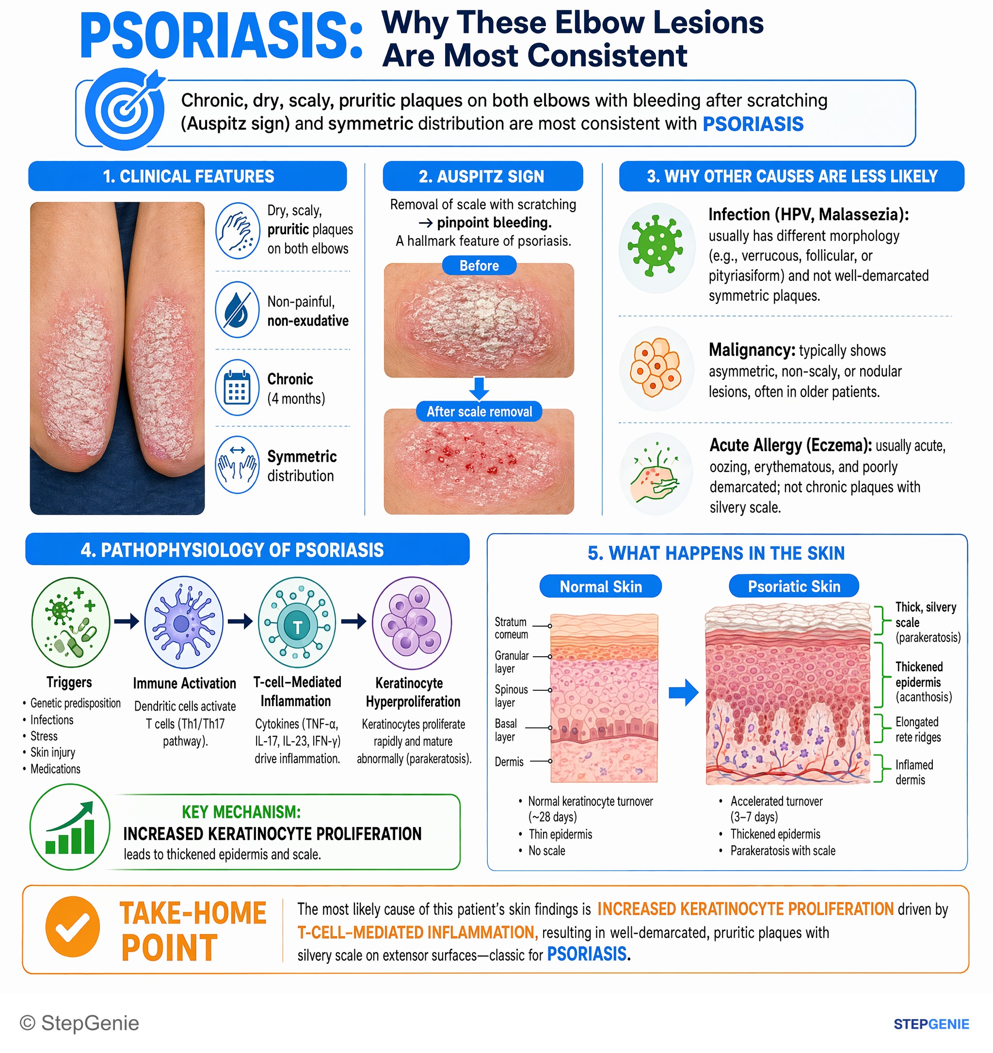
Psoriasis causes well-demarcated, pruritic plaques with silvery scale on extensor surfaces due to hyperproliferation of keratinocytes and…
USMLE Step 1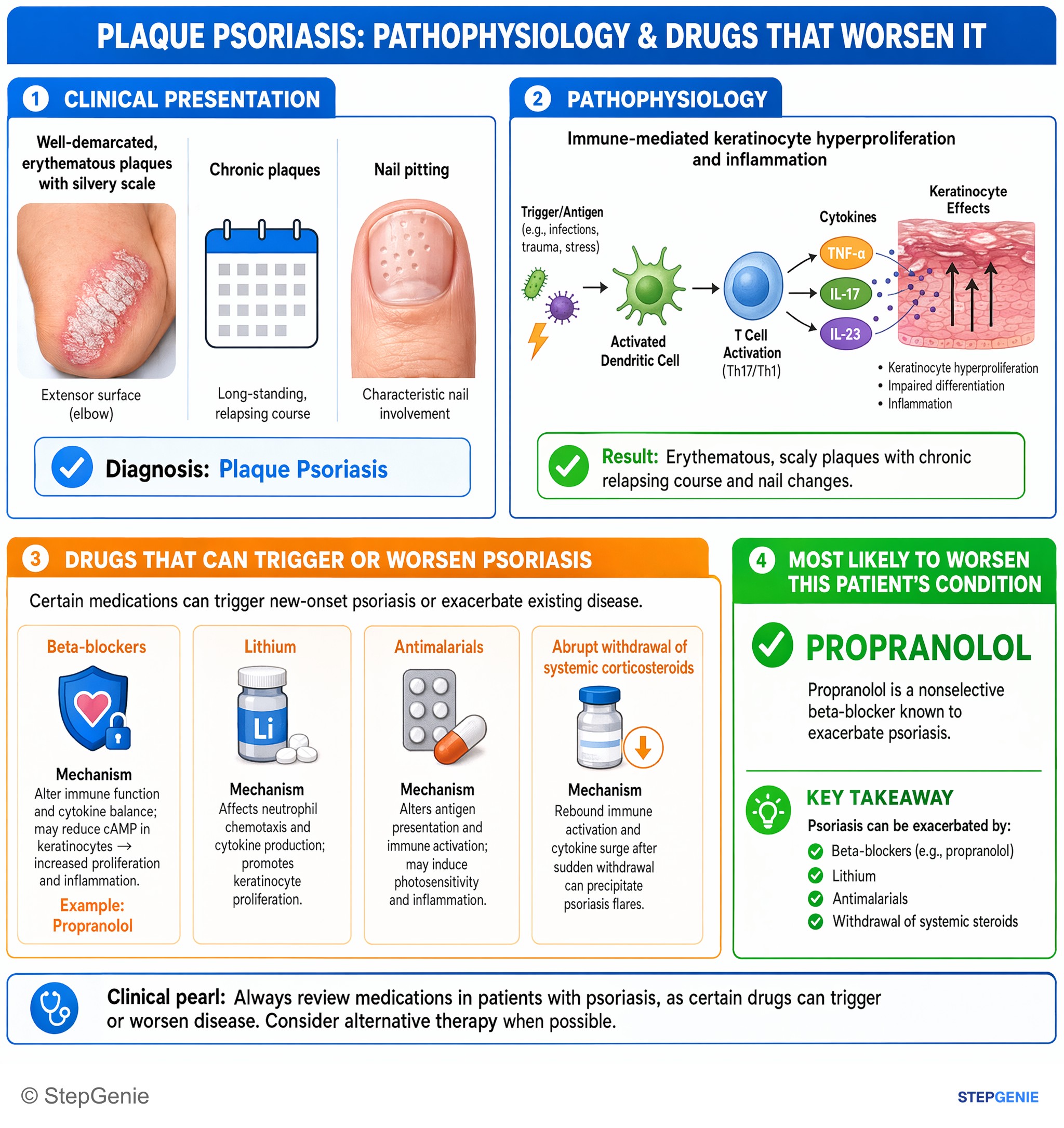
Psoriasis can be exacerbated by beta-blockers (e.g., propranolol), lithium, antimalarials, and withdrawal of systemic steroids.
USMLE Step 1Frequently Asked Questions
Why do psoriasis plaques have thick silvery scale and erythema?
Psoriasis plaques are thick and scaly because T-cell cytokines drive rapid keratinocyte proliferation, causing excessive epidermal turnover with a thickened stratum corneum and retained nuclei in the stratum corneum. They are erythematous because the same inflammatory pathway promotes angiogenesis and increased dermal blood flow, with dilated capillaries contributing to the red appearance.
How do you tell psoriasis from atopic dermatitis or seborrheic dermatitis on an exam?
Psoriasis is suggested by well-demarcated erythematous plaques with silvery scale on extensor surfaces, often with Auspitz sign, Koebner phenomenon, and nail pitting. Atopic dermatitis more often presents with pruritus, allergy or asthma history, lichenification, and a flexural distribution in older children and adults. Seborrheic dermatitis typically has greasy yellow scale on the scalp and nasolabial folds.
What causes Auspitz sign in psoriasis?
Auspitz sign is caused by dilated capillaries close to the skin surface, so scraping off scale leads to pinpoint bleeding. In psoriasis, histology shows dilated capillaries in the dermal papillae and thinning of the suprapapillary plates, which explains why removing the scale can expose these superficial vessels and produce the classic exam finding.
What histology findings are classic for psoriasis?
Classic psoriasis histology includes parakeratosis, acanthosis, hypogranulosis, and neutrophils in the stratum corneum called Munro microabscesses. You may also see thinning of the suprapapillary plates and dilated dermal capillaries. These findings reflect the underlying rapid epidermal turnover, epidermal thickening, and inflammatory changes driven by the Th17/Th1-mediated immune response.
What is the first-line treatment approach for psoriasis, and when do you use biologics?
Localized mild to moderate psoriasis is typically treated first with topical corticosteroids and vitamin D analogs such as calcipotriene, with topical retinoids also used. More widespread or refractory disease is treated with phototherapy or systemic agents. Biologics are high-yield options for moderate to severe disease because they target the core inflammatory pathway, including TNF-α, IL-17, IL-12/23, and IL-23.