Melanoma questions love to look “too easy”—a pigmented lesion, a worried patient, and the tempting urge to click “melanoma” and move on. But the real points on Step 1/2 come from knowing exactly why the correct choice fits and why each distractor doesn’t. This post walks through a classic ABCDE melanoma vignette, then dismantles the answer choices like a high-yield Q-bank review.
Tag
Dermatology > Skin Disorders
Clinical Vignette (Q-Bank Style)
A 52-year-old man presents for evaluation of a mole on his upper back that his partner noticed has been “changing” over the past 6 months. He has a history of intermittent intense sun exposure and multiple blistering sunburns as a teenager. Exam shows a 1.1 cm darkly pigmented lesion with irregular borders, variegated color (tan, brown, black), and asymmetry. A few areas appear slightly raised with recent itching. No palpable lymphadenopathy.
Which of the following is the best next step in management?
A. Shave biopsy of the lesion
B. Excisional biopsy with narrow margins
C. Wide local excision with 2-cm margins without biopsy
D. Cryotherapy of the lesion
E. Reassurance and routine follow-up in 12 months
The Correct Answer: B. Excisional biopsy with narrow margins
When melanoma is suspected clinically, the best next step is full-thickness excisional biopsy (elliptical excision) with narrow margins (often 1–3 mm). This allows accurate measurement of Breslow depth, which is the single most important prognostic factor and guides definitive management (including margin size and whether to consider sentinel lymph node biopsy).
Why this stem screams melanoma (ABCDE)
Melanoma classically presents with one or more of:
- Asymmetry
- Border irregularity
- Color variation (multiple shades)
- Diameter mm (not required, but common)
- Evolving (changing size/shape/color, symptoms like itching/bleeding)
This lesion has A, B, C, D, and E—it’s basically the ABCDE poster child.
High-Yield Melanoma Pearls (USMLE Favorite)
1) Breslow depth drives prognosis and next steps
- Breslow depth = measured in mm from the granular layer of epidermis down to deepest tumor cell.
- Deeper = worse prognosis and higher metastatic risk.
- Don’t confuse with Clark level (older, anatomic invasion levels; less emphasized now).
2) Best initial diagnostic test
- Excisional biopsy (full thickness) with narrow margins
- Avoid techniques that may truncate depth (especially shave biopsy when melanoma is strongly suspected).
3) Risk factors they love to test
- Intermittent intense UV exposure (especially blistering childhood sunburns)
- Fair skin, light hair/eyes, inability to tan
- Multiple nevi, atypical nevi
- Family history (e.g., CDKN2A)
- Immunosuppression
4) Classic melanoma subtypes (quick pattern recognition)
| Subtype | Hallmark | Common Location |
|---|---|---|
| Superficial spreading (most common) | Asymmetric, variegated, radial growth phase | Anywhere, often trunk in men |
| Nodular | Rapid vertical growth, can be uniformly dark or amelanotic | Trunk/head/neck |
| Lentigo maligna | Slowly enlarging, sun-damaged skin | Face/elderly |
| Acral lentiginous | Palms/soles, nail beds; more common in darker skin | Subungual, plantar |
Board trap: darker skin ≠ immune to melanoma; acral lentiginous melanoma is a key exception.
Why Each Distractor Is Wrong (and What It Is Used for)
A. Shave biopsy of the lesion
Why it’s wrong here:
- Shave biopsies can miss the deepest portion of the tumor, causing underestimation of Breslow depth, which can change staging and management.
When it might be acceptable:
- Low suspicion lesions or situations where a deep saucerization technique is used by experienced clinicians—but on exams, suspected melanoma → excisional biopsy.
Test-taking rule: If they’re pushing ABCDE and evolution, don’t shave.
C. Wide local excision with 2-cm margins without biopsy
Why it’s wrong:
- You don’t jump straight to definitive wide excision without diagnosis and staging.
- Margin size depends on Breslow depth, which you don’t know yet.
High-yield management sequence:
- Excisional biopsy (narrow margins) → get Breslow depth
- Wide local excision with margin based on depth
- Consider sentinel lymph node biopsy for higher-risk tumors (commonly tested concept: deeper lesions → more likely)
D. Cryotherapy of the lesion
Why it’s wrong:
- Cryotherapy destroys tissue and prevents histologic assessment—dangerous when malignancy is on the table.
What cryotherapy is for (common Step associations):
- Actinic keratoses (premalignant rough scaly lesions on sun-exposed skin)
- Viral warts
- Some benign lesions (selected cases)
Rule: If you must rule out cancer, you need tissue.
E. Reassurance and routine follow-up in 12 months
Why it’s wrong:
- The lesion is evolving and hits multiple ABCDE criteria—this is not a “watchful waiting” situation.
What reassurance fits instead (compare the look-alikes):
- Seborrheic keratosis: “stuck-on,” waxy, verrucous; can be pigmented but typically has a more superficial, pasted-on appearance
- Cherry angioma: red/purple papules (vascular)
- Stable symmetric nevi without change
Board mindset: “Changing pigmented lesion” + ABCDE = biopsy now.
Rapid-Fire Differential: Melanoma vs Common Pigmented Distractors
| Condition | Typical Look | Key Clue | Management |
|---|---|---|---|
| Melanoma | Asymmetric, irregular border, variegated color, evolving | ABCDE, “ugly duckling” lesion | Excisional biopsy |
| Basal cell carcinoma (BCC) | Pearly papule, rolled borders, telangiectasias; may ulcerate | “Rodent ulcer,” sun-exposed, slow-growing | Shave/punch biopsy → excision/Mohs |
| Squamous cell carcinoma (SCC) | Scaly erythematous plaque/nodule; may ulcerate | Sun-exposed; can arise from actinic keratosis | Biopsy → excision |
| Seborrheic keratosis | “Stuck-on,” waxy, warty | Benign, often multiple | Reassure or remove if symptomatic |
| Actinic keratosis | Rough, gritty papule on sun-exposed skin | Premalignant | Cryotherapy/topicals |
Exam-Grade Takeaways (What You Should Click Under Pressure)
- Suspicious pigmented lesion with ABCDE → excisional biopsy with narrow margins.
- Breslow depth is crucial; don’t pick an option that could truncate depth (shave biopsy).
- Don’t “treat” (cryotherapy) or “observe” (reassurance) a lesion that needs histology.
- Don’t skip diagnosis and jump to wide excision without knowing staging.
Visual explanations from the StepGenie QBank
Infographics StepGenie drew to explain questions in its own QBank, matched to this article's topic. Each caption is the takeaway of the question it explains.
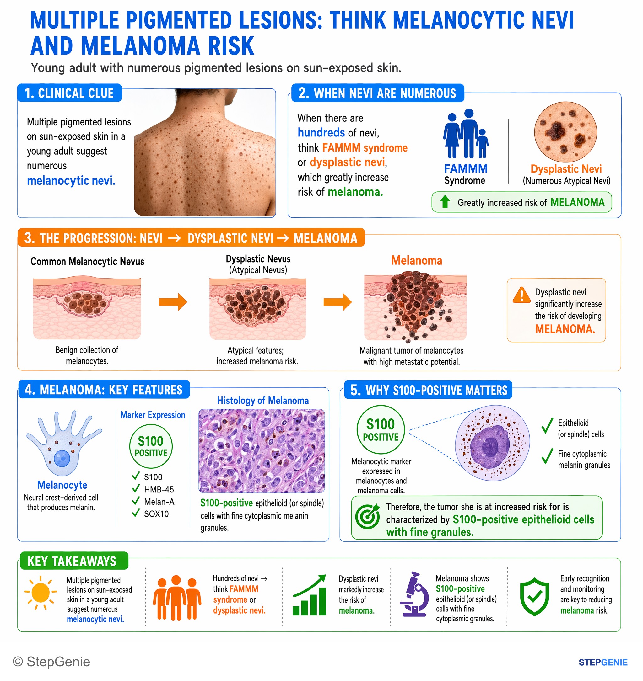
Dysplastic nevi (numerous atypical nevi) increase risk of melanoma, a tumor of S100-positive melanocytes with epithelioid cells and fine…
USMLE Step 1Frequently Asked Questions
What is the best next step when a pigmented lesion has ABCDE features of melanoma?
The best next step is a full-thickness excisional biopsy with narrow margins. When melanoma is suspected clinically, this approach allows accurate measurement of Breslow depth, which is the single most important prognostic factor. That depth then guides definitive management, including how wide the later excision should be and whether sentinel lymph node biopsy should be considered.
Why is shave biopsy a bad choice when melanoma is strongly suspected?
Shave biopsy is a poor choice because it can truncate the lesion and miss the deepest portion of the tumor. That can lead to underestimation of Breslow depth, which matters because Breslow depth drives prognosis, staging, and treatment decisions. In a lesion with classic ABCDE features and evolution, exams favor excisional biopsy rather than shaving the lesion.
Why can’t you do a wide local excision before getting a biopsy in suspected melanoma?
You should not go straight to wide local excision because definitive margin size depends on Breslow depth, which is not known until tissue is obtained. The correct sequence is excisional biopsy with narrow margins first, then wide local excision based on the measured depth. Higher-risk or deeper lesions may also prompt consideration of sentinel lymph node biopsy.
What features in a mole make a melanoma vignette obvious on exams?
A melanoma vignette becomes highly suspicious when the lesion shows the ABCDE pattern: asymmetry, border irregularity, color variation, diameter of at least 6 mm, and evolution over time. Symptoms such as itching or bleeding also support evolution. A changing pigmented lesion that hits multiple ABCDE criteria should push you toward biopsy now rather than reassurance or routine follow-up.
How do you tell melanoma from a benign pigmented lesion like seborrheic keratosis?
Melanoma is suggested by an asymmetric lesion with irregular borders, variegated color, and recent change, often described with the ABCDE criteria. Seborrheic keratosis, in contrast, is typically stuck-on, waxy, verrucous, and more superficial in appearance. A stable symmetric nevus also fits reassurance better, but an evolving pigmented lesion with ABCDE features should be biopsied.