Ovarian tumors are classified by the cell they arise from: surface epithelial tumors (85-90% of ovarian cancers, mostly in older women, tracked with CA-125), germ cell tumors (girls and women under about 30, with AFP, β-hCG or LDH as markers), sex cord-stromal tumors (the hormone makers: estrogen from granulosa and theca cells, androgens from Sertoli-Leydig cells) and metastases such as the bilateral Krukenberg tumor. High-grade serous carcinoma is the most common ovarian cancer and often starts in the fallopian tube; the mature cystic teratoma (dermoid) is the most common germ cell tumor. The biggest inherited risks are BRCA1/BRCA2 and Lynch syndrome, while oral contraceptives, pregnancy and breastfeeding lower risk.
The memory palace: the Ovary Hotel
Picture a hotel shaped like an ovary. Each floor is one cell of origin; each room holds a prop that triggers a tumor's buzzword, marker or hormone.
- Lobby: surface epithelial tumors. The busiest floor, full of older guests.
- Second floor: germ cell tumors. The family floor: young guests, and a tumor marker on every door.
- Third floor: sex cord-stromal tumors. The hormone spa.
- Basement: metastases. Luggage shipped in from the stomach.
Lobby: surface epithelial tumors
| Room and prop | Tumor | What to say on test day |
|---|---|---|
| Sea-view suite with sand on the floor | Serous | High-grade serous carcinoma is the most common subtype (more than 70% of epithelial carcinomas): TP53-mutated in about 96%, BRCA-linked, usually stage III-IV at diagnosis, and often born in the fallopian tube (serous tubal intraepithelial carcinoma). Psammoma bodies (laminated calcifications; psammos is Greek for sand) are typical of serous tumors, especially low-grade serous carcinoma (a slower-growing, KRAS- or BRAF-mutated tumor) |
| Kitchen stacked with giant honey jars | Mucinous | Mucin-filled cystic tumors; about 80% of mucinous carcinomas are diagnosed early, usually at stage I. A mucinous ovarian mass may be a metastasis from the appendix or GI tract, so surgeons often remove the appendix and CEA is checked |
| Twin rooms with chocolates on the pillows | Endometrioid and clear cell | Both linked to endometriosis ("chocolate cysts"). Endometrioid carcinoma looks like endometrial cancer; clear cell carcinoma has a hobnail pattern and a worse prognosis |
| Bathroom with a bladder-shaped tub and a bowl of coffee beans | Brenner | Nests of transitional (bladder-like) cells with grooved "coffee-bean" nuclei in dense fibrous stroma; usually benign and unilateral; about 2% of ovarian tumors |
Benign epithelial tumors include serous and mucinous cystadenomas and Brenner tumors. Borderline tumors tend to affect younger women and grow slowly.
Second floor: germ cell tumors
| Room and prop | Tumor | Marker | What to say on test day |
|---|---|---|---|
| Hair salon with teeth in the sink and a TV showing a thyroid | Mature cystic teratoma (dermoid) | None | Most common germ cell tumor (95% of ovarian teratomas, about 20% of all ovarian neoplasms). Tissue from all 3 germ layers, mostly ectoderm: skin, hair, sebum, fat, teeth, often in a Rokitansky protuberance. Struma ovarii (a teratoma made mainly of thyroid tissue) can cause hyperthyroidism. Like other benign masses over 4 cm, it can twist (adnexal torsion). Malignant change in 0.3-2% |
| Nursery with a half-built brain model | Immature teratoma | — | Malignant because of immature neuroepithelium, graded 1-3 by how much is present; usually in the first 3 decades of life |
| Disco with "LDH" in lights | Dysgerminoma | LDH (hCG in 3-5%) | Classically the most common malignant germ cell tumor (about a third; in SEER data immature teratoma is about as common); the ovarian counterpart of testicular seminoma; adolescents and young women; bilateral in 10-15%; seen with gonadal dysgenesis; radiosensitive, but chemotherapy is preferred because radiation causes ovarian failure |
| Kitchen with a cracked egg yolk | Yolk sac (endodermal sinus) tumor | AFP | Schiller-Duval bodies (a central blood vessel wrapped by tumor cells) are pathognomonic; girls and young women (median age about 19); grows fast but is chemosensitive |
| Room with a giant pregnancy test | Choriocarcinoma | β-hCG | Syncytiotrophoblast cells make the hCG; rare in the ovary. The gestational form is covered in gestational trophoblastic disease |
Embryonal carcinoma often raises both AFP and hCG. Malignant germ cell tumors are treated with fertility-sparing unilateral salpingo-oophorectomy plus BEP chemotherapy (bleomycin, etoposide, cisplatin); stage IA dysgerminoma and stage I grade 1 immature teratoma need surgery alone. More than 9 in 10 patients live at least 5 years. Compare the testicular tumors in seminoma vs nonseminoma.
Third floor: sex cord-stromal tumors (about 8% of ovarian neoplasms)
| Room and prop | Tumor | Hormone or marker | What to say on test day |
|---|---|---|---|
| Greenhouse with coffee beans and Call-Exner peepholes | Granulosa cell, adult type (95%) | Estrogen; inhibin B and AMH | Most common malignant sex cord-stromal tumor. Pelvic mass with abnormal or postmenopausal bleeding; endometrial hyperplasia in 29% and carcinoma in 5-7.5%. Call-Exner bodies (eosinophilic spaces ringed by granulosa cells), coffee-bean nuclei, FOXL2 C402G mutation. 47% of recurrences come more than 5 years out, so follow-up is long. The juvenile type (5%, average age 15) recurs within about 3 years |
| Steam sauna | Thecoma | Estrogen | Postmenopausal bleeding, endometrial hyperplasia or carcinoma |
| Fountain flooding the lobby and the balcony | Fibroma | None | Most common benign sex cord-stromal tumor (about 4% of ovarian neoplasms). Meigs syndrome = fibroma + ascites + pleural effusion, classically right-sided (about 1% of fibromas) |
| Gym with a bodybuilder and crystal trophies | Sertoli-Leydig cell | Androgens | Average age 25; virilization in 40-60% (hirsutism, deepening voice, clitoromegaly, amenorrhea); Reinke crystals; DICER1 mutations in about 60% |
In a prepubertal girl, a juvenile granulosa cell tumor causes isosexual (GnRH-independent) precocious puberty: estrogen-driven breast development and vaginal bleeding.
Basement: metastases
The Krukenberg tumor is a metastatic, mucin-rich signet-ring cell adenocarcinoma in the ovaries. The stomach is the primary in about 70% of cases (stomach plus colon nearly 90%), and it is bilateral in 80%. Breast cancer also spreads to the ovary. Review the primary in the gastric cancer sketch.
Presentation, work-up and treatment in brief
- Epithelial cancer is usually advanced when found, because early symptoms are vague: bloating, early satiety, dyspepsia, pelvic pain, a change in bowel habits, urinary frequency. A pelvic mass with ascites usually means cancer.
- Work-up: ultrasonography first, then CT or MRI if advanced disease is suspected, plus CA-125. Every woman with ovarian, tubal or peritoneal cancer should have a genetic risk evaluation; germline BRCA1/2 mutations are found in about 14-18% of high-grade serous cancers.
- Treatment: surgical cytoreduction, then carboplatin plus paclitaxel, with bevacizumab or a PARP inhibitor such as olaparib as maintenance in selected patients (especially with homologous recombination deficiency).
Tumor markers at a glance
| Marker | Points to | Caveat |
|---|---|---|
| CA-125 | Epithelial cancer, especially serous | Raised in about 80% of advanced cases but often normal in early disease; also raised in endometriosis, pregnancy, other cancers and inflammation. Used to monitor, not to screen average-risk women |
| CEA | Mucinous tumor or a GI primary | Checked with AFP and hCG to exclude GI and germ cell tumors |
| AFP | Yolk sac tumor; embryonal carcinoma | — |
| β-hCG | Choriocarcinoma; embryonal carcinoma; 3-5% of dysgerminomas | — |
| LDH | Dysgerminoma | — |
| Inhibin B, AMH | Granulosa cell tumor | Useful in follow-up because recurrences come late |
| Estrogen | Granulosa cell tumor, thecoma | Drives endometrial hyperplasia and cancer |
| Testosterone | Sertoli-Leydig cell tumor | Virilization |
More pairings are in the tumor markers guide.
Risk factors and protective factors
- Raise risk: BRCA1 (about 39-58% lifetime risk) and BRCA2 (13-29%) variants, mainly high-grade serous carcinoma; Lynch syndrome (mismatch repair genes MLH1, MSH2, MSH6, PMS2, EPCAM; see Lynch syndrome vs FAP); family history; older age; nulliparity, delayed childbearing, early menarche and late menopause; endometriosis; postmenopausal hormone therapy; obesity; tall height.
- Lower risk: oral contraceptives (about 50% lower risk after 5 years of use and 80% after 10 or more), multiparity, breastfeeding, and bilateral tubal ligation or salpingectomy. ACOG advises risk-reducing salpingo-oophorectomy at 35-40 for BRCA1 carriers and by 40-45 for BRCA2 carriers.
- No screening for average-risk women: CA-125 is not recommended as a screening test.
How USMLE tests it
- Postmenopausal bleeding + solid adnexal mass → granulosa cell tumor (and sample the endometrium).
- Virilization + adnexal mass → Sertoli-Leydig cell tumor. PCOS is the most common cause of hirsutism, but abrupt onset, virilization or a pelvic mass suggests a tumor, and Merck advises imaging when total testosterone is above 150 ng/dL (above 100 ng/dL after menopause). Compare the PCOS breakdown.
- Girl or young woman + rapidly growing mass: AFP → yolk sac tumor; LDH → dysgerminoma; β-hCG → choriocarcinoma.
- Pelvic mass + ascites + right pleural effusion with a benign fibroma → Meigs syndrome, the classic mimic of ovarian cancer.
- Bilateral ovarian masses in a patient with gastric cancer → Krukenberg tumor.
- Psammoma bodies → serous ovarian tumors, papillary thyroid carcinoma, meningioma, mesothelioma.
- Coffee-bean nuclei appear in both Brenner and granulosa cell tumors; let the hormones decide.
- Hyperthyroidism + ovarian teratoma → struma ovarii.
Practice question
A 58-year-old woman has had vaginal bleeding for 2 months; her last menstrual period was 6 years ago. Pelvic examination reveals a firm, mobile right adnexal mass. Ultrasound shows a 9-cm solid right ovarian mass and a thickened endometrium, and endometrial biopsy shows hyperplasia without atypia. CA-125 is normal. Microscopic examination of the ovarian mass is most likely to show which of the following?
A. Call-Exner bodies B. Schiller-Duval bodies C. Psammoma bodies D. Signet-ring cells E. Reinke crystals
Answer: A. Estrogen excess (postmenopausal bleeding, endometrial hyperplasia) plus a solid ovarian mass points to an adult granulosa cell tumor, and Call-Exner bodies are small eosinophilic spaces ringed by granulosa cells. Inhibin B is the marker to follow.
- B: Schiller-Duval bodies mark a yolk sac tumor, an AFP-secreting tumor of girls and young women.
- C: psammoma bodies point to a serous epithelial tumor, which would not explain estrogen excess.
- D: signet-ring cells in the ovary mean a Krukenberg tumor, usually bilateral and from the stomach.
- E: Reinke crystals belong to Leydig cells, so a Sertoli-Leydig cell tumor, which causes virilization rather than estrogen effects.
Key takeaways
- Sort by cell of origin: epithelial (85-90% of cancers; older women; CA-125), germ cell (young; AFP, β-hCG, LDH), sex cord-stromal (hormones) and metastatic (Krukenberg).
- High-grade serous is the most common ovarian cancer: BRCA-linked and often tubal in origin. Psammoma bodies = serous.
- Endometriosis → endometrioid and clear cell (hobnail). Brenner = transitional cells, coffee-bean nuclei, benign.
- Dermoid = most common germ cell tumor; dysgerminoma (LDH) = the classic most common malignant one; yolk sac (AFP, Schiller-Duval); choriocarcinoma (β-hCG).
- Granulosa = estrogen, Call-Exner bodies, inhibin B, late recurrence; Sertoli-Leydig = androgens; fibroma = Meigs syndrome.
- Krukenberg = signet-ring cells, usually from the stomach, bilateral.
- BRCA and Lynch raise risk; oral contraceptives, pregnancy, breastfeeding and tubal surgery lower it.
Sources
- Ovarian Epithelial, Fallopian Tube, and Primary Peritoneal Cancer Treatment (PDQ®)–Health Professional Version — National Cancer Institute (2025)
- Ovarian Germ Cell Tumors Treatment (PDQ®)–Health Professional Version — National Cancer Institute (2024)
- Ovarian, Fallopian Tube, and Peritoneal Cancer — Merck Manual Professional Edition (2023)
- Benign Adnexal Masses — Merck Manual Professional Edition (2026)
- Epithelial Ovarian Cancer — StatPearls, NCBI Bookshelf (2024)
- Rare Ovarian Tumors (in Ovarian Cancer, Exon Publications) — NCBI Bookshelf (2022)
- Krukenberg Tumor — StatPearls, NCBI Bookshelf (2024)
- What Is Ovarian Cancer? — American Cancer Society (2025)
- Brenner's tumour of the ovary — BMJ Case Reports (2022)
- BRCA Gene Changes: Cancer Risk and Genetic Testing — National Cancer Institute (2024)
- Psammoma bodies — Radiopaedia.org
- Hirsutism — Merck Manual Professional Edition
Visual explanations from the StepGenie QBank
Infographics StepGenie drew to explain questions in its own QBank, matched to this article's topic. Each caption is the takeaway of the question it explains.
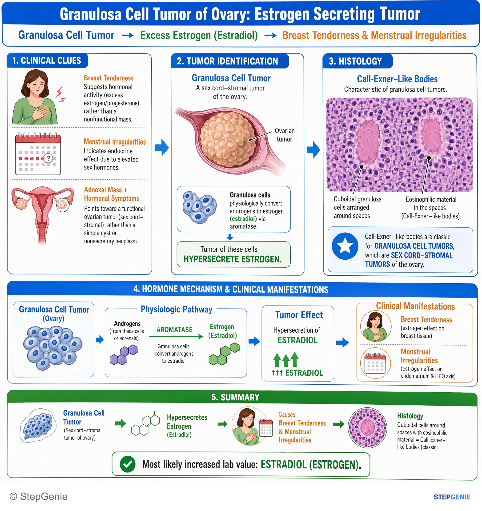
Granulosa cell tumor of ovary secretes estrogen (estradiol) → breast tenderness, menstrual irregularities; histology shows Call-Exner–like…
USMLE Step 1Frequently Asked Questions
How are ovarian tumors classified?
Ovarian tumors are grouped by the cell they come from. Surface epithelial tumors (serous, mucinous, endometrioid, clear cell and Brenner) make up 85 to 90 percent of ovarian cancers and mostly affect older women. Germ cell tumors (teratomas, dysgerminoma, yolk sac tumor, choriocarcinoma) affect girls and young women. Sex cord-stromal tumors (granulosa cell, thecoma, fibroma, Sertoli-Leydig) often make hormones. Metastases, such as the Krukenberg tumor from the stomach, form the fourth group.
Which tumor markers go with which ovarian tumors?
CA-125 goes with epithelial ovarian cancer, especially serous carcinoma, and is used to follow disease rather than to screen average-risk women. AFP points to a yolk sac tumor, beta-hCG to choriocarcinoma and LDH to dysgerminoma; embryonal carcinoma can raise both AFP and hCG. Inhibin B and anti-Müllerian hormone are markers for granulosa cell tumors, which also make estrogen. Sertoli-Leydig cell tumors make androgens such as testosterone, and CEA helps flag a mucinous or gastrointestinal primary.
What are Call-Exner bodies and Schiller-Duval bodies?
Both are histology buzzwords for ovarian tumors. Call-Exner bodies are small eosinophilic spaces surrounded by granulosa cells and identify a granulosa cell tumor, which typically makes estrogen and causes abnormal or postmenopausal bleeding. Schiller-Duval bodies have a central blood vessel wrapped by tumor cells; they are pathognomonic for a yolk sac tumor, which secretes AFP and affects girls and young women. Psammoma bodies, which are laminated calcifications, point to serous tumors.
What is Meigs syndrome?
Meigs syndrome is the combination of a benign ovarian fibroma, ascites and a pleural effusion, classically on the right side. It matters because a pelvic mass with ascites usually suggests ovarian cancer, and Meigs syndrome is the classic benign exception. Fibromas are the most common benign sex cord-stromal tumors, and Meigs syndrome occurs in only about 1 percent of them.
What is a Krukenberg tumor?
A Krukenberg tumor is a metastasis to the ovary made of mucin-rich signet-ring cells. The primary cancer is in the stomach in about 70 percent of cases, and the stomach and colon together account for nearly 90 percent. Krukenberg tumors are bilateral in about 80 percent of cases, so bilateral ovarian masses in a patient with a history of gastric cancer should suggest this diagnosis.
What are the risk and protective factors for ovarian cancer?
The strongest risk factors are inherited: about 39 to 58 percent of women with a harmful BRCA1 variant and 13 to 29 percent with a BRCA2 variant develop ovarian cancer, and Lynch syndrome also raises risk. Other factors include family history, older age, nulliparity, early menarche, late menopause, endometriosis and obesity. Oral contraceptives lower risk by about half after 5 years of use, and multiparity, breastfeeding and tubal ligation or salpingectomy are also protective.