Gestational trophoblastic disease (GTD) is one of those Step 1 topics that looks small on the outline but shows up everywhere: OB bleeding differentials, tumors, physiology, ultrasound patterns, and even hyperthyroidism. If you can quickly recognize the “snowstorm uterus,” know what to do with a rising - after evacuation, and distinguish complete vs partial mole vs choriocarcinoma, you’ll pick up easy points.
What is Gestational Trophoblastic Disease (GTD)?
GTD is a spectrum of disorders caused by abnormal proliferation of trophoblastic tissue (placental tissue). It ranges from benign molar pregnancy to malignant neoplasia.
Key entities you must know
- Hydatidiform mole
- Complete mole
- Partial mole
- Gestational choriocarcinoma
- (Less commonly tested, but part of spectrum: Placental site trophoblastic tumor)
First Aid Cross-References (high-yield)
In First Aid for the USMLE Step 1 (Reproductive section), GTD content is usually clustered with:
- Gestational trophoblastic disease: complete vs partial mole
- Choriocarcinoma
- Classic ultrasound + - associations
- “No fetal parts” vs “fetal parts present”
- Theca-lutein cysts (ovarian hyperstimulation from )
Use FA as your anchor, then layer in the “why” below to make the questions feel automatic.
Pathophysiology: the genetics behind the patterns
Complete hydatidiform mole
Core idea: All paternal genome, no maternal nuclear DNA.
- Most commonly:
- An “empty” ovum (no maternal chromosomes) is fertilized by one sperm that duplicates → 46,XX (most common)
- Or two sperm fertilize an empty ovum → 46,XX or 46,XY
- No embryo/fetus forms because there’s no maternal genome contribution to embryonic development.
- Trophoblast proliferates wildly → very high -.
Partial hydatidiform mole
Core idea: Triploidy with some maternal DNA—some fetal development can occur.
- A normal ovum is fertilized by two sperm → 69,XXX / 69,XXY / 69,XYY
- Fetal parts may be present, but nonviable.
- - elevated, but typically less dramatic than complete moles.
Clinical Presentation (how it shows up on exams)
Shared features of molar pregnancy
- Vaginal bleeding in early pregnancy
- Uterus size larger than gestational age (esp. complete mole)
- Very high -
- Absent fetal heart tones (especially complete)
- Hyperemesis gravidarum (from high )
- Preeclampsia < 20 weeks = red flag for molar pregnancy (preeclampsia this early is unusual otherwise)
High-yield systemic effects of high
- Hyperthyroidism
- can weakly stimulate the TSH receptor
- Step-style clue: tachycardia + heat intolerance + high - in pregnancy
- Theca-lutein cysts (bilateral ovarian cysts)
- Ovaries respond to high → cystic enlargement
Diagnosis: what the stem gives you
Ultrasound findings (classic)
| Condition | Ultrasound | Fetal parts? |
|---|---|---|
| Complete mole | “Snowstorm” / “cluster of grapes” appearance due to swollen chorionic villi | Absent |
| Partial mole | Abnormal placenta; may show cystic spaces; often some fetal tissue | May be present |
Labs
- Markedly elevated - (especially complete mole)
Histology (Step 1 favorite)
| Feature | Complete mole | Partial mole |
|---|---|---|
| Chorionic villi | Diffuse hydropic swelling | Focal hydropic change |
| Trophoblastic proliferation | Diffuse, circumferential | Focal, mild |
| Fetal tissue | Absent | Present (often) |
| Karyotype | 46,XX (most common), paternal only | Triploid (69,XXX/XXY/XYY) |
High-yield pathology phrase:
- Hydropic swelling of chorionic villi = “grape-like” villi
Choriocarcinoma: when it’s malignant
Gestational choriocarcinoma is a malignant tumor of trophoblasts. It may occur after:
- Molar pregnancy (classic)
- Normal pregnancy
- Ectopic pregnancy
- Spontaneous abortion
Hallmarks
- Very high -
- No chorionic villi on pathology (important distinction from mole)
- Early hematogenous metastasis
- Lungs most common → cough, hemoptysis, dyspnea
- Also brain, liver
Histology (high-yield)
- Proliferation of cytotrophoblasts and syncytiotrophoblasts
- Hemorrhage and necrosis
- Absence of chorionic villi
Management (what you do next)
Hydatidiform mole (suspected/confirmed)
- Suction curettage (evacuation)
- Serial - monitoring until undetectable
- Contraception recommended during surveillance
- Rationale: you need to interpret - trends clearly (pregnancy would confuse the marker)
Why the monitoring matters: persistent or rising - after evacuation suggests persistent gestational trophoblastic neoplasia, which requires treatment.
Choriocarcinoma / persistent GTD
- Chemotherapy is highly effective
- Classic Step association: methotrexate (especially low-risk GTN)
- Multi-agent regimens for high-risk disease (more Step 2/clinical management nuance)
- Continue serial - to confirm response/remission
High-Yield Associations & “Buzz Phrases”
If you see these, think GTD
- “Snowstorm uterus” → complete mole
- “Uterus larger than dates + very high - + hyperemesis”
- Preeclampsia before 20 weeks
- Hyperthyroid symptoms in pregnancy with very high -
- Theca-lutein cysts
- Post-evacuation persistent bleeding + plateauing/rising - → persistent GTN/choriocarcinoma
- Hemoptysis + high - after pregnancy event → metastatic choriocarcinoma
Common USMLE-Style Differentials (quick sorting)
Molar pregnancy vs normal pregnancy
- Normal: fetal heart tones present, appropriate uterus size, normal placenta
- Mole: no viable fetus, abnormal ultrasound, very high -, systemic effects (hyperemesis, hyperthyroid)
Molar pregnancy vs ectopic pregnancy
- Ectopic: pain + bleeding + no intrauterine pregnancy on ultrasound; - rises abnormally (often slower), not typically sky-high
- Mole: intrauterine mass with characteristic ultrasound + markedly elevated -
Mole vs choriocarcinoma (path clue)
- Mole: chorionic villi present
- Choriocarcinoma: no villi, aggressive spread
Exam-Day Memory Anchors (fast recall)
- Complete mole: “Complete paternal takeover” → 46,XX, no fetus, diffuse villi swelling, very high -, “snowstorm”
- Partial mole: “Partial fetus” → triploid, some fetal parts, focal changes
- Choriocarcinoma: “Chemo-sensitive, spreads by blood” → lungs, no villi, very high -
Lightning Round: High-Yield Questions You Should Be Able to Answer
- What causes hyperthyroid symptoms in GTD?
cross-reacts with the TSH receptor. - What’s the classic ultrasound finding in complete mole?
Snowstorm/cluster of grapes. - Which has fetal parts: complete or partial?
Partial. - What do you do after suction curettage?
Serial quantitative - monitoring + contraception during follow-up. - How does choriocarcinoma spread?
Hematogenous (lungs classically). - Key histologic distinction between mole and choriocarcinoma?
Choriocarcinoma has no chorionic villi.
Visual explanations from the StepGenie QBank
Infographics StepGenie drew to explain questions in its own QBank, matched to this article's topic. Each caption is the takeaway of the question it explains.
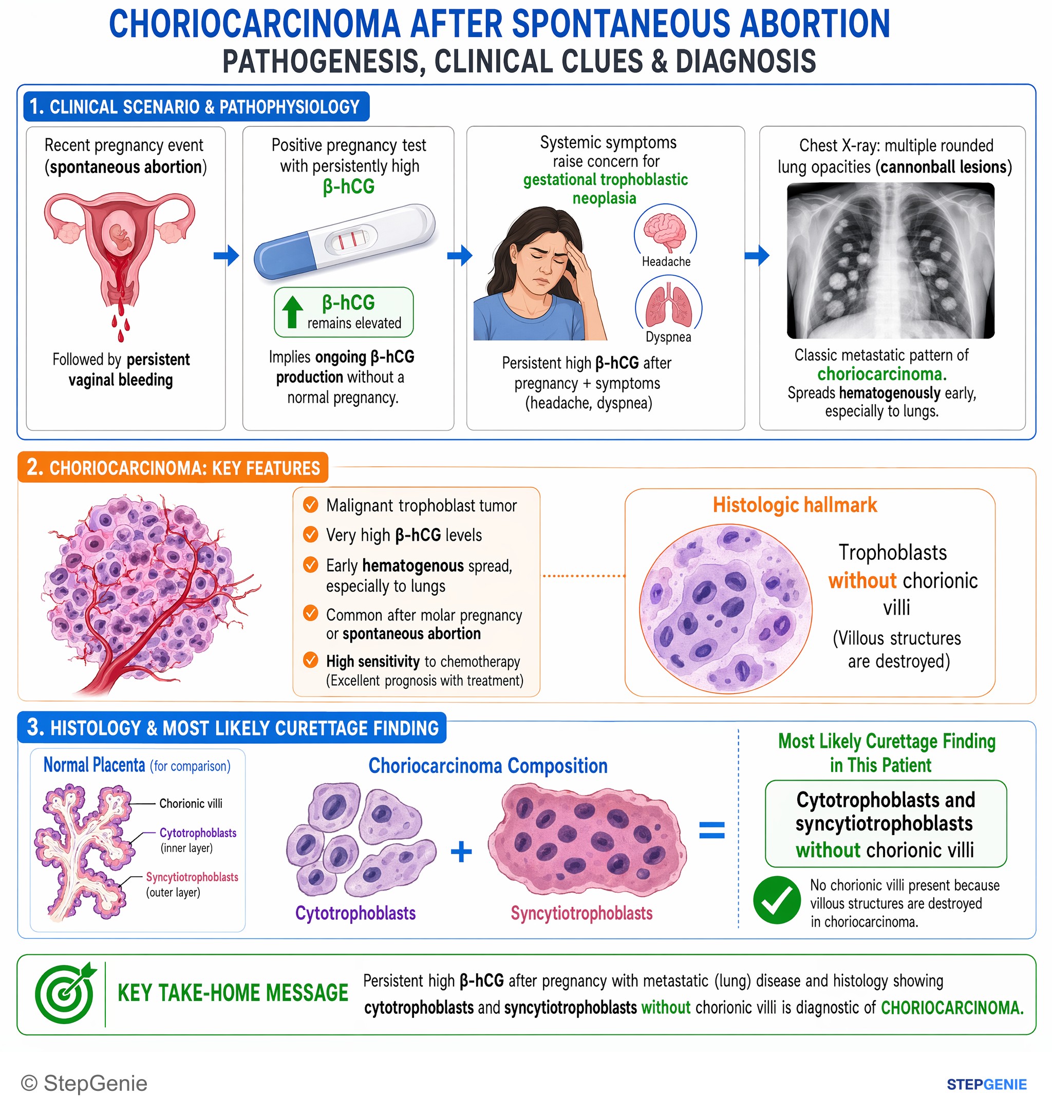
Choriocarcinoma after spontaneous abortion: malignant trophoblasts, very high hCG, early lung metastases, histology shows trophoblasts…
USMLE Step 1Frequently Asked Questions
How do you tell a complete mole from a partial mole on Step 1?
A complete mole has no fetal tissue and shows a classic snowstorm or cluster-of-grapes ultrasound pattern, while a partial mole may have some fetal tissue present. Complete moles are usually paternal only, most often 46,XX, with diffuse hydropic villi and diffuse circumferential trophoblastic proliferation. Partial moles are triploid and show focal hydropic change with milder, focal trophoblastic proliferation.
Why can a molar pregnancy cause hyperthyroid symptoms?
A molar pregnancy can cause hyperthyroid symptoms because very high hCG levels can weakly stimulate the TSH receptor. This is why exam stems may pair pregnancy with tachycardia, heat intolerance, and markedly elevated beta-hCG. The same excess hCG also explains other systemic findings, including hyperemesis gravidarum and theca-lutein cysts from ovarian stimulation.
What is the first-line management for a hydatidiform mole?
The first step is suction curettage followed by serial beta-hCG monitoring until the level becomes undetectable. Contraception is recommended during surveillance so a new pregnancy does not confuse interpretation of hCG trends. This follow-up matters because persistent or rising beta-hCG after evacuation suggests persistent gestational trophoblastic neoplasia, which requires further treatment.
What does rising beta-hCG after molar evacuation mean?
Rising or persistent beta-hCG after evacuation suggests persistent gestational trophoblastic neoplasia rather than simple resolution of a mole. A post-evacuation stem with persistent bleeding and plateauing or rising beta-hCG should raise concern for persistent GTN or choriocarcinoma. That is why serial beta-hCG monitoring is essential after treatment of a hydatidiform mole.
How do you distinguish choriocarcinoma from a hydatidiform mole?
Choriocarcinoma is distinguished by malignant trophoblastic proliferation with no chorionic villi on pathology, whereas moles have hydropic swelling of chorionic villi. Choriocarcinoma also tends to metastasize early through the bloodstream, especially to the lungs, causing cough, hemoptysis, or dyspnea. Both can have very high beta-hCG, so the pathology and metastatic pattern are key clues.