Guillain-Barré syndrome (GBS) is an acute, immune-mediated polyradiculoneuropathy: rapidly progressive, symmetric flaccid weakness that usually starts in the legs and climbs, with loss of reflexes, often beginning 5 days to 3 weeks after an infection such as Campylobacter jejuni. The classic CSF finding is albuminocytologic dissociation (high protein, normal cell count), but protein is still normal in 30-50% of patients in the first week. Treatment is IVIG or plasma exchange, not corticosteroids, with close tracking of vital capacity, because about 1 in 5 patients needs a ventilator.
How GBS happens: molecular mimicry
About two-thirds of patients had an infection, surgery or vaccination in the preceding weeks. The immune response cross-reacts with nerve roots and peripheral nerves and spares the brain and cord (contrast the CNS demyelination of multiple sclerosis).
- Triggers. C. jejuni is the most common antecedent infection; CDC estimates it is behind 5-41% of GBS cases (see Campylobacter jejuni). Others: CMV, EBV, Mycoplasma pneumoniae, hepatitis E and Zika; a COVID-19 link remains controversial.
- The mimicry. C. jejuni lipooligosaccharide carries ganglioside-like sugars, so antibodies made against the bacterium also bind the gangliosides GM1 and GD1a on motor axons: the textbook case of molecular mimicry.
- Two injury patterns. In demyelinating GBS (AIDP) the attack strips Schwann cell myelin, so conduction slows or blocks; in axonal forms antibodies hit the axon itself, so amplitudes fall.
- Vaccines are a rare trigger: about 1-2 extra cases per million influenza vaccinees, if any. A 2024 National Academies review found the evidence favors a causal link for the Ad26.COV2.S (adenovirus-vector) COVID-19 vaccine and favors rejecting one for the mRNA vaccines.
GBS variants: comparison table
| Variant | Core picture | Antibody | Key clue |
|---|---|---|---|
| AIDP (classic, demyelinating) | Symmetric ascending weakness, areflexia, paresthesias, back pain | No single antibody | Demyelinating nerve studies: slow conduction velocity, prolonged distal latencies and F waves, conduction block. 80-90% of cases in Europe and North America |
| AMAN (acute motor axonal neuropathy) | Pure motor weakness, no sensory loss | Anti-GM1, anti-GD1a | Axonal: low motor (CMAP) amplitudes with preserved velocities. Classic after C. jejuni; the dominant form in parts of Asia (Bangladesh, northern China) |
| AMSAN (motor-sensory axonal) | Motor plus sensory loss, often severe | Ganglioside antibodies (GM1, GD1a) | Low CMAP and sensory (SNAP) amplitudes; axonal subtypes carry a worse prognosis |
| Miller Fisher syndrome | Ophthalmoplegia + ataxia + areflexia, little limb weakness | Anti-GQ1b (up to 90%) | About 5% of GBS in Western countries vs 15-25% in Asia; usually recovers in 8-12 weeks, often without immunotherapy |
| Pharyngeal-cervical-brachial | Weak throat, neck and arms; legs largely spared | Anti-GT1a | Rare (under 5% of GBS) |
| Bickerstaff brainstem encephalitis | Ophthalmoparesis + ataxia + drowsiness, with brisk reflexes | Anti-GQ1b | Brainstem (CNS) overlap with Miller Fisher; rare |
Exam tip: ophthalmoplegia + ataxia + areflexia is Miller Fisher (anti-GQ1b). Add altered consciousness and hyperreflexia and it becomes Bickerstaff.
Presentation and time course
- Motor: symmetric flaccid weakness, usually legs first, then arms. Weakness always outweighs the sensory findings, and deep tendon reflexes are reduced or lost (a minority, mostly pure motor AMAN, keep normal or even brisk reflexes).
- Sensory and pain: distal tingling; low back or radicular pain from nerve-root inflammation is common and can come first.
- Cranial nerves: facial and oropharyngeal weakness in more than half of severe cases.
- Autonomic (about two-thirds, variable severity): blood pressure swings, arrhythmias, SIADH, gastrointestinal stasis, urinary retention and pupillary changes. Life-threatening dysautonomia is infrequent.
- Respiratory: about 20% develop respiratory failure needing mechanical ventilation.
- The clock: most patients reach maximum weakness (nadir) within 2 weeks, and 90% by 3-4 weeks. A nadir within 24 hours, or progression beyond 4 weeks, suggests another diagnosis. Progression beyond 8 weeks (or three or more treatment-related fluctuations) means acute-onset CIDP, the eventual diagnosis in about 5% of patients first labeled GBS.
Diagnosis: CSF, nerve studies and antibodies
GBS is a clinical diagnosis supported by tests: the 2023 European Academy of Neurology/Peripheral Nerve Society (EAN/PNS) guideline calls CSF valuable, especially when the diagnosis is less certain, and advises nerve conduction studies.
| Test | Classic finding | Trap |
|---|---|---|
| CSF protein | High protein, normal cells (albuminocytologic dissociation) | Protein is normal in 30-50% in week 1 and 10-30% in week 2: an early normal LP does not exclude GBS |
| CSF cells | Normal | 10-50 cells/µL is compatible but should prompt a search for alternatives; more than 50/µL suggests another disease (infection, leptomeningeal malignancy) |
| Nerve conduction | Demyelinating or axonal pattern | Demyelination slows velocity; axonal loss lowers amplitude |
| Anti-ganglioside antibodies | Anti-GQ1b in Miller Fisher | Of limited value in typical sensorimotor GBS (EAN/PNS 2023) |
| MRI or nerve ultrasound | Considered in atypical cases | Spine MRI also excludes cord compression or myelitis |
Respiratory and autonomic monitoring
Respiratory failure from diaphragm and bulbar weakness is the main acute danger, so the "next step" is a bedside measure of breathing strength, not pulse oximetry or a blood gas.
- Measure forced vital capacity (FVC) several times a day: every 6-8 hours (Merck), or 3-6 times a day depending on severity and about every 4 hours while FVC is still falling (EAN/PNS 2023). Add maximal inspiratory pressure (MIP, also called negative inspiratory force) and maximal expiratory pressure (MEP).
- 20/30/40 rule: VC under 20 mL/kg, MIP weaker than 30 cm H₂O (less negative than -30), or MEP under 40 cm H₂O signals impending respiratory failure. Merck advises intubation once VC falls below 15 mL/kg.
- ICU for imminent respiratory failure (breathlessness at rest or while talking, inability to count to 15 in one breath, VC under 15-20 mL/kg or under 1 L, abnormal blood gases), severe dysautonomia, severe swallowing problems or a weak cough, or rapidly progressing weakness. A single-breath count of 19 or less predicts mechanical ventilation.
- Neck-flexor weakness (cannot lift the head off the pillow) is a danger sign.
- Risk scores: EAN/PNS 2023 advises the modified Erasmus GBS scores, mEGRIS for ventilation risk and mEGOS for outcome.
- Autonomic: cardiac monitoring for arrhythmias and blood pressure lability; check sodium for SIADH.
Treatment: IVIG or plasma exchange, not steroids
| IVIG | Plasma exchange | |
|---|---|---|
| Regimen (EAN/PNS 2023) | 0.4 g/kg daily for 5 days (2 g/kg total) | 12-15 L in 4-5 exchanges over 1-2 weeks |
| Time window | Within 2 weeks of weakness onset (also reasonable at 2-4 weeks) | Within 4 weeks of weakness onset |
| Who | Patients unable to walk unaided | Patients unable to walk unaided |
- Corticosteroids do not work. The guideline recommends against oral steroids and weakly against IV steroids; they do not hasten recovery and may delay it.
- Equally effective, so pick one: plasma exchange followed immediately by IVIG is no better than either alone, and a second IVIG course for a poor prognosis is not recommended.
- Supportive care: low molecular weight heparin to prevent DVT in bed-bound patients; pain control with gabapentinoids, tricyclic antidepressants or carbamazepine; early passive range of motion, then active physiotherapy.
- Treatment-related fluctuations: 6-10% worsen again within 2 months of first improving; repeating the same therapy is common practice and often helps, though trial evidence is lacking. Three or more, or progression beyond 8 weeks, means acute-onset CIDP, which, unlike GBS, responds to corticosteroids.
Prognosis
- About 80% walk independently at 6 months, but residual weakness, pain and fatigue are common.
- Death occurs in about 3-7% overall (published estimates run from under 4% to 10%) and in about 20% of ventilated patients, from respiratory and cardiovascular complications, pulmonary embolism and infection.
- Worse outcome: older age, severe or rapidly progressive weakness, need for ventilation, preceding C. jejuni diarrhea and axonal subtypes.
- GBS is usually monophasic; true recurrences occur in 2-5%.
Look-alikes: how USMLE tries to trick you
| Condition | Where | Pattern | Clincher |
|---|---|---|---|
| GBS | Peripheral nerves and roots | Ascending symmetric weakness, areflexia | Albuminocytologic dissociation; abnormal nerve studies |
| Myasthenia gravis | Postsynaptic NMJ | Fatigable ptosis, diplopia and proximal weakness; reflexes and sensation normal | AChR antibodies (80-90% of generalized cases) or MuSK; see myasthenia gravis |
| Botulism | Presynaptic NMJ (blocks ACh release) | Descending paralysis starting with cranial nerves; no sensory loss, clear sensorium, no fever; pupillary light reflex reduced or lost | Home-canned food or wounds; honey and constipation in infants; antitoxin |
| Tick paralysis | Neurotoxin in tick saliva (Dermacentor, Amblyomma) | Ascending flaccid paralysis, can reach bulbar and respiratory muscles | Remove the tick: recovery starts within hours, though weakness can progress for 24-48 h |
| Acute cord lesion (transverse myelitis, compression) | Spinal cord | Sensory level, early bladder and bowel dysfunction, UMN signs below the lesion | MRI of the spine; review the spinal cord tracts |
| CIDP | Peripheral nerves and roots | GBS-like, but progresses for more than 8 weeks and can be asymmetric | Same CSF picture; steroids, IVIG or plasma exchange all work |
How the stems read:
- Steroids are the wrong answer for GBS; they are right for CIDP and MS relapses.
- Molecular mimicry pairs: C. jejuni → GBS; group A strep → rheumatic fever, including Sydenham chorea (the post-infectious chorea to keep separate from the genetic chorea of Huntington disease).
Practice question
A 34-year-old man has 4 days of tingling feet and progressive leg weakness; today he cannot climb stairs or walk without help. Two weeks ago he had 3 days of fever and bloody diarrhea after a barbecue. Strength is reduced symmetrically, legs more than arms, and patellar and ankle reflexes are absent. There is no sensory level, and eye movements and pupils are normal. Forced vital capacity is 40 mL/kg. CSF shows protein 95 mg/dL (normal under 45), 2 white cells/µL and normal glucose. Which is the most appropriate treatment?
A. IVIG 0.4 g/kg daily for 5 days B. IV methylprednisolone 1 g daily for 5 days C. Oral azithromycin D. Pyridostigmine E. Plasma exchange followed immediately by IVIG
Answer: A. Post-Campylobacter ascending weakness, areflexia and albuminocytologic dissociation is GBS. He cannot walk unaided and is within 2 weeks of onset, so IVIG (or plasma exchange alone) is indicated, with admission for serial FVC monitoring.
- B: corticosteroids do not improve GBS and may delay recovery; the guideline recommends against them.
- C: the diarrhea has resolved, and antibiotics do not treat the antibody-mediated neuropathy that follows it.
- D: pyridostigmine treats myasthenia gravis, which causes fatigable weakness with normal reflexes and no sensory symptoms.
- E: plasma exchange followed immediately by IVIG is no more effective than either treatment alone, so the guideline does not recommend it.
Key takeaways
- GBS = acute ascending, symmetric, flaccid weakness with areflexia, often 5 days to 3 weeks after an infection; C. jejuni is the classic trigger.
- Mimicry: C. jejuni lipooligosaccharide → anti-GM1/GD1a → AMAN. Miller Fisher = ophthalmoplegia, ataxia, areflexia, anti-GQ1b.
- CSF: high protein, normal cells, but normal in up to half of patients in week 1; more than 50 cells/µL suggests another diagnosis.
- Track FVC several times a day (every 4-8 h; 20/30/40 rule); ICU if VC is under 15-20 mL/kg, the patient can't count to 15, or dysautonomia or bulbar weakness is severe.
- IVIG 0.4 g/kg/day × 5 days (within 2 weeks) or plasma exchange (within 4 weeks) if the patient can't walk unaided; not both in sequence, no steroids.
- Progression beyond 8 weeks = acute-onset CIDP, which does respond to steroids.
Sources
- Guillain-Barré Syndrome (GBS) — Merck Manual Professional Edition (2026)
- Guillain-Barre Syndrome — StatPearls, NCBI Bookshelf (2026)
- European Academy of Neurology/Peripheral Nerve Society Guideline on Diagnosis and Treatment of Guillain-Barré Syndrome — European Journal of Neurology, full text via Erasmus University Rotterdam repository (2023)
- Diagnosis and Management of Guillain-Barré Syndrome in Ten Steps — Nature Reviews Neurology, PMC (2019)
- Guillain-Barré Syndrome — National Institute of Neurological Disorders and Stroke (2026)
- Miller Fisher Syndrome — StatPearls, NCBI Bookshelf (2023)
- Neurologic Conditions and COVID-19 Vaccines (Guillain-Barré Syndrome) — National Academies, NCBI Bookshelf (2024)
- Clinical Overview of Campylobacter — CDC (2026)
- Chronic Inflammatory Demyelinating Polyneuropathy (CIDP) — Merck Manual Professional Edition (2026)
- Myasthenia Gravis — Merck Manual Professional Edition (2026)
- Botulism — Merck Manual Professional Edition (2026)
- Tick Paralysis — Merck Manual Professional Edition (2025)
Visual explanations from the StepGenie QBank
Infographics StepGenie drew to explain questions in its own QBank, matched to this article's topic. Each caption is the takeaway of the question it explains.
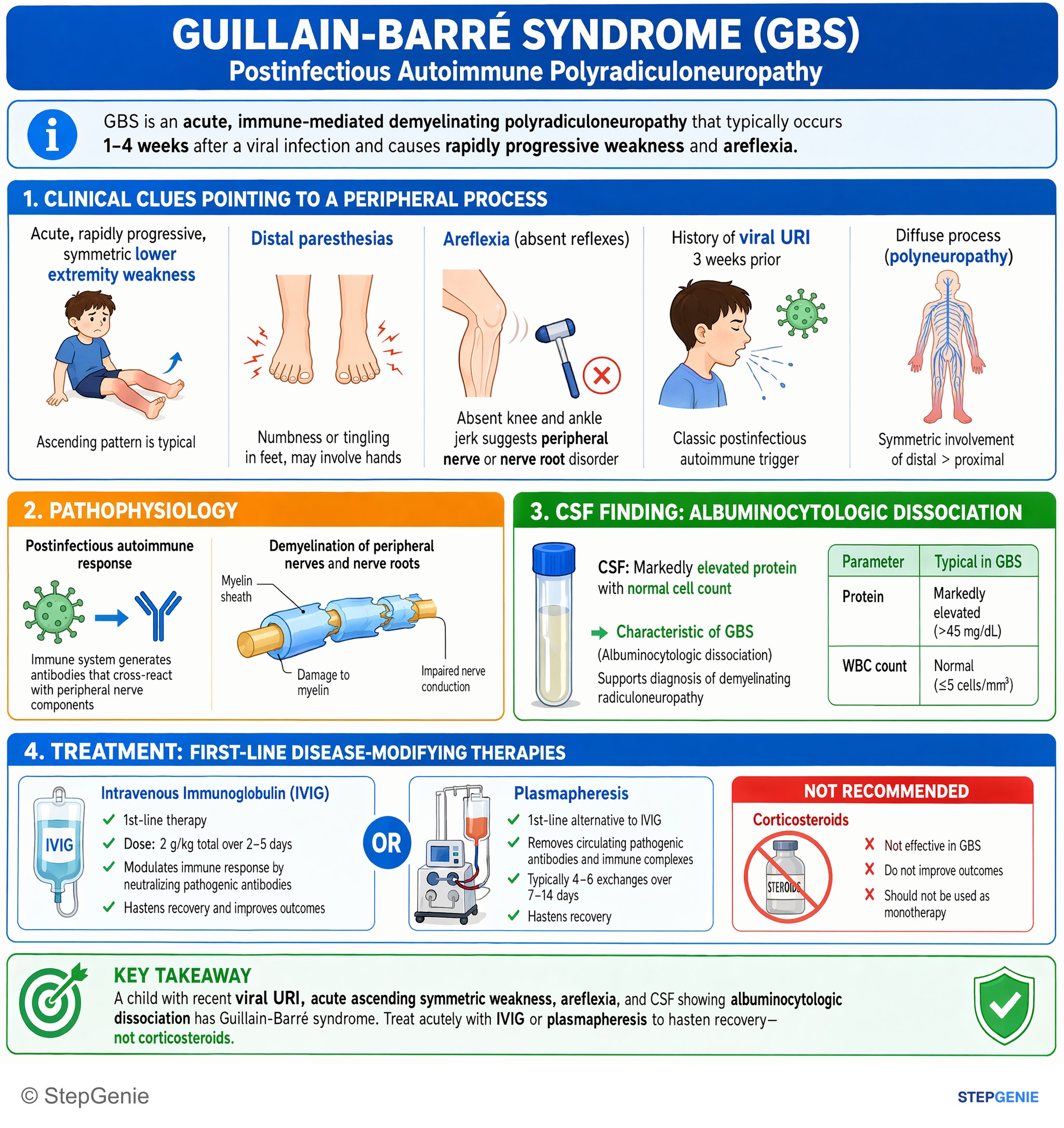
Guillain-Barré syndrome after viral URI with acute ascending weakness and albuminocytologic dissociation is treated acutely with IVIG or…
USMLE Step 2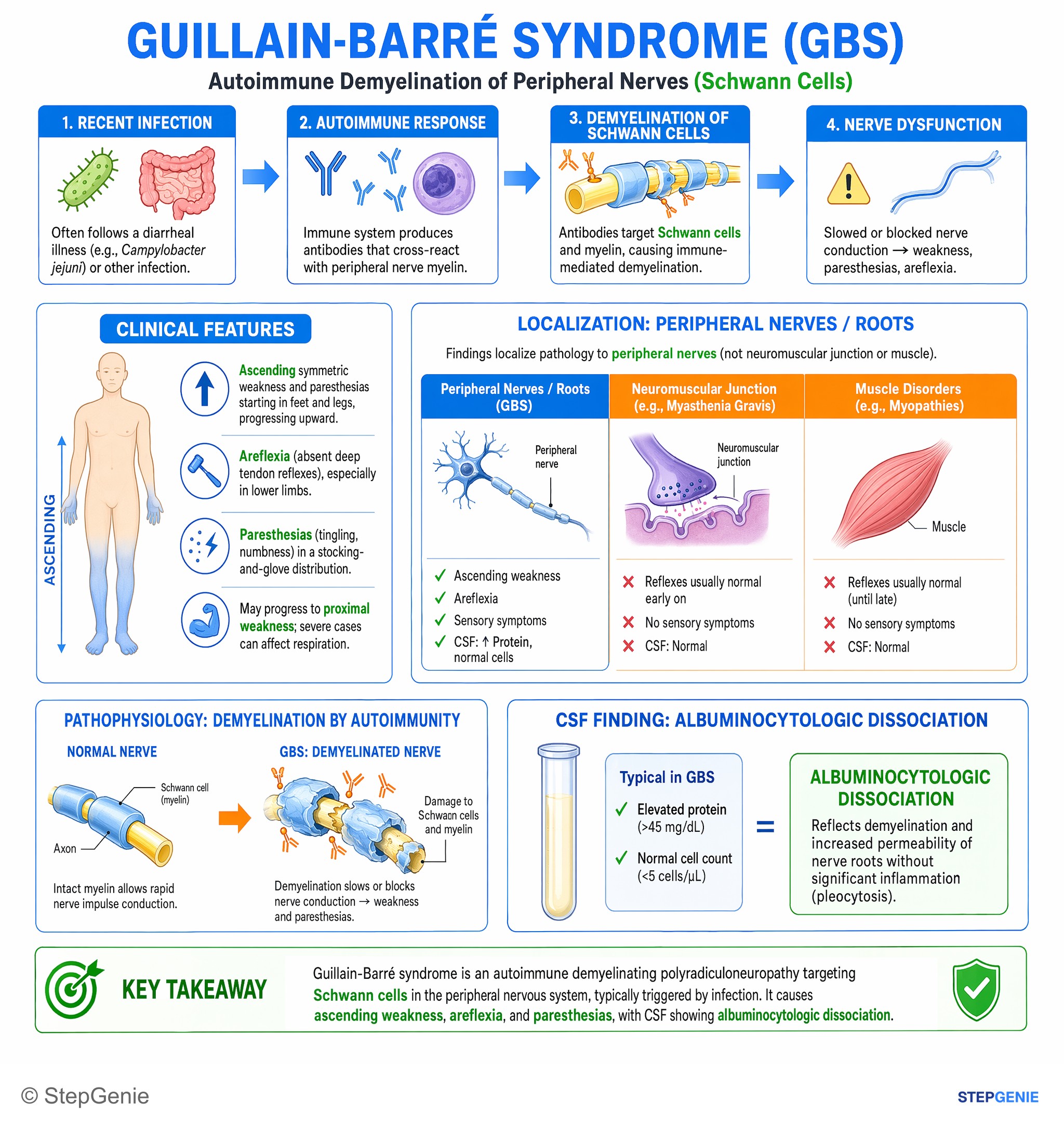
Guillain-Barré syndrome causes autoimmune demyelination of peripheral nerves (Schwann cells) → ascending weakness, areflexia…
USMLE Step 1Frequently Asked Questions
What is the classic CSF finding in Guillain-Barré syndrome, and when does it appear?
The classic finding is albuminocytologic dissociation: raised CSF protein with a normal white cell count, reflecting nerve-root inflammation rather than infection of the CSF. The rise takes time. Protein is still normal in 30-50% of patients in the first week and 10-30% in the second, so an early normal lumbar puncture does not rule out GBS. More than 50 cells/µL suggests another diagnosis, such as an infection or leptomeningeal malignancy.
Which antibodies go with which Guillain-Barré syndrome variants?
Classic demyelinating GBS (AIDP) has no single diagnostic antibody. Acute motor axonal neuropathy (AMAN), the pure motor form classically seen after Campylobacter jejuni, is linked to anti-GM1 and anti-GD1a antibodies. Miller Fisher syndrome (ophthalmoplegia, ataxia and areflexia) has anti-GQ1b antibodies in up to 90% of patients, and anti-GQ1b is also found in Bickerstaff brainstem encephalitis. The pharyngeal-cervical-brachial variant is linked to anti-GT1a. Antibody testing adds little in typical GBS but helps when Miller Fisher syndrome is suspected.
Why are steroids not used to treat Guillain-Barré syndrome?
Corticosteroids do not speed recovery in GBS, and there is some evidence they may delay it. The 2023 EAN/PNS guideline recommends against oral steroids and weakly against IV steroids. The effective treatments are IVIG (0.4 g/kg daily for 5 days, ideally within 2 weeks of onset) or plasma exchange (within 4 weeks), which work equally well. Steroids do help chronic inflammatory demyelinating polyneuropathy (CIDP), diagnosed when progression continues beyond 8 weeks.
When does a patient with Guillain-Barré syndrome need ICU care or intubation?
ICU care is advised for imminent respiratory failure (breathlessness at rest or while talking, inability to count to 15 in one breath, a vital capacity below 15-20 mL/kg or 1 L, or abnormal blood gases), severe dysautonomia, severe swallowing problems or a weak cough, and rapidly progressing weakness. The 20/30/40 rule flags impending failure: vital capacity under 20 mL/kg, maximal inspiratory pressure weaker than 30 cm H2O, or maximal expiratory pressure under 40 cm H2O. Merck advises intubation once vital capacity falls below 15 mL/kg. Check vital capacity several times a day, about every 4 hours while it is still falling.
How is Campylobacter jejuni linked to Guillain-Barré syndrome?
Campylobacter jejuni is the most common infection preceding GBS. Its lipooligosaccharide carries ganglioside-like sugars, so antibodies against the bacterium cross-react with gangliosides such as GM1 and GD1a on motor axons (molecular mimicry), classically causing the axonal variant AMAN. Only about 0.2-1.7 per 1,000 Campylobacter illnesses lead to GBS, but CDC estimates Campylobacter is behind 5-41% of GBS cases. Weakness typically starts 1-3 weeks after the diarrhea, and preceding diarrhea predicts a worse outcome.
How do you tell Guillain-Barré syndrome from myasthenia gravis and botulism?
GBS causes ascending, symmetric weakness with lost reflexes and tingling that evolves over days to weeks. Myasthenia gravis causes fatigable weakness, often with ptosis and double vision, while reflexes and sensation stay normal. Botulism causes a descending paralysis that starts in the cranial nerves (blurred or double vision, drooping eyelids, trouble swallowing), with no sensory loss, a clear mind, no fever and a reduced pupillary light reflex. A sensory level with bladder problems points to the spinal cord instead.