Multiple sclerosis (MS) is one of those Step 1 topics that keeps showing up because it ties together immunology, neuroanatomy, pathology, imaging, and pharmacology in one classic package. If you can recognize the vignette and understand why lesions happen where they do, you’ll pick up easy points—and avoid common traps.
What Is Multiple Sclerosis?
Multiple sclerosis is a chronic autoimmune demyelinating disease of the CNS characterized by dissemination in time and space (separate neurologic episodes affecting different CNS locations).
Key definition points (Step-style):
- CNS only (brain, spinal cord, optic nerves)
- Immune-mediated demyelination + inflammation → eventual axonal loss and neurodegeneration
- Classically relapsing-remitting early; progressive disability may develop later
First Aid cross-reference: Neurology → Demyelinating diseases (Multiple sclerosis); Immunology → Type IV hypersensitivity; Pharm → MS disease-modifying therapies
Epidemiology & Risk Factors (High-Yield)
Who gets MS?
- Often presents in young adults, classically women
- More common in populations farther from the equator (vitamin D/latitude association is frequently mentioned)
Risk factors & associations:
- HLA-DRB1 associations (classically tied to autoimmune risk)
- EBV exposure association (commonly tested as an epidemiologic link)
- Smoking increases risk and worsens progression
Testable vignette clue: young woman with episodes of neuro deficits that resolve and recur.
Pathophysiology: What’s Actually Happening?
Immune Mechanism (Classic Step 1 framing)
MS is primarily T-cell–mediated (Type IV hypersensitivity) targeting CNS myelin.
Core idea:
- Autoreactive CD4+ T cells cross the BBB → inflammatory cascade
- Macrophages + cytokines damage myelin and oligodendrocytes
- Over time: incomplete remyelination + axonal degeneration
What Gets Damaged?
- Oligodendrocytes (CNS myelin)
- Myelin → slowed conduction, conduction block, ephaptic transmission
- Chronic inflammation → gliosis and plaques
Pathology Buzzwords
- Periventricular plaques with gliosis
- Lesions can occur in:
- Optic nerve (optic neuritis)
- Brainstem/cerebellum
- Spinal cord (sensory level, weakness)
- Dawson fingers: ovoid periventricular lesions oriented perpendicular to ventricles (radiology pearl)
First Aid cross-reference: Demyelinating diseases; oligodendrocytes vs Schwann cells; Type IV hypersensitivity
Clinical Presentation (Recognize the Pattern)
The Core Step 1 Phrase
“Dissemination in time and space.”
- Time: separate attacks separated by weeks/months
- Space: different CNS regions affected over time
Common Symptoms (High-Yield)
- Optic neuritis
- Pain with eye movement + decreased visual acuity
- Often afferent pupillary defect
- Internuclear ophthalmoplegia (INO)
- Lesion of medial longitudinal fasciculus (MLF)
- Impaired adduction of ipsilateral eye + abducting nystagmus of contralateral eye
- Sensory symptoms
- Paresthesias, numbness, “pins and needles”
- Motor symptoms
- Weakness, spasticity (UMN signs)
- Cerebellar dysfunction
- Ataxia, intention tremor, dysmetria, scanning speech
Two Classic Eponyms/Associations
- Uhthoff phenomenon: symptoms worsen with heat (hot shower, fever, exercise) due to impaired conduction in demyelinated axons
- Lhermitte sign: electric shock-like sensation down the spine with neck flexion (often cervical spinal cord involvement)
The “Charcot Triad” (often taught; know it, but don’t over-rely)
- Nystagmus
- Intention tremor
- Scanning speech
First Aid cross-reference: INO/MLF lesions; optic neuritis; UMN signs; cerebellar findings
Diagnosis: How MS Is Confirmed
Clinical Framework
Diagnosis requires evidence of:
- Dissemination in time
- Dissemination in space
- And excluding better explanations
MRI Findings (Most Tested)
MRI brain/spine shows:
- Periventricular white matter lesions
- Often contrast-enhancing during active inflammation (gadolinium enhancement)
High-yield imaging logic:
- Enhancing lesions = active/new
- Non-enhancing lesions = older
- Both present = supports dissemination in time
CSF Findings (Classic Step 1)
Lumbar puncture may show:
- Oligoclonal IgG bands
- Increased IgG index
- Mild lymphocytic pleocytosis (variable)
Visual Evoked Potentials
- Can show delayed conduction (supports demyelination), especially with optic pathway involvement
Quick Table: MS Diagnostic Clues
| Modality | High-yield finding | Why it matters |
|---|---|---|
| MRI | Periventricular plaques; enhancing + non-enhancing lesions | Dissemination in space/time |
| CSF | Oligoclonal IgG bands, ↑ IgG index | Intrathecal Ig production |
| Exam | INO, optic neuritis, UMN signs | Localizes CNS demyelination |
| History | Relapses/remissions; heat sensitivity | Classic pattern recognition |
First Aid cross-reference: Oligoclonal bands; periventricular plaques; Dawson fingers
Clinical Course Types (Know the Big Ones)
- Relapsing-remitting MS (RRMS): most common initially; attacks with recovery
- Secondary progressive MS (SPMS): starts as RRMS → transitions to steady progression
- Primary progressive MS (PPMS): progressive from onset (less classic Step 1, but fair game)
Test-taking tip: Many vignettes describe RRMS because it’s the easiest to pattern-match.
Treatment: Acute vs Long-Term (Very Testable)
Acute Relapse Treatment
Goal: reduce inflammation quickly.
- High-dose corticosteroids (e.g., IV methylprednisolone)
- Plasmapheresis can be used for severe relapses refractory to steroids
Disease-Modifying Therapy (DMT): Reduce Relapses/Progression
These target immune activity to reduce new lesions and disability accumulation.
High-yield Step 1 DMT list (recognize names + core concept):
- Interferon-beta
- Glatiramer acetate
- Natalizumab (big association: PML risk)
- Fingolimod
- Ocrelizumab (anti-CD20; also used in PPMS)
Table: Key Drugs & Board-Relevant Associations
| Drug | Mechanism (board-level) | High-yield adverse effect / association |
|---|---|---|
| Interferon-β | Immunomodulation; decreases T-cell activation | Flu-like symptoms, depression (commonly tested) |
| Glatiramer | Myelin basic protein “decoy”; shifts immune response | Flushing, chest tightness (transient reaction) |
| Natalizumab | Blocks leukocyte adhesion/migration (anti-α4 integrin) | PML (JC virus) risk |
| Fingolimod | Sequesters lymphocytes in lymph nodes (S1P modulator) | Bradycardia, macular edema (core associations) |
| Ocrelizumab | Anti-CD20 B-cell depletion | Infection risk; infusion reactions |
First Aid cross-reference: MS pharmacology; natalizumab → PML/JC virus
Symptomatic Management (Step 2-ish but shows up)
- Spasticity: baclofen, tizanidine
- Neuropathic pain: gabapentin, duloxetine
- Fatigue: amantadine/modafinil (varies by source)
High-Yield Differentials (Don’t Get Tricked)
MS vs Neuromyelitis Optica (NMO)
NMO can look similar but is a different disease.
| Feature | MS | NMO (Devic disease) |
|---|---|---|
| Primary target | CNS myelin | Aquaporin-4 astrocyte water channel (often) |
| Hallmark | Dissemination in time/space | Optic neuritis + transverse myelitis, often longitudinally extensive |
| CSF | Oligoclonal bands common | May be absent; different pattern |
| Treatment approach | MS DMTs | Immunosuppression (e.g., rituximab); avoid some MS meds |
MS vs Guillain-Barré Syndrome (GBS)
- MS = CNS demyelination
- GBS = PNS demyelination (Schwann cells), ascending weakness, areflexia
HY Anatomy Connections (Where Symptoms Come From)
- Optic neuritis → optic nerve demyelination → painful monocular vision loss
- INO → MLF lesion → impaired conjugate gaze
- UMN signs (spasticity, hyperreflexia, Babinski) → corticospinal tract involvement
- Sensory level/Lhermitte → spinal cord plaques
- Cerebellar signs → cerebellar pathway involvement
This localization thinking is how Step vignettes go from “random symptoms” to “oh, that’s MS.”
Rapid-Fire High-Yield Facts (Exam Quick Recall)
- MS is CNS demyelination due to Type IV hypersensitivity
- Oligoclonal IgG bands in CSF are classic
- MRI: periventricular plaques (“Dawson fingers”)
- Uhthoff phenomenon: heat worsens symptoms
- INO strongly suggests MS in a young patient
- Treat acute flares with high-dose steroids
- Natalizumab → PML (JC virus) is a classic association
Mini-Vignette Pattern (What You Should Instantly Recognize)
Young woman with episodic neurologic deficits (vision changes, sensory symptoms, weakness) that come and go, worse with heat, MRI with periventricular white matter lesions, and CSF with oligoclonal bands → Multiple sclerosis.
Visual explanations from the StepGenie QBank
Infographics StepGenie drew to explain questions in its own QBank, matched to this article's topic. Each caption is the takeaway of the question it explains.
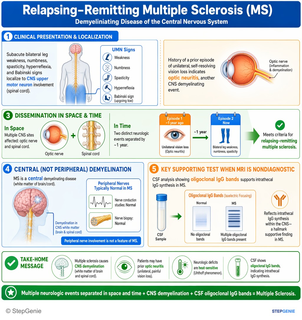
Multiple sclerosis causes CNS demyelination with heat-sensitive neurologic deficits and prior optic neuritis; CSF shows oligoclonal IgG…
USMLE Step 1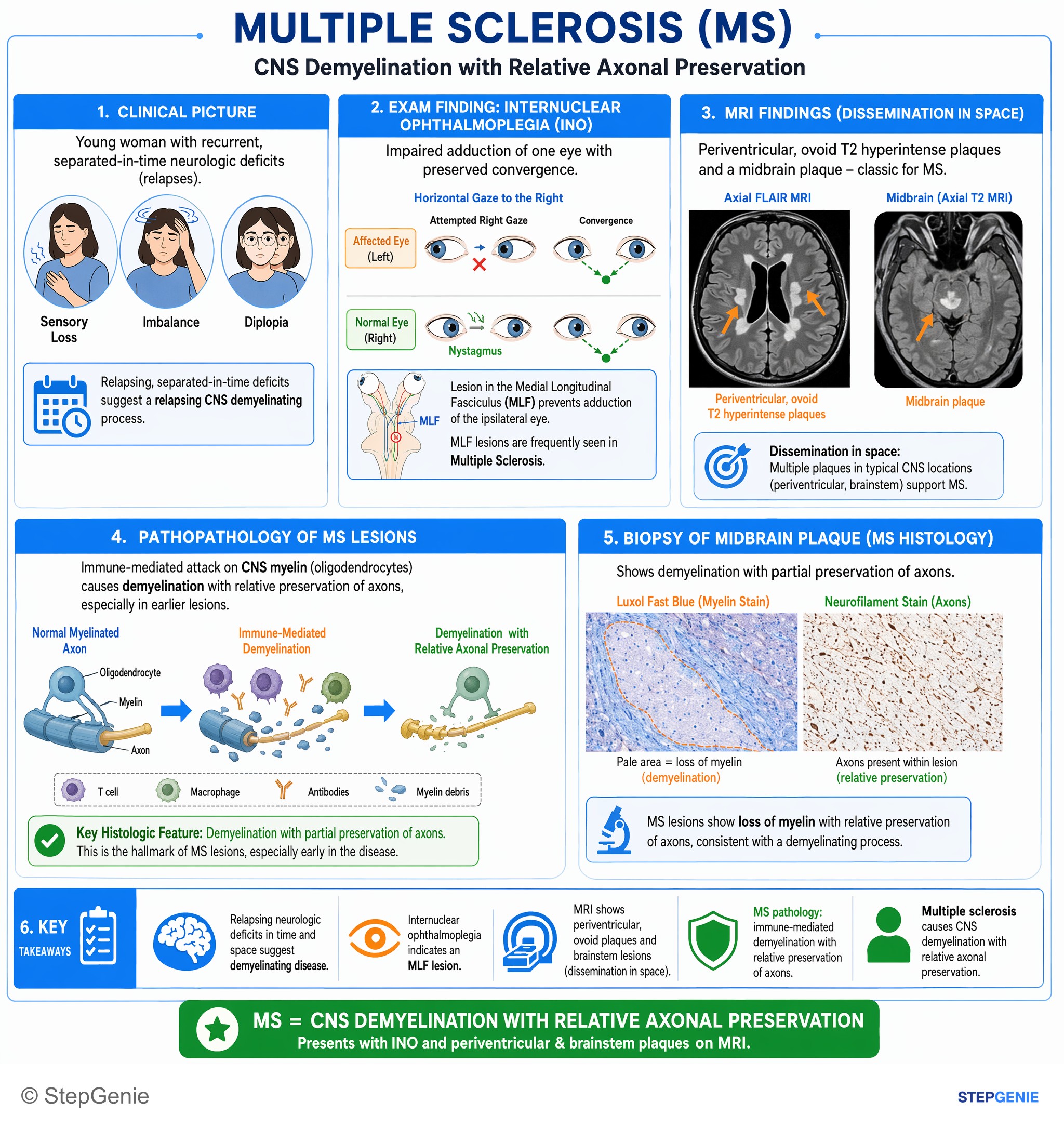
Multiple sclerosis causes CNS demyelination with relative axonal preservation, presenting with internuclear ophthalmoplegia and…
USMLE Step 1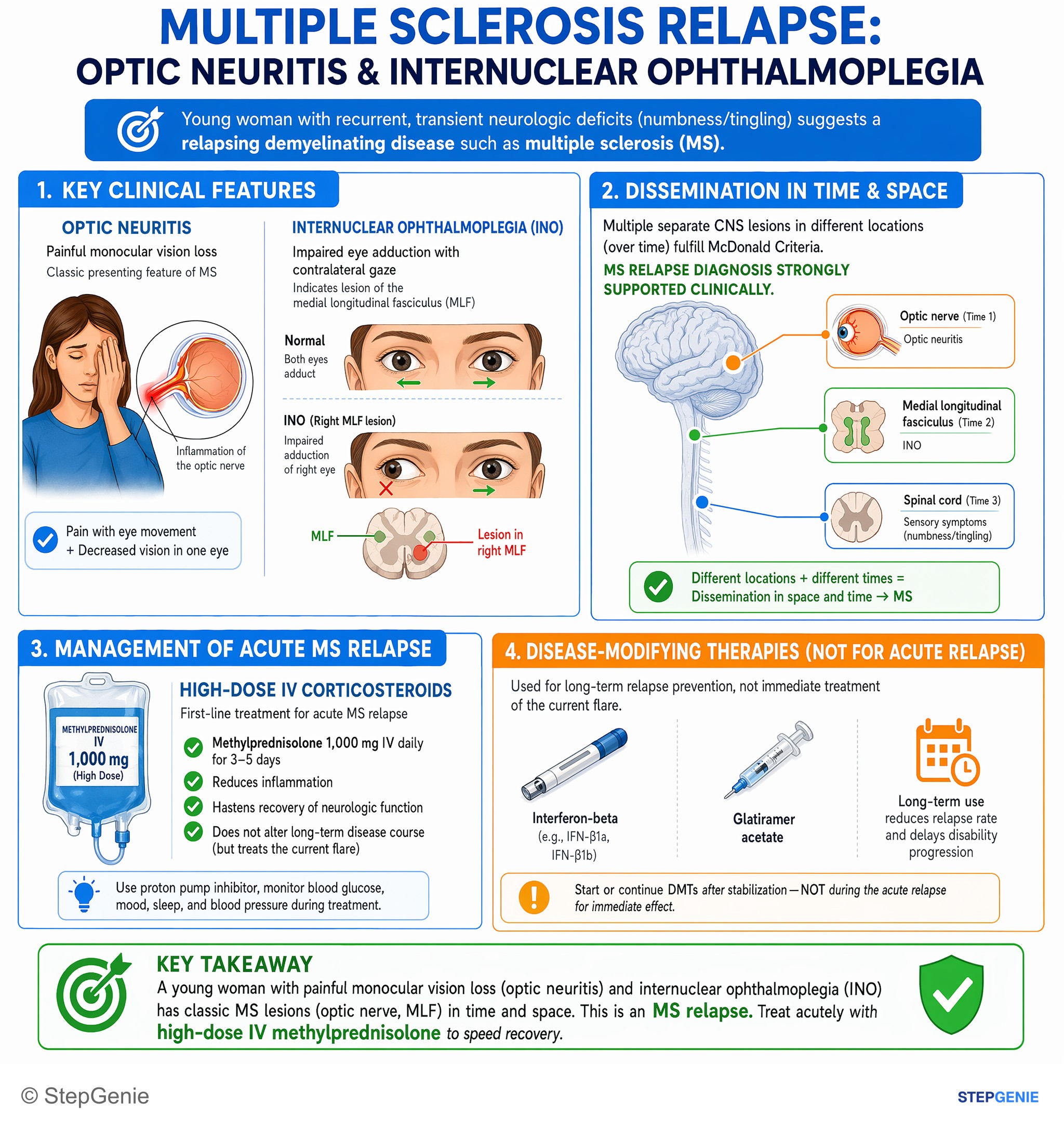
Multiple sclerosis relapse with optic neuritis and internuclear ophthalmoplegia is treated acutely with high-dose IV methylprednisolone.
USMLE Step 2Frequently Asked Questions
Why does multiple sclerosis cause neurologic symptoms that come and go in different parts of the CNS?
Multiple sclerosis causes this pattern because it is a chronic autoimmune demyelinating disease of the CNS with dissemination in time and space. Separate inflammatory attacks occur at different times and in different locations such as the optic nerve, brainstem, cerebellum, or spinal cord. Demyelination slows or blocks conduction, so symptoms can resolve and recur as different CNS regions are affected.
How do you recognize multiple sclerosis in a classic Step 1 vignette?
A classic vignette is a young woman with episodes of neurologic deficits that resolve and later recur. The most important clue is dissemination in time and space, meaning separate attacks over weeks or months affecting different CNS regions. Common findings include optic neuritis, internuclear ophthalmoplegia, sensory symptoms, weakness or spasticity, and cerebellar signs such as ataxia or intention tremor.
Why do heat and exercise make multiple sclerosis symptoms worse?
Heat can worsen multiple sclerosis symptoms because demyelinated axons already conduct impulses poorly. This is called Uhthoff phenomenon and is classically triggered by a hot shower, fever, or exercise. Since myelin loss causes slowed conduction or conduction block, increasing body temperature can further impair signal transmission and temporarily make existing neurologic deficits more noticeable.
How is multiple sclerosis confirmed on MRI and CSF?
Multiple sclerosis is supported by MRI showing periventricular white matter lesions, often with gadolinium enhancement during active inflammation. Enhancing lesions indicate active or new disease, while non-enhancing lesions indicate older lesions, and seeing both supports dissemination in time. CSF may show oligoclonal IgG bands and an increased IgG index, which point to intrathecal immune activity.
What is the first-line treatment for an acute multiple sclerosis relapse?
An acute multiple sclerosis relapse is treated with high-dose corticosteroids such as IV methylprednisolone. The goal is to reduce inflammation quickly during an attack. If a relapse is severe and does not respond to steroids, plasmapheresis can be used. Long-term disease-modifying therapy is aimed instead at reducing relapses, new lesions, and disability progression.