A hydrocele is serous fluid between the parietal and visceral layers of the tunica vaginalis: a painless, smooth scrotal swelling around the testis that transilluminates and that you can usually "get above". In infants it comes from a patent processus vaginalis and usually resolves on its own by 12-18 months; persistence beyond about 12-24 months (sources differ), a coexisting inguinal hernia, or symptoms lead to repair (inguinal herniotomy). In adults, a new hydrocele needs a scrotal ultrasound to look for a cause such as a testicular tumor or epididymitis, and a symptomatic one is treated with hydrocelectomy.
Hydrocele cheat sheet: the one-minute version
| Topic | What to know |
|---|---|
| Where the fluid is | Between the two layers of the tunica vaginalis, the distal remnant of the processus vaginalis |
| Classic exam | Painless, smooth, "water balloon" swelling; testis often impalpable because fluid surrounds it; normal cord above the swelling |
| Transillumination | Positive (light from the side, so the testis does not block it) |
| Best test when unsure | Scrotal ultrasound with Doppler: anechoic fluid around the testis, and a look at the testis itself |
| Infants | Patent processus vaginalis; most resolve by 12-18 months |
| Adults | Idiopathic, or secondary to epididymitis, trauma, surgery or tumor; filariasis worldwide |
| Red flags | Swelling you cannot get above (hernia); a solid or non-transilluminating mass in the testis (cancer); sudden severe pain (torsion) |
Communicating vs noncommunicating hydrocele
The processus vaginalis is the peritoneal tube that leads the testis into the scrotum. At birth about 80% of term boys still have a patent processus; that falls to 25-40% by age 2 and about 20% in adults (higher in preterm infants).
| Communicating | Noncommunicating | |
|---|---|---|
| Processus vaginalis | Open: peritoneal fluid flows in and out | Closed proximally; fluid is trapped in the distal sac |
| Size | Changes: bigger with crying, straining or standing, smaller lying down or overnight | Stable |
| Typical patient | Infants and children; usually congenital | Infants, adolescents and adults |
| Linked problem | Same defect as an indirect inguinal hernia; it becomes a hernia if bowel or other tissue enters | In adults, often a coexisting scrotal disorder (tumor, epididymitis) |
| Key move | Watch, but repair if it persists or a hernia appears | Infants: observe. Adults: ultrasound for the cause |
Exam framing: questions usually label the infant hydrocele "congenital (communicating)" and the adult one "acquired (noncommunicating)". That is the default, but either type can occur at either age; an open processus vaginalis is what defines communicating, and a change in size is its bedside sign.
Infant vs adult hydrocele
| Infant or child | Adult | |
|---|---|---|
| Cause | Patent processus vaginalis | Imbalance of fluid production and absorption; or secondary to infection, trauma, surgery, tumor or filariasis |
| Natural history | Most resolve by 12-18 months | Usually does not go away on its own |
| When to operate | Persists beyond 12-18 months (AAP) or 18-24 months (StatPearls); an inguinal hernia is present; symptomatic or complicated | Symptomatic (size, discomfort) or complicated, once the cause has been checked |
| Operation | Herniotomy (open groin incision or laparoscopic): ligate the patent processus at the internal ring | Scrotal hydrocelectomy: plication for thin-walled sacs, excision or eversion (Jaboulay) for thick-walled ones |
| Not recommended | Waiting on a coexisting hernia | Aspiration alone (recurrence is very frequent, sometimes within a week); aspiration with sclerotherapy works but is painful and rarely used |
| Must not miss | Incarcerated hernia, torsion | Testicular cancer |
- The AAP's patient guidance: most infant hydroceles go away by around the first birthday; most "can be watched until 12 to 18 months of age", after which they are unlikely to resolve.
- StatPearls (2026): observation "until at least 1 to 2 years of age"; herniotomy if the hydrocele has not resolved by 18 to 24 months.
- A hernia is different: once an infant inguinal hernia is diagnosed, repair is scheduled, and an incarcerated or strangulated hernia needs to be dealt with immediately.
Exam and imaging: transillumination and ultrasound
Transilluminates (cystic): hydrocele, spermatocele, and the diffuse scrotal edema of fluid overload.
Does not transilluminate: a solid testicular tumor and, classically, a varicocele (a tangle of veins) or a hematocele (blood).
Three traps:
- Infant hernias can glow. The thin bowel wall in an infant hernia may let light through and mimic a hydrocele, so transillumination does not settle "hydrocele or hernia" in a baby. Check reducibility and whether you can get above the swelling.
- Transillumination does not exclude cancer. A tumor can sit behind a transilluminating hydrocele, and the fluid makes the testis hard to examine. Merck's rule: the cause of a hydrocele must be determined.
- Use the side. Shine the light from the side of the scrotum, not the front or back.
Order scrotal ultrasound with Doppler when the diagnosis is uncertain, a hydrocele is present in an adult (to find the cause), the mass does not transilluminate, or the testis cannot be felt. In acute pain, Doppler helps only when torsion is equivocal; a clear clinical picture of torsion goes straight to treatment.
Scrotal mass differential: the comparison table
| Condition | Pain | Key exam clue | Transilluminates? | Next step |
|---|---|---|---|---|
| Hydrocele | No | Smooth sac around the testis; normal cord above it | Yes | Ultrasound if adult or unsure |
| Spermatocele | No | Cystic nodule at the upper pole of the testis, beside the epididymis, separate from the testis | Yes | Clinical; ultrasound if unsure |
| Varicocele | Dull ache or fullness when standing | "Bag of worms", usually left; larger standing or with Valsalva, empties lying down | No | Clinical; repair for infertility with abnormal semen |
| Indirect inguinal hernia | Usually none; pain if incarcerated | Cannot get above it; may reduce; cough impulse, bowel sounds | No (an infant's bowel may) | Surgical repair |
| Testicular torsion | Sudden, severe; nausea, vomiting | High-riding, horizontal testis; absent cremasteric reflex | No | Immediate surgery (manual detorsion may be tried first); fix both testes |
| Epididymitis | Gradual | Tender epididymis; urethritis or UTI symptoms | No | NAAT, urine culture, antibiotics |
| Testicular tumor | Usually none | Firm mass within the testis | No | Ultrasound, AFP, β-hCG, LDH, radical inguinal orchiectomy |
| Hematocele | Often tender | After trauma, surgery, or with anticoagulants | No | Ultrasound |
High-yield details for the look-alikes:
- Varicocele: present in about 15% of adult men and 40% of infertile men; about 85% are on the left side only, because of asymmetric gonadal vein anatomy (the left gonadal vein drains into the left renal vein). AUA/ASRM (2020, amended 2024) advises considering varicocelectomy for men trying to conceive with a palpable varicocele, infertility and abnormal semen parameters, and not routinely imaging the abdomen for an isolated small or moderate right varicocele. Imaging is considered for a new-onset or non-reducible varicocele, especially a large one. The classic exam framing still applies: Merck notes that a varicocele that persists lying down, or a right-sided one, suggests IVC obstruction. See the varicocele breakdown.
- Torsion: predisposed by a bell-clapper deformity; peaks at ages 12-18, with a second peak in infancy. Testicular salvage is 80-100% within about 6-8 hours and drops steeply after that (it is rare beyond 24 hours), and the other testis is fixed too. More in the testicular torsion breakdown.
- Epididymitis: under 35, mostly N. gonorrhoeae or C. trachomatis; over 35, mostly gram-negative coliforms. Torsion must be excluded unless the findings are clearly limited to the epididymis (see the epididymitis breakdown).
- Testicular cancer: the most common solid cancer in males aged 15-35. Most intratesticular masses are malignant, most extratesticular ones are not. Transscrotal biopsy is not appropriate because of the risk of seeding the scrotum or inguinal nodes; the diagnosis is made by radical inguinal orchiectomy with early high ligation of the cord (seminoma vs nonseminoma).
How USMLE tests it
- Infant with a scrotal swelling that transilluminates and changes size during the day: communicating hydrocele. Reassure and recheck; refer for repair if it persists past about 12-24 months or a hernia is felt.
- Infant groin bulge that reduces and that you cannot get above: indirect inguinal hernia, which needs surgery. Do not let a glowing scrotum talk you out of it.
- Young man with a new painless hydrocele: scrotal ultrasound before anything else; a reactive hydrocele can hide a tumor.
- Firm, non-transilluminating testicular mass: ultrasound, tumor markers, then radical inguinal orchiectomy. Needle aspiration or transscrotal biopsy is the classic wrong answer.
- Embryology link: the processus vaginalis and gubernaculum guide testicular descent; failure of descent is cryptorchidism, while failure of the processus to close gives hydrocele and indirect hernia.
Practice question
A 32-year-old man has 2 months of painless enlargement of the left hemiscrotum. He has no fever, dysuria or trauma. Examination shows a smooth, nontender, fluctuant swelling that surrounds the testis, which cannot be palpated separately. The spermatic cord above the swelling feels normal, the swelling does not change with Valsalva or lying down, and it transilluminates. Which is the most appropriate next step?
A. Scrotal ultrasonography with Doppler B. Reassurance and observation C. Needle aspiration of the fluid D. Scrotal hydrocelectomy E. Transscrotal biopsy of the testis
Answer: A. This is a new noncommunicating hydrocele in an adult, and the fluid hides the testis. Ultrasound confirms the fluid and, more importantly, examines the testis for a tumor or epididymo-orchitis, because adult hydroceles are often secondary.
- B: observation is reasonable only after ultrasound shows a normal testis and the hydrocele is not bothering him.
- C: aspiration gives temporary relief, the fluid usually returns, and it does nothing to exclude a tumor.
- D: hydrocelectomy is for symptomatic hydroceles once the cause has been checked; operating first could miss a cancer that needs an inguinal approach.
- E: a suspected testicular cancer is never biopsied through the scrotum; it is removed by radical inguinal orchiectomy after ultrasound and tumor markers.
Key takeaways
- Hydrocele = fluid in the tunica vaginalis: painless, smooth, transilluminates, normal cord above it.
- Communicating (patent processus, changes size) vs noncommunicating (closed processus, stable size).
- Infants: most resolve by 12-18 months; repair (inguinal herniotomy) if it persists beyond about 12-24 months, if a hernia is present, or if symptomatic.
- Adults: new hydrocele means ultrasound to find the cause; hydrocelectomy if symptomatic; aspiration alone recurs.
- Transilluminates: hydrocele and spermatocele. Does not: tumor, varicocele, hematocele. An infant's hernia may glow.
- Testicular tumor: firm intratesticular mass, ultrasound and markers, radical inguinal orchiectomy, never transscrotal biopsy.
- Torsion is a clinical diagnosis: detorse and operate within hours.
Sources
- Hydrocele — StatPearls, NCBI Bookshelf (2026)
- Painless Scrotal Mass — Merck Manual Professional Edition (2024)
- Scrotal Swelling in Children: What Is a Hydrocele? — American Academy of Pediatrics, HealthyChildren.org (2023)
- Inguinal Hernia in Infants & Children — American Academy of Pediatrics, HealthyChildren.org (2023)
- Hydrocele — MedlinePlus Medical Encyclopedia (2025)
- Testicular Torsion — Merck Manual Professional Edition (2025)
- Testicular Torsion — StatPearls, NCBI Bookshelf (2023)
- Epididymitis — Merck Manual Professional Edition (2026)
- Testicular Cancer — Merck Manual Professional Edition (2025)
- Testicular Cancer Treatment (PDQ), Health Professional Version — National Cancer Institute (2025)
- Diagnosis and Treatment of Infertility in Men: AUA/ASRM Guideline — American Urological Association (2020, amended 2024)
Visual explanations from the StepGenie QBank
Infographics StepGenie drew to explain questions in its own QBank, matched to this article's topic. Each caption is the takeaway of the question it explains.
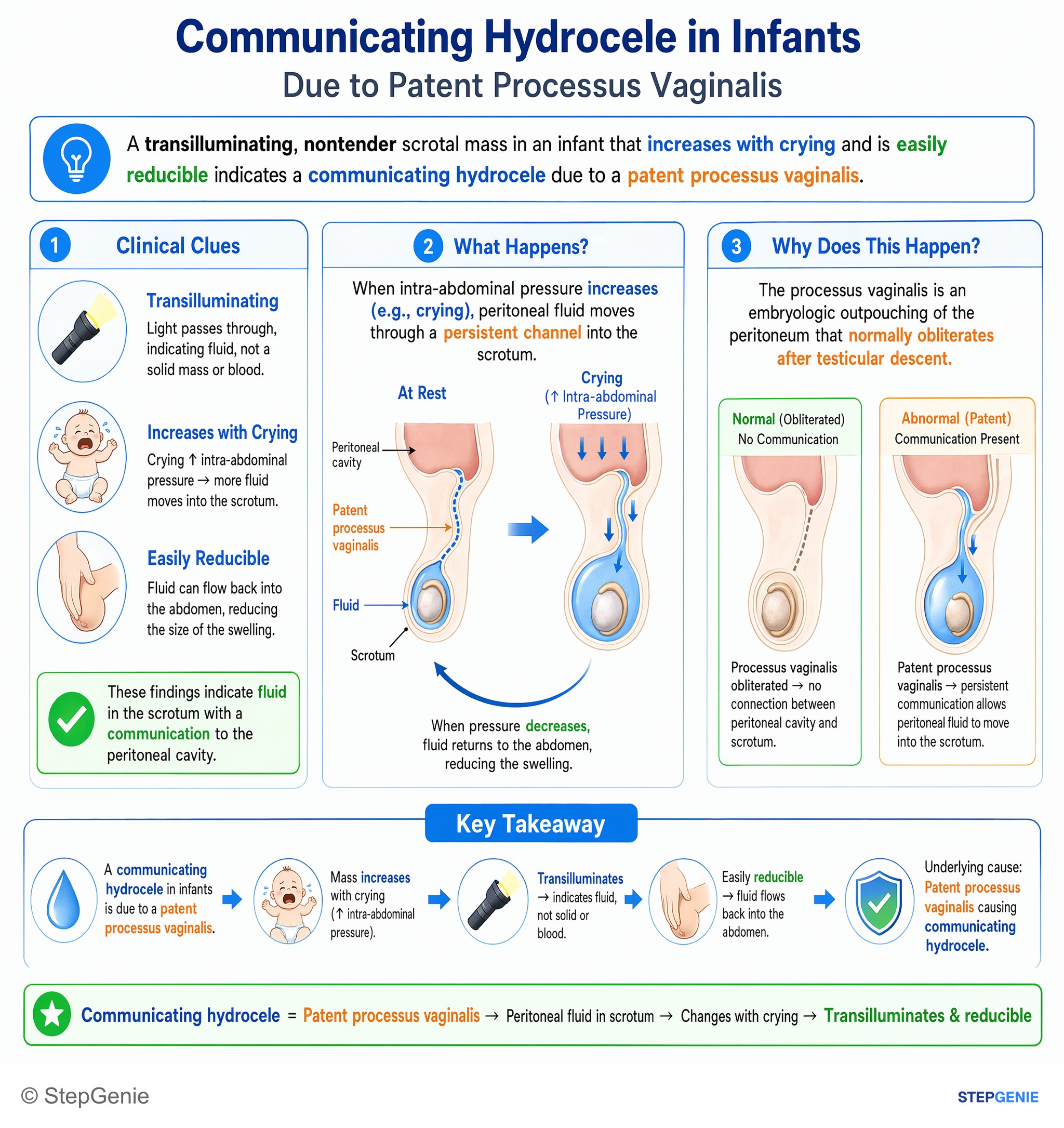
Communicating hydrocele in infants is due to a patent processus vaginalis, causing peritoneal fluid in scrotum; mass transilluminates and…
USMLE Step 1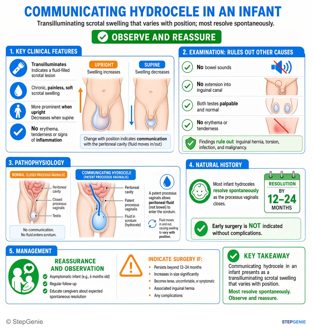
Communicating hydrocele in an infant presents as transilluminating scrotal swelling that varies with position; most resolve spontaneously…
USMLE Step 2Frequently Asked Questions
What is the difference between a communicating and a noncommunicating hydrocele?
In a communicating hydrocele the processus vaginalis stays open, so peritoneal fluid moves between the abdomen and the scrotum and the swelling changes size, growing with crying, straining or standing and shrinking when lying down. It is usually congenital and shares its defect with an indirect inguinal hernia. In a noncommunicating hydrocele the processus has closed and fluid is trapped around the testis, so the size stays stable. In adults it is often secondary to epididymitis, trauma or a testicular tumor.
When does a hydrocele in a baby need surgery?
Most infant hydroceles resolve without treatment, usually by 12 to 18 months of age. The American Academy of Pediatrics says most can be watched until 12 to 18 months, after which they are unlikely to resolve, and StatPearls advises repair if the hydrocele persists beyond 18 to 24 months. Surgery is done sooner if an inguinal hernia is present, or if the hydrocele is symptomatic or complicated. Repair is a herniotomy, done through a small groin incision or laparoscopically, that closes the patent processus vaginalis.
Which scrotal masses transilluminate?
Cystic collections of clear fluid transilluminate: hydroceles, spermatoceles and the diffuse scrotal edema of fluid overload. Solid testicular tumors do not, and varicoceles and hematoceles classically do not either. There are two traps. In infants, the thin wall of bowel in an inguinal hernia can let light through and mimic a hydrocele. In adults, a hydrocele can transilluminate while a tumor sits in the testis behind it, so a new adult hydrocele still needs a scrotal ultrasound.
Why does an adult with a new hydrocele need an ultrasound?
Adult hydroceles are often secondary to another scrotal problem, such as epididymitis, trauma or a testicular tumor, and the fluid can make the testis impossible to feel. Scrotal ultrasound with Doppler confirms the fluid collection and examines the testis and epididymis. If it shows a solid intratesticular mass, the next steps are tumor markers (AFP, beta-hCG and LDH) and radical inguinal orchiectomy, not a transscrotal biopsy. A normal testis on ultrasound allows observation or elective repair.
How do you tell a hydrocele from an inguinal hernia on exam?
With a hydrocele you can usually get above the swelling, because the spermatic cord above it feels normal, and the mass transilluminates and does not reduce. An indirect inguinal hernia extends up into the inguinal canal, so you cannot get above it; it may reduce, enlarge when the child strains or cries, and have a cough impulse or bowel sounds. A communicating hydrocele also changes size and an infant's bowel can transilluminate, so ultrasound helps when the examination is unclear.
How is a hydrocele treated in adults?
An adult hydrocele that causes no symptoms can be observed once ultrasound has shown a normal testis, because adult hydroceles usually persist but are not harmful. Symptomatic hydroceles, from size or discomfort, are treated with scrotal hydrocelectomy: plication of the tunica vaginalis for thin-walled sacs, or excision or eversion (the Jaboulay procedure) for thick-walled ones. Aspiration gives only temporary relief because the fluid usually returns, and aspiration with sclerotherapy is effective but painful and rarely used.