Collagen types I-IV map to four mechanical jobs. Type I resists pulling in bone, skin, tendon, ligament, dentin, cornea and scar, makes up over 90% of the body's collagen, and is the defect in osteogenesis imperfecta (COL1A1/COL1A2). Type II resists compression in hyaline cartilage, intervertebral discs and the vitreous (COL2A1: Stickler syndrome); type III forms reticular fibers, the stretchy walls of vessels, bowel and uterus, and early granulation tissue (COL3A1: vascular Ehlers-Danlos syndrome); and type IV forms the basement-membrane sheet of the glomerulus, inner ear and lens (COL4A3/4/5: Alport syndrome; the α3 chain is the Goodpasture antigen). Two classic traps: classical Ehlers-Danlos is a type V collagen disease, and achondroplasia is FGFR3, not a collagen gene.
The visual hack: one rope, two knees, three tubes, four on the floor
Count from one to four:
| Type | Picture | What it resists | Where to put it | Disease hook |
|---|---|---|---|---|
| I | One rope | Pulling (tension) | Bone, skin, tendon, ligament, dentin, cornea, mature scar | Osteogenesis imperfecta: brittle bones, blue sclerae |
| II | Two knees, two eyes | Squeezing (compression) | Hyaline and articular cartilage (knees), vitreous (eyes), intervertebral discs | Stickler syndrome: high myopia, retinal detachment, early arthritis |
| III | Three tubes | Stretch; a soft mesh | Arteries, bowel wall, uterus; skin, lungs; reticular fibers; granulation tissue | Vascular EDS tears all three tubes: arteries, colon, uterus |
| IV | Four on the floor | Filtering; a flat sheet | Basement membranes: glomerulus, inner ear, lens and retina, lung | Alport (can't pee, can't hear, can't see); Goodpasture (lungs + kidneys) |
Say it as one line: "One rope, two knees, three tubes, four on the floor."
Collagen types I-IV at a glance
| Type | Built as | Key tissues | Classic disease (gene) | Vignette clue |
|---|---|---|---|---|
| I | Fibrils; two α1(I) chains + one α2(I) chain | Bone, skin, tendon, ligament, dentin, cornea, sclera, scar | Osteogenesis imperfecta (COL1A1/COL1A2 in more than 85%) | Fractures after minimal trauma, blue sclerae, hearing loss, dentinogenesis imperfecta |
| II | Fibrils | Hyaline/articular cartilage, nucleus pulposus, vitreous, inner ear | Type II collagenopathies (COL2A1): Stickler, SED congenita, Kniest dysplasia, achondrogenesis type 2 | Flat midface, cleft palate, high myopia, retinal detachment, hearing loss, early arthritis |
| III | Fibrils; three identical α1(III) chains | Reticular fibers (liver, marrow, spleen, lymph nodes), vessel walls, bowel, uterus, skin, lungs, granulation tissue | Vascular EDS (COL3A1, autosomal dominant) | Arterial dissection or rupture, sigmoid perforation, uterine rupture, thin translucent skin |
| IV | Flexible network (sheet); not cleaved after secretion | Basement membranes: glomerulus, lung, cochlea, lens, retina | Alport (COL4A5 X-linked; COL4A3/COL4A4 autosomal); Goodpasture (antibodies to the α3 chain) | Hematuria + sensorineural hearing loss + anterior lenticonus; or hemoptysis + rapidly progressive GN |
Type by type: the one-liners to say out loud
Type I: the rope
- Most abundant collagen: over 90% of the body's collagen.
- Built for tensile strength: bone matrix, dermis, tendons, ligaments, dentin, cornea, sclera and the mature scar.
- Osteogenesis imperfecta: more than 85% of patients carry a COL1A1 or COL1A2 variant. Mild (type I) OI means less normal collagen; the severe types make structurally abnormal chains, usually a glycine swapped for a bulkier residue, which breaks the triple helix. Hearing loss affects up to 70%, and bisphosphonates are the main drug therapy. Full workup: osteogenesis imperfecta guide.
Type II: two knees, two eyes
- The collagen of hyaline cartilage (joints, growth plates, ends of long bones), the nucleus pulposus, the vitreous and part of the inner ear.
- COL2A1 variants cause a family of skeletal dysplasias. The one to know is Stickler syndrome (COL2A1 in about 80%): flat midface, Pierre Robin sequence with cleft palate, high myopia and retinal detachment, cataracts or glaucoma, hearing loss, and early-onset arthritis.
- Trap: achondroplasia is FGFR3, an overactive receptor that disturbs the conversion of cartilage to bone; about 80% of cases are new variants.
Type III: three tubes
- The main component of reticular fibers, the fine supporting mesh of liver, bone marrow, spleen and lymph nodes, and a major collagen of vessel walls, skin, lungs and bowel.
- First collagen of wound repair: fibroblasts lay down thin type III in granulation tissue, then replace it with type I as the scar matures. A wound regains only about 80% of its original strength by 3 months.
- Vascular EDS (COL3A1, autosomal dominant): arterial dissection or rupture, bowel perforation (especially the sigmoid colon), uterine rupture in pregnancy, thin translucent skin, easy bruising and a characteristic face. The skin is barely hyperextensible and hypermobility is limited to small joints. Median survival was 50 years in a retrospective series; arteriography and collision sports are avoided. Subtype-by-subtype breakdown: Ehlers-Danlos USMLE question.
Type IV: four on the floor
- A network-forming collagen: its helix is interrupted in many places so it bends into a mesh, and unlike the fibrillar types it is not cleaved after secretion.
- The α3-α4-α5 network lines the glomerular basement membrane, inner ear, lens and retina: exactly the organs Alport syndrome hits.
- Alport syndrome: COL4A5 (X-linked) in most families, COL4A3/COL4A4 (autosomal recessive or dominant) in the rest. Hematuria comes first, then proteinuria and kidney failure (about half of X-linked males reach kidney failure by 35). Sensorineural hearing loss is often detected in late childhood or early adolescence in boys, and the eye signs are anterior lenticonus (virtually pathognomonic) and fleck retinopathy. Electron microscopy shows a thin GBM early, then a split, layered "basket-weave" lamina densa. ACE inhibitors or ARBs delay kidney failure. More: Alport syndrome question.
- Goodpasture (anti-GBM) disease: a type II hypersensitivity in which autoantibodies bind the non-collagenous (NC1) domain of the α3 chain. Immunofluorescence shows bright linear IgG (and C3) along the GBM; the result is crescentic glomerulonephritis plus pulmonary hemorrhage. Smoking and exposure to hydrocarbons or solvents are recognized triggers, and lung injury makes pulmonary bleeding more likely; treatment is plasmapheresis with glucocorticoids and cyclophosphamide.
- A single COL4A3 or COL4A4 variant can cause thin basement membrane nephropathy: persistent hematuria that rarely progresses to kidney failure.
Beyond I-IV: the extra proteins that still get tested
| Protein | Job | Disease (gene) | Clue |
|---|---|---|---|
| Type V collagen | Co-assembles with type I and sets fibril diameter | Classical EDS (COL5A1/COL5A2; rarely COL1A1) | Hyperextensible, soft, doughy skin; atrophic scars; generalized joint hypermobility; easy bruising |
| Type VII collagen | Anchoring fibrils that pin epidermis to dermis | Dystrophic epidermolysis bullosa (COL7A1) | Blisters after minor friction, scarring, nail loss; squamous cell carcinoma risk in severe recessive forms |
| Fibrillin-1 (not a collagen) | Microfibrils of connective and elastic tissue | Marfan syndrome (FBN1) | Tall, long fingers, dislocated lens, aortic aneurysm and dissection |
Step-by-step flowchart: name the collagen type from a stem
- What force does the tissue handle? Pulling → I. Squeezing → II. Stretching, or a soft scaffold → III. Filtering or separating layers → IV.
- Confirm the location. Bone, tendon, dentin, cornea, scar → I. Cartilage, disc, vitreous → II. Reticular fibers, vessels, bowel, uterus, granulation tissue → III. Glomerulus, cochlea, lens, lung basement membrane → IV.
- Match the disease. Blue sclerae + fractures → I. Retinal detachment + cleft palate + early arthritis → II. Young adult with arterial or bowel rupture → III. Hematuria + deafness + lenticonus, or hemoptysis + RPGN → IV.
- Check whether the stem is really about synthesis. A missing vitamin (C), metal (copper) or processing enzyme weakens many collagen types at once, so the answer is a step, not a type. Map those steps with the collagen synthesis steps breakdown; vitamin C deficiency itself is covered in scurvy: vitamin C, collagen hydroxylation and mimics.
How USMLE tests it
- "EDS = type III" is only half true. Vascular EDS is type III (COL3A1), classical EDS is type V (COL5A1/COL5A2), and hypermobile EDS, the most common type, has no known gene. The 2017 classification listed 13 types; Merck now counts 14 clinical types caused by variants in 20 genes.
- Three eye findings, three proteins: anterior lenticonus = type IV (Alport); high myopia and retinal detachment = type II (Stickler); dislocated lens = fibrillin-1 (Marfan).
- Alport vs Goodpasture: both involve type IV, but Alport is an inherited structural defect (kidneys, ears, eyes; no lung bleeding), while Goodpasture is an autoantibody (lungs + kidneys, linear IgG). After a kidney transplant, people with Alport are monitored for anti-GBM nephritis.
- Wound age and cartilage: granulation tissue is type III, a mature scar is type I; a "cartilage" stem wants type II, while achondroplasia wants FGFR3.
Practice question
A 27-year-old woman comes to the emergency department with sudden, severe left lower quadrant pain. CT shows free air from a perforated sigmoid colon. At age 24 she had a spontaneous carotid artery dissection. She bruises easily, and the veins of her chest show through thin, translucent skin. Her skin is not hyperextensible, she is of average height, and only her fingers are hypermobile. Her mother died of a ruptured abdominal artery at 41. A defect in which protein is most likely?
A. Type I collagen B. Type II collagen C. Type III collagen D. Type IV collagen E. Type V collagen
Answer: C. Spontaneous arterial dissection, sigmoid perforation, thin translucent skin and an autosomal dominant family history of arterial rupture add up to vascular EDS from a COL3A1 variant; type III collagen gives vessel walls and hollow organs their strength.
- A: type I defects mainly cause osteogenesis imperfecta (fractures, blue sclerae, hearing loss). Rare COL1A1 variants can mimic vascular or classical EDS, but this classic pattern points to type III.
- B: type II defects hit cartilage and the vitreous (Stickler: myopia, retinal detachment, early arthritis), not arteries or bowel.
- D: type IV defects present through basement membranes: hematuria, hearing loss and lenticonus (Alport), or hemoptysis with glomerulonephritis (Goodpasture).
- E: type V defects cause classical EDS: hyperextensible skin, atrophic scars and generalized joint hypermobility. Arterial rupture has been reported in only a few patients with classical EDS, and this patient's skin is not hyperextensible.
- Look-alike: add tall stature, long fingers and a dislocated lens, and the answer becomes fibrillin-1, not a collagen (see the Marfan syndrome guide).
Key takeaways
- One rope, two knees, three tubes, four on the floor: I = tension (bone, skin, tendon, scar), II = compression (cartilage, vitreous, disc), III = stretch (vessels, bowel, uterus, reticular fibers, granulation tissue), IV = basement-membrane sheet.
- Type I is over 90% of body collagen; osteogenesis imperfecta = COL1A1/COL1A2.
- Vascular EDS = type III (COL3A1); classical EDS = type V (COL5A1/COL5A2); hypermobile EDS has no known gene.
- Alport = inherited type IV defect (COL4A5, X-linked, in most): hematuria, deafness, anterior lenticonus. Goodpasture = antibodies to the α3 chain of type IV: hemoptysis + RPGN with linear IgG.
- Stickler (COL2A1) is the type II disease to know; achondroplasia is FGFR3.
- Granulation tissue is type III; a mature scar is type I and regains only about 80% of normal strength.
- Vitamin C or copper problems are synthesis-step questions, not type questions.
Sources
- Physiology, Connective Tissue — StatPearls, NCBI Bookshelf (2022)
- Biochemistry, Collagen Synthesis — StatPearls, NCBI Bookshelf (2026)
- The Extracellular Matrix of Animals — Molecular Biology of the Cell, 4th ed., NCBI Bookshelf (2002)
- Osteogenesis Imperfecta — Merck Manual Professional Edition (2025)
- Stickler syndrome — MedlinePlus Genetics (2024)
- Vascular Ehlers-Danlos Syndrome — GeneReviews, NCBI Bookshelf (2025)
- Classic Ehlers-Danlos Syndrome — GeneReviews, NCBI Bookshelf (2024)
- Ehlers-Danlos Syndromes — Merck Manual Professional Edition (2026)
- Alport Syndrome — GeneReviews, NCBI Bookshelf (2025)
- COL4A3 gene — MedlinePlus Genetics (2023)
- Goodpasture Syndrome — StatPearls, NCBI Bookshelf (2024)
- Wound Physiology — StatPearls, NCBI Bookshelf (2023)
Visual explanations from the StepGenie QBank
Infographics StepGenie drew to explain questions in its own QBank, matched to this article's topic. Each caption is the takeaway of the question it explains.
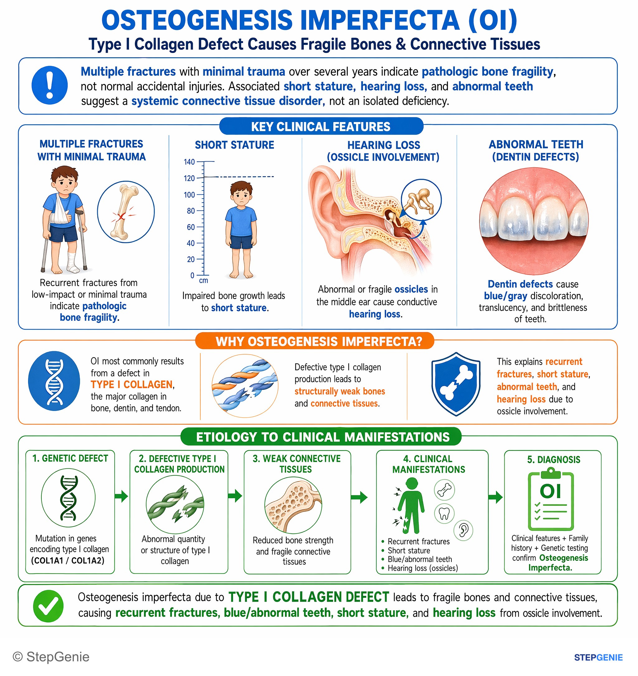
Osteogenesis imperfecta from type I collagen defect → recurrent fractures, blue/abnormal teeth, short stature, and hearing loss due to…
USMLE Step 1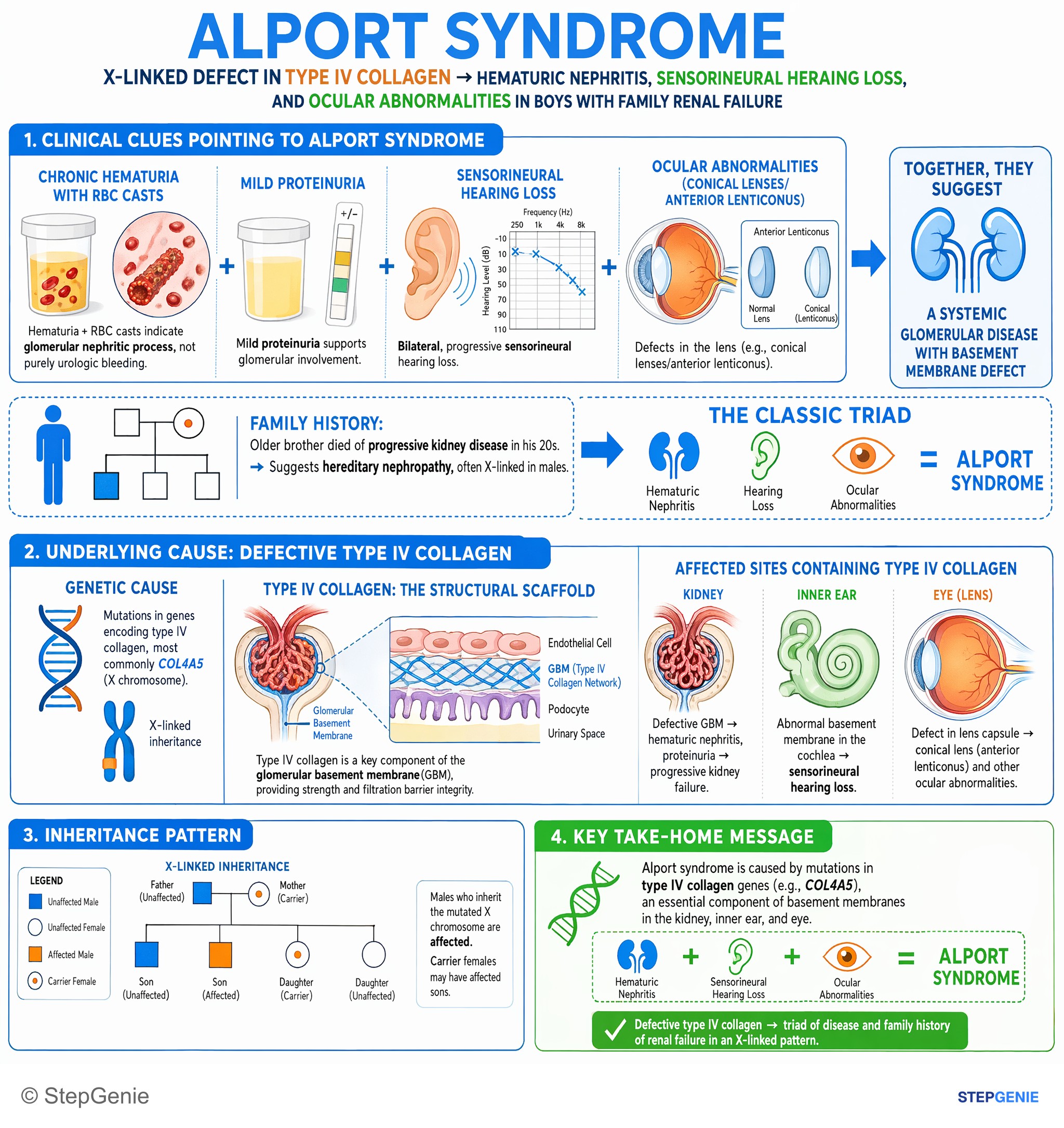
Alport syndrome: X-linked defect in type IV collagen → hematuric nephritis, sensorineural hearing loss, and ocular abnormalities in boys…
USMLE Step 1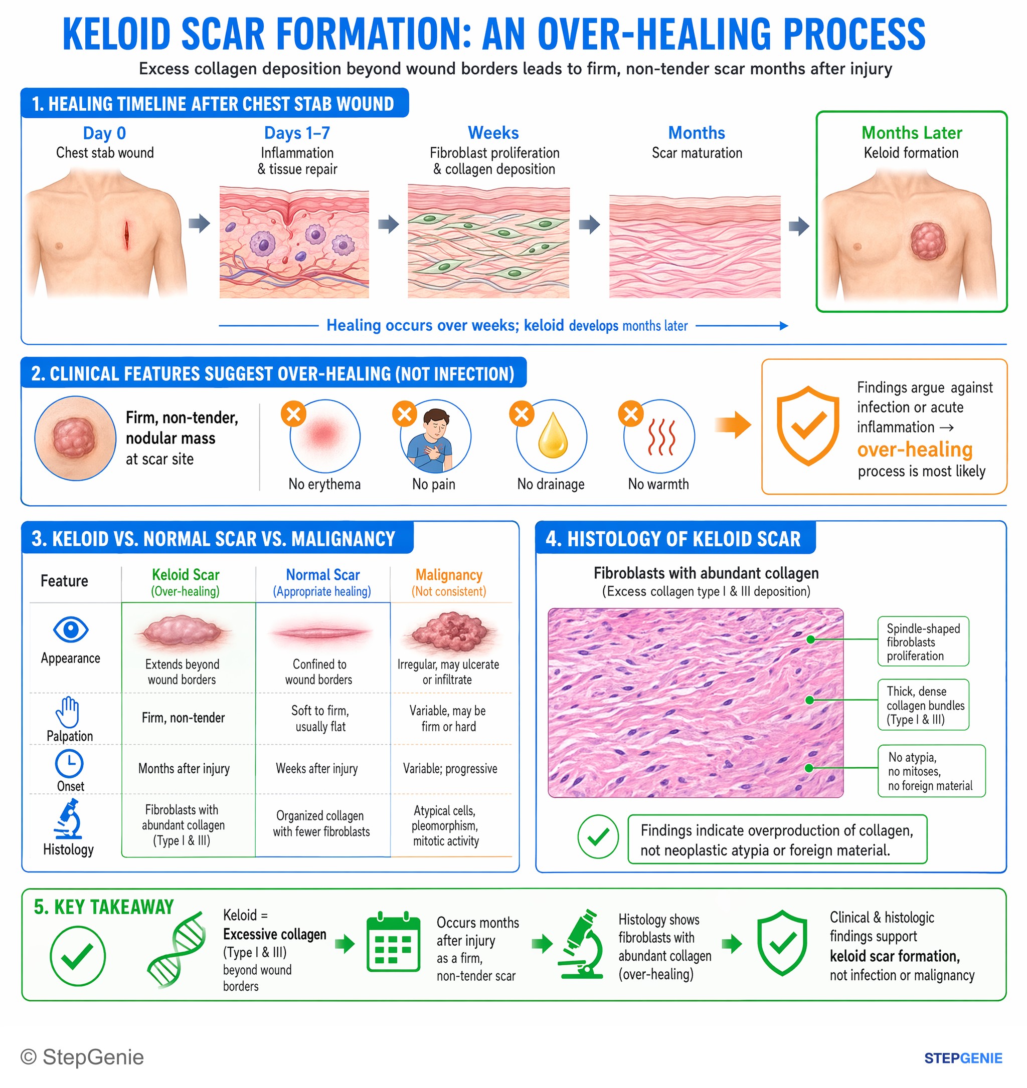
Keloid = excessive collagen (type I & III) beyond wound borders → firm, non-tender scar months after injury; histology shows fibroblasts…
USMLE Step 1Frequently Asked Questions
What are collagen types I, II, III and IV, and where is each found?
Type I is the tension-resisting collagen of bone, skin, tendon, ligament, dentin, cornea and mature scar, and it makes up over 90% of the body's collagen. Type II resists compression in hyaline cartilage, intervertebral discs and the vitreous of the eye. Type III forms reticular fibers and the stretchy walls of blood vessels, bowel and uterus, and it is the first collagen laid down in granulation tissue. Type IV forms the sheet-like network of basement membranes, including the glomerulus, inner ear and lens.
What is an easy way to remember collagen types I to IV?
Count from one to four: one rope, two knees, three tubes, four on the floor. Type I is the rope that resists pulling in bone, skin and tendon. Type II sits in the two knees and two eyes, meaning cartilage and vitreous. Type III lines the three tubes that rupture in vascular Ehlers-Danlos syndrome: arteries, bowel and uterus. Type IV is the floor, the basement membrane under the kidney filter, inner ear and lens.
Which collagen is defective in Ehlers-Danlos syndrome?
It depends on the type. Vascular Ehlers-Danlos syndrome is caused by COL3A1 variants affecting type III collagen and leads to arterial, bowel and uterine rupture. Classical Ehlers-Danlos syndrome is usually caused by COL5A1 or COL5A2 variants affecting type V collagen, with hyperextensible skin, atrophic scars and joint hypermobility. Hypermobile Ehlers-Danlos syndrome, the most common type, has no known gene. Rarer types involve type I collagen or collagen-processing enzymes.
What is the difference between Alport syndrome and Goodpasture syndrome?
Both involve type IV collagen in basement membranes. Alport syndrome is inherited, most often X-linked through COL4A5, and causes hematuria that progresses to kidney failure, sensorineural hearing loss and eye findings such as anterior lenticonus. Goodpasture (anti-GBM) disease is autoimmune: antibodies against the alpha-3 chain of type IV collagen cause rapidly progressive glomerulonephritis and pulmonary hemorrhage, with linear IgG along the glomerular basement membrane on immunofluorescence.
Which collagen type is in granulation tissue, and which is in a mature scar?
Granulation tissue is rich in type III collagen, the thinner collagen that fibroblasts lay down first during repair. As the wound matures, type I collagen replaces type III and the scar forms. A healed wound regains only about 80% of its original strength by around 3 months and never returns to full strength, which is why a scar stays weaker than intact skin.
Is achondroplasia caused by a type II collagen defect?
No. Achondroplasia is caused by variants in FGFR3 that make the receptor overly active, which disturbs the conversion of cartilage to bone in the limbs. About 80% of cases are new variants in children of average-height parents. Type II collagen is the collagen of cartilage and vitreous, and its gene, COL2A1, causes different disorders such as Stickler syndrome and spondyloepiphyseal dysplasia congenita.