Collagen synthesis happens in two halves. Inside the fibroblast, pre-pro-α chains built on Gly-X-Y repeats are made on the rough ER, where proline and lysine are hydroxylated (vitamin C), some hydroxylysines are glycosylated, and three chains wind into a triple-helical procollagen that the Golgi packages and secretes. Outside the cell, peptidases cut off the propeptides to make tropocollagen, which self-assembles into fibrils that copper-dependent lysyl oxidase cross-links. Scurvy breaks hydroxylation, osteogenesis imperfecta lowers type I chain output or breaks the helix, a few Ehlers-Danlos types break processing or fibril assembly, and Menkes disease breaks cross-linking.
Collagen synthesis steps: the map
| # | Step | Where | Needs | Fails in |
|---|---|---|---|---|
| 1 | Pre-pro-α chains translated; glycine at every third residue (Gly-X-Y) | Ribosomes on the RER (genes transcribed in the nucleus) | Intact collagen genes | Osteogenesis imperfecta: too little type I chain, or glycine replaced by a bulkier residue |
| 2 | Hydroxylation of selected proline and lysine residues | RER lumen | Prolyl and lysyl hydroxylases + vitamin C | Scurvy; kyphoscoliotic EDS (PLOD1 = lysyl hydroxylase 1) |
| 3 | Glycosylation of some hydroxylysines (glucose, galactose) | RER | Hydroxylysine from step 2 | Rarely the tested defect |
| 4 | Three chains zip into a triple helix = procollagen | RER | Hydroxyproline and glycine; propeptides guide assembly | OI (glycine substitutions); vascular EDS (COL3A1) |
| 5 | Packaging and exocytosis | Golgi → secretory vesicles | Intact secretory pathway | Rarely tested |
| 6 | Propeptides cleaved → tropocollagen | Extracellular | Procollagen peptidases (for example ADAMTS2, the procollagen I N-proteinase) | Dermatosparaxis EDS (ADAMTS2); arthrochalasia EDS (COL1A1/COL1A2) |
| 7 | Tropocollagen self-assembles into fibrils | Extracellular | Type V collagen sets type I fibril diameter | Classical EDS (COL5A1/COL5A2) |
| 8 | Cross-linking of lysine and hydroxylysine on adjacent molecules | Extracellular | Lysyl oxidase + copper | Menkes disease (ATP7A); acquired copper deficiency |
Inside vs outside in one line: steps 1-5 are intracellular (RER, then Golgi); steps 6-8 are extracellular.
Why cut the propeptides outside? The propeptides guide the three chains into a helix, and because they come off only after secretion, they stop large collagen fibrils from forming inside the cell, which would be catastrophic for it. Basement-membrane (type IV) collagen is not cleaved after secretion at all; it forms a network instead of fibrils.
Exam lore vs current sources: some review tables put glycosylation in the Golgi or say "problems with cleavage cause EDS." StatPearls (2026) and the Alberts cell biology text place hydroxylation, glycosylation and helix formation in the ER, and only the rare dermatosparaxis and arthrochalasia types of EDS are true processing defects; classical EDS is a type V fibril problem.
Question 1: the child who stopped walking
A 7-year-old boy is brought to the clinic because he has refused to walk for 2 weeks and cries when his legs are moved. His gums are swollen and bleed easily, and his legs show perifollicular hemorrhages and coiled, corkscrew hairs. For the past year he has eaten only plain pasta, white bread and milk. His hemoglobin is low. Which step of collagen synthesis is most directly impaired?
A. Cleavage of N- and C-terminal propeptides in the extracellular space B. Hydroxylation of proline and lysine residues in the rough ER C. Glycosylation of hydroxylysine residues in the Golgi apparatus D. Triple-helix formation of procollagen in the rough ER E. Cross-linking of collagen fibrils by lysyl oxidase
Answer: B. This is scurvy. Vitamin C is the cofactor for prolyl and lysyl hydroxylases in the rough ER; without it, the pro-α chains that are made cannot form a stable triple helix and are degraded inside the cell. As older normal collagen is lost, vessels become fragile (bleeding gums, perifollicular and subperiosteal hemorrhages) and wounds heal poorly. Children typically have bone pain, and X-rays can show the white line of Fraenkel at the metaphyses. Signs can appear within a month of intake below about 10 mg/day, and oral vitamin C reverses them. The adult version of this stem (a man with alcohol use disorder, bleeding gums, corkscrew hairs and a cut that will not heal) has the same answer. Full workup and mimics: scurvy: vitamin C, collagen hydroxylation and mimics.
- A, propeptide cleavage: a real extracellular step, but it needs no vitamin C. Its defects are rare inherited EDS types: dermatosparaxis (ADAMTS2: soft, fragile, sagging skin) and arthrochalasia (COL1A1/COL1A2: severe hypermobility with both hips dislocated at birth).
- C, glycosylation "in the Golgi": wrong compartment (glycosylation happens in the ER) and no vitamin C requirement; it is almost never the tested defect.
- D, triple-helix formation: it does fail in scurvy, but only as a consequence of missing hydroxylation. "Most directly impaired" asks for the step that lost its cofactor. The helix is the answer when glycine is substituted, as in osteogenesis imperfecta.
- E, cross-linking: lysyl oxidase needs copper, not vitamin C. Pick it for Menkes disease or acquired copper deficiency (Question 2).
Question 2: the infant with kinky hair
A 3-month-old boy has hypotonia, seizures and poor weight gain. His hair is sparse, pale and twisted (pili torti under the microscope), his skin is loose, and ultrasound shows bladder diverticula. Serum copper and ceruloplasmin are low. Decreased activity of which enzyme best explains his loose skin and bladder diverticula?
A. Dopamine β-hydroxylase B. Lysyl oxidase C. Prolyl hydroxylase D. ADAMTS2 (procollagen peptidase) E. Tyrosinase
Answer: B. This is Menkes disease: X-linked recessive variants in ATP7A, a copper-transport protein, leave the brain and other tissues short of copper, so copper-dependent enzymes fail. Lysyl oxidase cross-links collagen and elastin outside the cell; without it, connective tissue is weak, giving loose skin and joints, bladder diverticula, fragile tortuous vessels and bone abnormalities. Children with the classic form often do not live past age 3, although copper injections started early can improve outcomes.
- A, dopamine β-hydroxylase: also copper-dependent and also low in Menkes, but it makes norepinephrine; its deficit shows up as abnormal plasma catecholamines, not lax connective tissue.
- C, prolyl hydroxylase: needs vitamin C (scurvy), not copper; serum copper would be normal.
- D, ADAMTS2: its loss causes dermatosparaxis EDS (autosomal recessive, very fragile skin) with normal copper and no neurodegeneration.
- E, tyrosinase: a copper enzyme of melanin synthesis whose deficit explains the hypopigmentation, not the loose skin or diverticula.
- Don't swap the copper diseases: Menkes is ATP7A (X-linked, too little copper reaching tissues); Wilson disease is ATP7B (autosomal recessive, copper builds up in liver, brain and cornea, with Kayser-Fleischer rings). Compare them in the copper deficiency mnemonic and the Wilson disease question. Acquired copper deficiency (excess zinc, bariatric surgery, malabsorption, unsupplemented TPN) shows up mainly as anemia, neutropenia and a B12-like myeloneuropathy.
Which disease breaks which step
| Disease | Cause | Broken step | Classic clue |
|---|---|---|---|
| Scurvy | Dietary vitamin C deficiency | Hydroxylation (RER) | Bleeding gums, perifollicular hemorrhages, corkscrew hairs, poor wound healing |
| Kyphoscoliotic EDS | PLOD1 (lysyl hydroxylase 1); autosomal recessive | Lysine hydroxylation | Hypotonia, progressive kyphoscoliosis, joint hypermobility |
| Osteogenesis imperfecta | COL1A1/COL1A2; usually autosomal dominant | Chain amount, or triple helix (glycine substitutions) | Fractures, blue sclerae, hearing loss, dentinogenesis imperfecta |
| Vascular EDS | COL3A1; autosomal dominant | Type III chains and helix | Arterial, bowel or uterine rupture |
| Dermatosparaxis EDS | ADAMTS2; autosomal recessive | Propeptide cleavage | Soft, fragile, sagging skin; easy bruising |
| Arthrochalasia EDS | COL1A1/COL1A2; autosomal dominant | Processing of type I procollagen | Severe hypermobility; both hips dislocated at birth |
| Classical EDS | COL5A1/COL5A2; autosomal dominant | Fibril assembly (type V sets fibril diameter) | Hyperextensible skin, atrophic scars, joint hypermobility |
| Menkes disease | ATP7A; X-linked recessive | Cross-linking (lysyl oxidase) | Kinky hair, neurodegeneration, loose skin, bladder diverticula |
OI types and management live in the osteogenesis imperfecta guide, every EDS subtype in the Ehlers-Danlos question, and the tissue side of each defect in collagen types I-IV made easy.
How USMLE tests it
- "Most directly impaired" = the enzyme step that lost its cofactor, not the downstream damage (unstable helix, weak fibrils).
- Location traps: nucleus = transcription only; RER = hydroxylation, glycosylation and the triple helix; Golgi = packaging and secretion; extracellular = cleavage, fibril assembly and cross-linking.
- Vitamin C vs copper: vitamin C feeds the hydroxylases inside the cell; copper feeds lysyl oxidase outside it.
- Glycine is the OI amino acid: it is the smallest residue, and sitting at every third position lets three chains pack tightly; a bulkier substitute buckles the helix.
- Procollagen vs tropocollagen: procollagen still carries its propeptides; tropocollagen is what is left after they are cut off, ready to build fibrils.
- Lysyl oxidase cross-links elastin too, which is why Menkes arteries are tortuous. Marfan syndrome (fibrillin-1) is not a collagen-synthesis step.
Key takeaways
- Inside the cell (RER → Golgi): translation, hydroxylation (vitamin C), glycosylation, triple helix (procollagen), secretion.
- Outside the cell: propeptide cleavage (tropocollagen), fibril assembly, cross-linking by lysyl oxidase (copper).
- Scurvy = hydroxylation: under-hydroxylated chains cannot form a stable helix and are degraded.
- Menkes (ATP7A, X-linked) = cross-linking: kinky hair, neurodegeneration, loose skin, bladder diverticula, low copper and ceruloplasmin.
- OI = too little type I collagen or glycine substitutions; vascular EDS = COL3A1; classical EDS = COL5A1/COL5A2.
- Only the rare dermatosparaxis (ADAMTS2) and arthrochalasia types of EDS are true processing (cleavage) defects.
- For "most directly impaired," choose the step that lost its cofactor.
Sources
- Biochemistry, Collagen Synthesis — StatPearls, NCBI Bookshelf (2026)
- The Extracellular Matrix of Animals — Molecular Biology of the Cell, 4th ed., NCBI Bookshelf (2002)
- Vitamin C Deficiency — Merck Manual Professional Edition (2026)
- Vitamin C: Fact Sheet for Health Professionals — NIH Office of Dietary Supplements (2025)
- ATP7A-Related Copper Transport Disorders — GeneReviews, NCBI Bookshelf (2021)
- Copper Deficiency — Merck Manual Professional Edition (2025)
- Menkes Disease — StatPearls, NCBI Bookshelf (2023)
- Ehlers-Danlos syndrome — MedlinePlus Genetics (2022)
- ADAMTS2 gene — MedlinePlus Genetics (2017)
- PLOD1 gene — MedlinePlus Genetics (2017)
- COL1A2 gene — MedlinePlus Genetics (2019)
- Classic Ehlers-Danlos Syndrome — GeneReviews, NCBI Bookshelf (2024)
Visual explanations from the StepGenie QBank
Infographics StepGenie drew to explain questions in its own QBank, matched to this article's topic. Each caption is the takeaway of the question it explains.
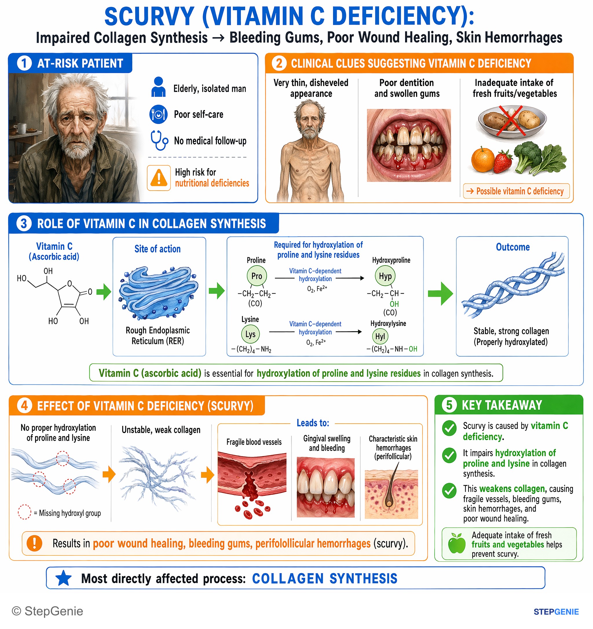
Scurvy (vitamin C deficiency) impairs hydroxylation of proline and lysine in collagen, causing poor wound healing, bleeding gums, and…
USMLE Step 1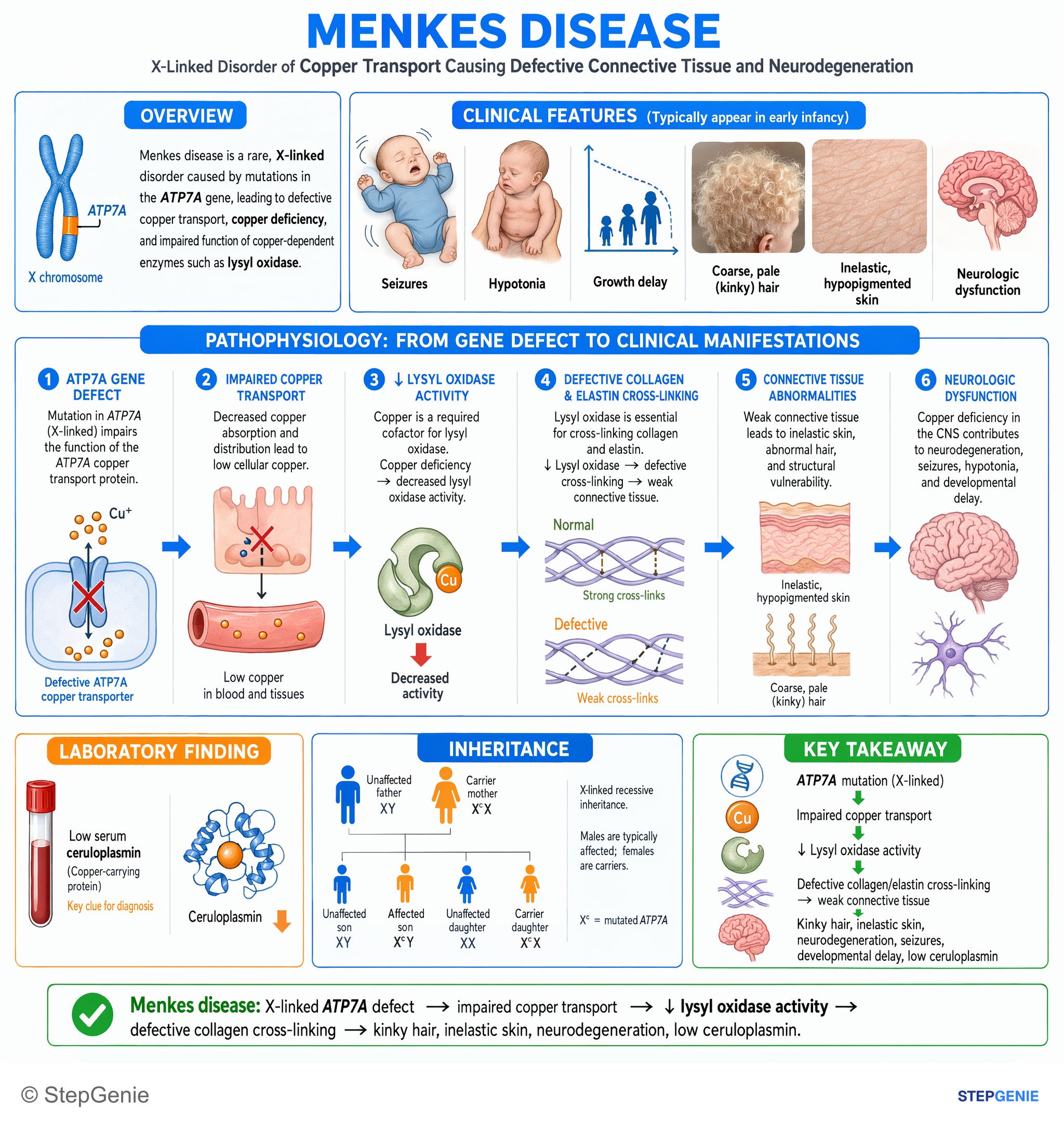
Menkes disease: X-linked ATP7A defect → impaired copper transport → ↓ lysyl oxidase activity → defective collagen cross-linking, kinky…
USMLE Step 1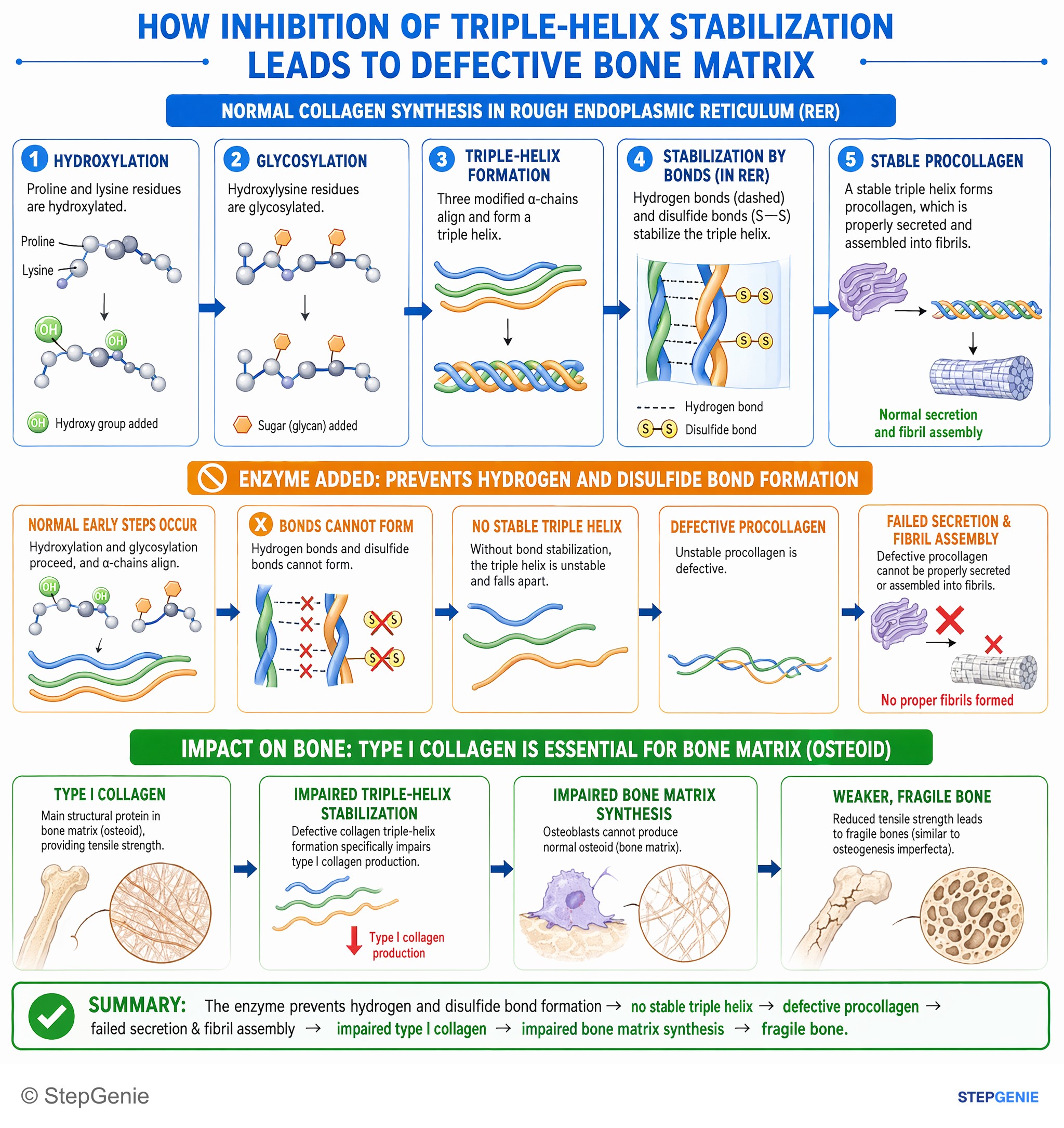
Defective collagen triple-helix formation impairs type I collagen production, leading to impaired bone matrix synthesis (similar to…
USMLE Step 1Frequently Asked Questions
What are the steps of collagen synthesis in order?
Collagen synthesis starts in the rough ER, where glycine-rich pre-pro-alpha chains are translated, selected proline and lysine residues are hydroxylated with vitamin C as a cofactor, some hydroxylysines are glycosylated, and three chains form a triple-helical procollagen. The Golgi packages and secretes procollagen. Outside the cell, peptidases remove the end propeptides to make tropocollagen, which self-assembles into fibrils that lysyl oxidase, a copper-dependent enzyme, strengthens with covalent cross-links.
Which steps of collagen synthesis are intracellular and which are extracellular?
Translation of the pro-alpha chains, hydroxylation of proline and lysine, glycosylation of hydroxylysine and formation of the triple helix all happen inside the cell in the rough endoplasmic reticulum, followed by packaging and secretion through the Golgi. Cleavage of the propeptides to form tropocollagen, assembly of fibrils and cross-linking by lysyl oxidase happen outside the cell. Removing the propeptides only after secretion prevents large fibrils from forming inside the fibroblast.
Which step of collagen synthesis is impaired in scurvy?
Scurvy most directly impairs hydroxylation of proline and lysine residues in the rough endoplasmic reticulum, because vitamin C is the cofactor for prolyl and lysyl hydroxylases. The chains that are made cannot form a stable triple helix and are degraded inside the cell, so collagen is gradually lost. The result is bleeding gums, perifollicular hemorrhages, corkscrew hairs, easy bruising and poor wound healing, and in children bone pain and refusal to walk.
What does copper do in collagen synthesis, and what goes wrong in Menkes disease?
Copper is the cofactor for lysyl oxidase, the extracellular enzyme that forms covalent cross-links between lysine and hydroxylysine residues of neighboring collagen molecules; it also cross-links elastin. In Menkes disease, X-linked recessive variants in the ATP7A copper-transport gene leave copper enzymes underactive. Weak cross-linking explains loose skin, bladder diverticula and tortuous arteries, alongside sparse kinky hair, neurodegeneration, and low serum copper and ceruloplasmin.
What is the difference between procollagen and tropocollagen?
Procollagen is the triple-helical molecule assembled in the rough endoplasmic reticulum and secreted through the Golgi. It still carries propeptides at both ends, which help the chains align and stop fibrils from forming inside the cell. Tropocollagen is the molecule left after peptidases cut off those propeptides outside the cell. Tropocollagen molecules then self-assemble into fibrils, which lysyl oxidase cross-links into strong collagen fibers.
Which types of Ehlers-Danlos syndrome are defects in collagen processing?
Only a few are true processing defects. Dermatosparaxis Ehlers-Danlos syndrome, caused by ADAMTS2 variants, prevents normal trimming of procollagen and produces very fragile, sagging skin. Arthrochalasia Ehlers-Danlos syndrome, caused by COL1A1 or COL1A2 variants that remove a segment needed for processing, causes severe joint hypermobility with dislocated hips at birth. Kyphoscoliotic Ehlers-Danlos syndrome is a lysyl hydroxylase (PLOD1) defect, classical type involves type V collagen, and vascular type involves type III.