Cutaneous squamous cell carcinoma (SCC) is a malignant tumor of epidermal keratinocytes that invades the dermis. It is the second most common skin cancer after basal cell carcinoma (BCC) and the one most tied to cumulative sun damage: the classic stem is an older, fair-skinned patient with a firm, scaly or crusted, often tender papule or plaque that may ulcerate or bleed, on sun-exposed skin such as the face, ears, lip or back of the hand, usually among actinic keratoses. SCC is more likely than BCC to metastasize (3.7% over 10 years in one cohort study, mostly to regional lymph nodes). Diagnosis is a biopsy that reaches the dermis; treatment is excision with 4-6 mm margins for low-risk lesions or Mohs surgery for high-risk ones, with PD-1 or PD-L1 inhibitors for advanced disease.
This page covers SCC of the skin. Squamous cell carcinomas of other organs are covered elsewhere, for example esophageal SCC vs adenocarcinoma and HPV and cervical cancer.
The case
A 72-year-old man who worked outdoors for decades as a golf course groundskeeper has a lesion on the back of his right hand that has slowly enlarged over 6 months. It is a 1.5-cm red, scaly, firm (indurated) plaque with a central crust; it is tender and bleeds when scraped. Several rough, gritty, pink papules on his forehead and balding scalp are easier to feel than to see. There is no lymphadenopathy.
What is the most likely diagnosis of the lesion on his hand?
A. Basal cell carcinoma B. Squamous cell carcinoma C. Seborrheic keratosis D. Keratoacanthoma E. Melanoma F. Actinic keratosis
Answer: B, squamous cell carcinoma
| Clue in the stem | What it tells you |
|---|---|
| Older man, decades outdoors | Cumulative UV damage, which is more strongly linked to SCC than to BCC |
| Back of the hand | Sun-exposed skin; the hands are also a high-risk location for SCC |
| Slow growth over months | SCC grows over weeks to months; a keratoacanthoma typically appears within a few weeks |
| Red, scaly, firm, crusted, bleeds | A keratinizing tumor invading the dermis; it may become nodular, hyperkeratotic or ulcerated |
| Tender | SCC is often tender or painful |
| Rough papules on the forehead and scalp | Actinic keratoses: precursor lesions and a sign of widespread sun damage |
Pathology: nests of atypical squamous cells with abundant eosinophilic cytoplasm extend from the epidermis into the dermis, often forming keratin pearls. When the atypical cells stay within the epidermis, it is SCC in situ (Bowen disease).
Next step: a biopsy that extends into the dermis. Because the hand is a high-risk site, Mohs surgery would be preferred if SCC is confirmed.
Why every other choice is wrong
A. Basal cell carcinoma. The most common skin cancer (about 2 million US cases a year). It starts as a shiny papule that slowly develops a pearly border with telangiectases and a central dell or ulcer; 80% arise on the head and neck. Most BCCs are driven by Hedgehog pathway activation, usually loss-of-function variants in PTCH1, and intermittent intense UV exposure (especially in childhood) is the main environmental risk. Metastasis is rare, but local growth can be highly destructive. Wrong here: the lesion is scaly, crusted and keratotic, not pearly and telangiectatic. See the basal cell carcinoma breakdown.
C. Seborrheic keratosis. A benign, "stuck-on" growth with a verrucous, velvety, waxy, scaling or crusted surface in flesh, brown or black tones, usually on the trunk and temples from middle age onward. It is not premalignant. Large, multiple or rapidly appearing seborrheic keratoses (the Leser-Trélat sign) can be a paraneoplastic clue to an internal cancer, such as a gastrointestinal malignancy or lymphoma (see paraneoplastic syndromes). Wrong here: an indurated, bleeding lesion growing on sun-damaged skin is not a stuck-on plaque.
D. Keratoacanthoma. A sharply demarcated, firm nodule with a central keratin plug that grows rapidly, within a few weeks, to 1-2 cm, usually on the sun-exposed face or arms of people over 60. It often regresses on its own over 4-6 months, leaving a depressed scar, but it can be indistinguishable from well-differentiated SCC (Merck calls keratoacanthomas "probably squamous cell carcinomas themselves"), so a keratoacanthoma-like lesion is usually excised and managed as SCC. Wrong here: 6 months of slow growth and no central keratin plug.
E. Melanoma. A pigmented lesion recognized by ABCDE: asymmetry, irregular borders, color variation, diameter over 6 mm and evolution. Its sun-exposure risk centers on repeated blistering sunburns, especially in childhood or adolescence (SCC tracks cumulative exposure), and its most common type, superficial spreading melanoma, favors the legs in women and the trunk in men. Breslow thickness, with ulceration, sets the T stage, and stage drives prognosis. Wrong here: a nonpigmented, scaly, keratotic lesion with no ABCDE features. See the melanoma ABCDE breakdown.
F. Actinic keratosis. Pink or red (occasionally gray or brown) papules with adherent scale that are often easier to feel than to see, on the balding scalp, face, lateral neck and distal arms and legs. They are precursors of SCC; estimates of progression for any single lesion range from under 1% to 10%, and most never become SCC. Wrong here: the forehead papules are actinic keratoses, but the hand lesion is indurated, larger than 1 cm and bleeding, exactly the atypical features that call for a biopsy to exclude SCC.
SCC and its look-alikes at a glance
| Lesion | Typical look | Key clue | Behavior |
|---|---|---|---|
| SCC | Red, scaly or crusted papule or plaque; may turn nodular, hyperkeratotic or ulcerated | Sun-damaged skin among actinic keratoses; often tender | Metastasized in 3.7% over 10 years in one cohort |
| BCC | Shiny, pearly papule with telangiectases and a central dell or ulcer | Head and neck in 80% | Metastasis rare; locally destructive |
| Actinic keratosis | Pink-red papule with adherent scale, felt more than seen | Balding scalp, face, distal limbs | Premalignant; under 1% to 10% per lesion progress |
| Keratoacanthoma | Firm nodule with a central keratin plug | Reaches 1-2 cm within weeks | Often regresses in 4-6 months; managed as SCC |
| Seborrheic keratosis | Stuck-on, waxy or verrucous; flesh, brown or black | Trunk and temples; middle age onward | Not premalignant |
| Melanoma | Pigmented lesion with ABCDE features | Blistering sunburns, changing mole | Breslow thickness and ulceration set the T stage |
Risk factors and high-risk features
- Cumulative UV exposure and light skin: people with lightly pigmented skin are far more susceptible.
- Immunosuppression: organ transplant recipients on immunosuppressive drugs have a higher risk of skin cancer, particularly SCC. In people who are not immunocompromised, BCC is at least 3 times more common than SCC.
- Precursors and settings: actinic keratosis, SCC in situ (Bowen disease), and chronic wounds or burn scars; SCC arising in a scar or chronic ulcer is a Marjolin ulcer.
- Other exposures: arsenic, therapeutic or ionizing radiation, certain beta-HPV types, smoking and xeroderma pigmentosum.
- High-risk tumors (Merck, DermNet): diameter over 2 cm, invasion deeper than 2 mm, perineural invasion, poor differentiation, location on the ear, lip vermilion, central face, hands, feet or genitalia, and tumors in immunosuppressed patients.
- Spread: when SCC metastasizes, regional lymph nodes are involved in 80%; lungs, liver, brain, bone and skin are other sites. SCCs that arise from actinic keratoses on sun-exposed skin are less likely to metastasize than SCCs arising de novo or on non-sun-exposed skin.
Management
- Biopsy that extends into the dermis.
- Low-risk lesions: standard excision with 4-6 mm clear margins. Curettage and electrodesiccation, cryosurgery, topical imiquimod or 5-fluorouracil and photodynamic therapy are other options Merck lists.
- High-risk lesions: Mohs micrographic surgery is preferred (or wider excision), and recurrences and large tumors are treated aggressively with Mohs. SCC on the lip or another mucocutaneous junction should be excised.
- Radiation: an adjunct to surgery or for nonsurgical candidates, including positive margins, recurrence or extensive perineural invasion.
- Advanced or metastatic disease: checkpoint inhibitors, namely the PD-1 inhibitors cemiplimab and pembrolizumab and the PD-L1 inhibitor cosibelimab (approved 2024) for disease that cannot be cured by surgery or radiation. Since 2025, cemiplimab is also approved as adjuvant therapy for SCC at high risk of recurrence after surgery and radiation. Cetuximab (anti-EGFR) has been moderately effective with surgery or radiation.
- Prevention: sun avoidance, protective clothing, broad-spectrum sunscreen and treatment of actinic keratoses (cryosurgery, or field therapy with 5-fluorouracil, imiquimod, diclofenac, tirbanibulin or photodynamic therapy). Oral retinoids such as acitretin are used to reduce new SCCs in patients who keep developing them.
- Prognosis: the 5-year survival for cutaneous SCC is about 98% overall, and lower with high-risk features.
How USMLE tests it
- Scaly, crusted, indurated, tender lesion on the hand, ear or lip of an older outdoor worker, among actinic keratoses: SCC.
- Pearly papule with telangiectases on the face: BCC, which rarely metastasizes.
- Nodule with a central keratin plug that grew within weeks: keratoacanthoma, excised and treated like SCC.
- Transplant recipient with multiple skin cancers: think SCC.
- Nonhealing ulcer in an old burn scar: Marjolin ulcer (SCC); biopsy it.
- Sudden crop of seborrheic keratoses: Leser-Trélat sign; look for an internal cancer.
- Biopsy showing keratin pearls: squamous differentiation.
- Atypical actinic keratosis (indurated, over 1 cm, rapidly enlarging, bleeding or ulcerated): biopsy to exclude SCC.
Key takeaways
- Cutaneous SCC = malignant keratinocytes invading the dermis, the second most common skin cancer, driven by cumulative UV.
- Look for a scaly, crusted, indurated, often tender lesion on sun-exposed skin with actinic keratoses nearby.
- Keratin pearls on histology; Bowen disease is SCC in situ.
- Metastasizes more than BCC (3.7% over 10 years in one cohort), mainly to regional nodes; transplant patients are at particular risk.
- Biopsy into the dermis, then excision with 4-6 mm margins (low risk) or Mohs (high risk); checkpoint inhibitors for advanced disease.
- Keratoacanthoma grows within weeks and is managed as SCC; seborrheic keratosis is benign; melanoma follows ABCDE.
Sources
- Squamous Cell Carcinoma — Merck Manual Professional Edition (2026)
- Skin Cancer Treatment (PDQ), Health Professional Version — National Cancer Institute (2025)
- Actinic Keratoses — Merck Manual Professional Edition (2026)
- Basal Cell Carcinoma — Merck Manual Professional Edition (2026)
- Seborrheic Keratoses — Merck Manual Professional Edition (2025)
- Melanoma — Merck Manual Professional Edition (2026)
- Cutaneous Squamous Cell Carcinoma — DermNet (2015)
- Keratoacanthoma — DermNet (2021)
- Squamous Cell Carcinoma Pathology — DermNet (2014)
- Libtayo (cemiplimab-rwlc) injection: prescribing information — DailyMed (2026)
- Unloxcyt (cosibelimab-ipdl) injection: prescribing information — DailyMed (2026)
- Keytruda (pembrolizumab) injection: prescribing information — DailyMed (2026)
Visual explanations from the StepGenie QBank
Infographics StepGenie drew to explain questions in its own QBank, matched to this article's topic. Each caption is the takeaway of the question it explains.
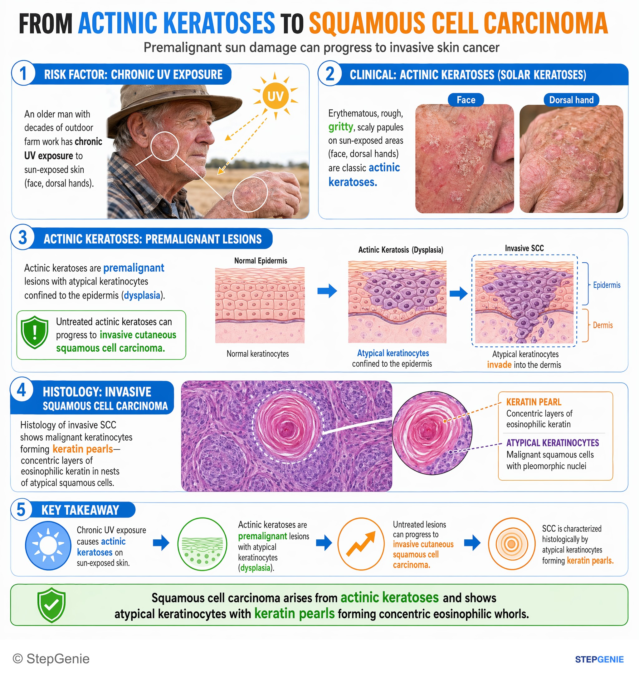
Squamous cell carcinoma arises from actinic keratoses and shows atypical keratinocytes with keratin pearls forming concentric eosinophilic…
USMLE Step 1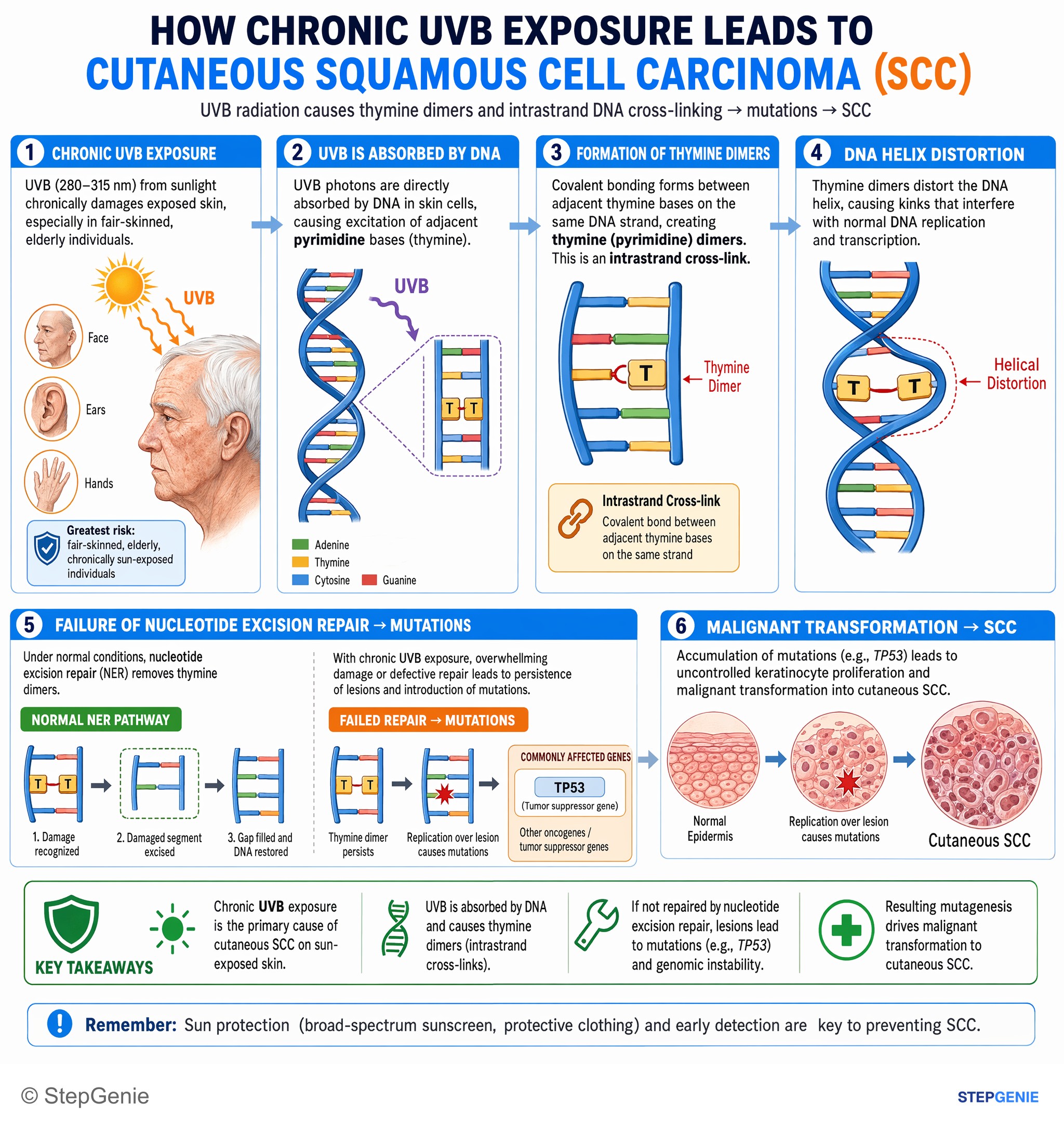
Cutaneous squamous cell carcinoma from chronic UVB exposure involves pyrimidine (thymine) dimers causing intrastrand DNA cross-linking and…
USMLE Step 1Frequently Asked Questions
How do you tell squamous cell carcinoma from basal cell carcinoma?
Squamous cell carcinoma usually looks like a red, scaly or crusted, firm papule or plaque that may become hyperkeratotic or ulcerate, is often tender, and arises on sun-damaged skin among actinic keratoses. Basal cell carcinoma is a shiny, pearly papule with telangiectases and a central dell or ulcer, 80% on the head and neck. SCC is linked more to cumulative sun exposure and metastasizes more often (3.7% over 10 years in one cohort study), whereas BCC rarely metastasizes but can be locally destructive.
Does actinic keratosis turn into squamous cell carcinoma?
Actinic keratoses are precursor lesions of squamous cell carcinoma. Estimates of progression for any single lesion range from under 1% to 10%, and most never become cancer; some regress. They are pink or red papules with adherent scale on sun-exposed skin that are often easier to feel than to see. A keratosis that becomes indurated, grows beyond 1 cm, enlarges quickly, bleeds or ulcerates should be biopsied to exclude SCC.
What is the difference between keratoacanthoma and squamous cell carcinoma?
Keratoacanthoma is a firm, sharply demarcated nodule with a central keratin plug that grows rapidly over a few weeks to 1 to 2 cm and often regresses on its own within 4 to 6 months. However, it can be indistinguishable from well-differentiated squamous cell carcinoma, and many consider it a form of SCC, so a keratoacanthoma-like lesion is usually excised and managed as SCC. Slow growth over months favors ordinary SCC.
What does squamous cell carcinoma look like under the microscope?
Invasive cutaneous squamous cell carcinoma shows nests of atypical squamous cells with abundant eosinophilic cytoplasm extending from the epidermis into the dermis, often forming keratin pearls, a sign of squamous differentiation. When the atypical cells stay within the epidermis, the lesion is squamous cell carcinoma in situ, also called Bowen disease or intraepidermal carcinoma. This is why a biopsy for suspected SCC must reach the dermis.
Why do organ transplant recipients get squamous cell carcinoma?
Organ transplant recipients take long-term immunosuppressive drugs, and immunosuppression raises the risk of skin cancer, particularly squamous cell carcinoma. In people who are not immunocompromised, basal cell carcinoma is at least three times more common than SCC, so multiple SCCs in a transplant patient are a classic exam clue. Immunosuppression is also a high-risk feature that makes a tumor more likely to recur or spread.
How is squamous cell carcinoma of the skin treated?
After a biopsy that reaches the dermis, low-risk tumors are usually excised with 4 to 6 mm clear margins. Mohs micrographic surgery is preferred for high-risk tumors, such as those over 2 cm, deeper than 2 mm, with perineural invasion, or on the ear, lip, hands or other high-risk sites. Radiation is used as an adjunct or for nonsurgical candidates. Advanced or metastatic disease is treated with checkpoint inhibitors such as cemiplimab, pembrolizumab or cosibelimab.